|
Cardiac Branches Of The Vagus Nerve
The cardiac branches of the vagus nerve are two sets of nerves found in the upper torso, in close proximity to the larynx. The specific branches are the cervical cardiac branches of vagus nerve and the thoracic cardiac branches of vagus nerve. Cervical cardiac branches of vagus nerve The cervical cardiac branches (sometimes ambiguously called superior cardiac branches) of vagus nerve, two or three in number, arise from the vagus, at the upper and lower parts of the neck. * The upper branches are small, and communicate with the cardiac branches of the sympathetic. They can be traced to the deep part of the cardiac plexus. * The lower branch arises at the root of the neck, just above the first rib. That from the right vagus passes in front or by the side of the innominate artery, and proceeds to the deep part of the cardiac plexus; that from the left runs down across the left side of the aortic arch, and joins the superficial part of the cardiac plexus. Thoracic cardiac branches of ... [...More Info...] [...Related Items...] OR: [Wikipedia] [Google] [Baidu] |
Larynx
The larynx (), commonly called the voice box, is an organ (anatomy), organ in the top of the neck involved in breathing, producing sound and protecting the trachea against food aspiration. The opening of larynx into pharynx known as the laryngeal inlet is about 4–5 centimeters in diameter. The larynx houses the vocal cords, and manipulates pitch (music), pitch and sound pressure, volume, which is essential for phonation. It is situated just below where the tract of the pharynx splits into the trachea and the esophagus. The word 'larynx' (: larynges) comes from the Ancient Greek word ''lárunx'' ʻlarynx, gullet, throatʼ. Structure The triangle-shaped larynx consists largely of cartilages that are attached to one another, and to surrounding structures, by muscles or by fibrous and elastic tissue components. The larynx is lined by a respiratory epithelium, ciliated columnar epithelium except for the vocal folds. The laryngeal cavity, cavity of the larynx extends from its tria ... [...More Info...] [...Related Items...] OR: [Wikipedia] [Google] [Baidu] |
Vagus
The vagus nerve, also known as the tenth cranial nerve (CN X), plays a crucial role in the autonomic nervous system, which is responsible for regulating involuntary functions within the human body. This nerve carries both sensory and motor fibers and serves as a major pathway that connects the brain to various organs, including the heart, lungs, and digestive tract. As a key part of the parasympathetic nervous system, the vagus nerve helps regulate essential involuntary functions like heart rate, breathing, and digestion. By controlling these processes, the vagus nerve contributes to the body's "rest and digest" response, helping to calm the body after stress, lower heart rate, improve digestion, and maintain homeostasis. The vagus nerve consists of two branches: the right and left vagus nerves. In the neck, the right vagus nerve contains approximately 105,000 fibers, while the left vagus nerve has about 87,000 fibers, according to one source. However, other sources report slig ... [...More Info...] [...Related Items...] OR: [Wikipedia] [Google] [Baidu] |
Cardiac Plexus
The cardiac plexus is a plexus of nerves situated at the base of the heart that innervates the heart. Structure The cardiac plexus is divided into a superficial part, which lies in the concavity of the aortic arch, and a deep part, between the aortic arch and the trachea. The two parts are, however, closely connected. The sympathetic component of the cardiac plexus comes from cardiac nerves, which originate from the sympathetic trunk. The parasympathetic component of the cardiac plexus originates from the cardiac branches of the vagus nerve. Superficial part The superficial part of the cardiac plexus lies beneath the aortic arch, in front of the right pulmonary artery. It is formed by the superior cervical cardiac branch of the left sympathetic trunk and the inferior cardiac branch of the left vagus nerve. A small ganglion, the ''cardiac ganglion of Wrisberg'', is occasionally found connected with these nerves at their point of junction. This ganglion, when present, is situate ... [...More Info...] [...Related Items...] OR: [Wikipedia] [Google] [Baidu] |
Innominate Artery
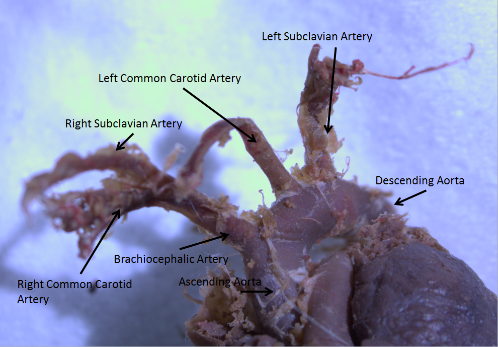
The brachiocephalic artery, brachiocephalic trunk, or innominate artery is an artery of the mediastinum that supplies blood to the right arm, head, and neck. It is the first branch of the aortic arch. Soon after it emerges, the brachiocephalic artery divides into the right common carotid artery and the right subclavian artery. There is no brachiocephalic artery for the left side of the body. The left common carotid artery and the left subclavian artery come directly off the aortic arch. Despite this, there are two brachiocephalic veins. Structure The brachiocephalic artery arises on a level with the upper border of the second right costal cartilage from the start of the aortic arch on a plane anterior to the origin of the left carotid artery. It ascends obliquely upward, backward, and to the right to the level of the upper border of the right sternoclavicular articulation, where it divides into the right common carotid artery and right subclavian arteries. The artery then cros ... [...More Info...] [...Related Items...] OR: [Wikipedia] [Google] [Baidu] |
Aortic Arch
The aortic arch, arch of the aorta, or transverse aortic arch () is the part of the aorta between the ascending and descending aorta. The arch travels backward, so that it ultimately runs to the left of the trachea. Structure The aorta begins at the level of the upper border of the second/third sternocostal articulation of the right side, behind the ventricular outflow tract and pulmonary trunk. The right atrial appendage overlaps it. The first few centimeters of the ascending aorta and pulmonary trunk lies in the same pericardial sheath and runs at first upward, arches over the pulmonary trunk, right pulmonary artery, and right main bronchus to lie behind the right second coastal cartilage. The right lung and sternum lies anterior to the aorta at this point. The aorta then passes posteriorly and to the left, anterior to the trachea, and arches over left main bronchus and left pulmonary artery, and reaches to the left side of the T4 vertebral body. Apart from T4 vertebral ... [...More Info...] [...Related Items...] OR: [Wikipedia] [Google] [Baidu] |
Vertebrate Trachea
The trachea (: tracheae or tracheas), also known as the windpipe, is a cartilaginous tube that connects the larynx to the bronchi of the lungs, allowing the passage of air, and so is present in almost all animals' lungs. The trachea extends from the larynx and branches into the two primary bronchi. At the top of the trachea, the cricoid cartilage attaches it to the larynx. The trachea is formed by a number of horseshoe-shaped rings, joined together vertically by overlying ligaments, and by the trachealis muscle at their ends. The epiglottis closes the opening to the larynx during swallowing. The trachea begins to form in the second month of embryo development, becoming longer and more fixed in its position over time. Its epithelium is lined with column-shaped cells that have hair-like extensions called cilia, with scattered goblet cells that produce protective mucins. The trachea can be affected by inflammation or infection, usually as a result of a viral illness affectin ... [...More Info...] [...Related Items...] OR: [Wikipedia] [Google] [Baidu] |
Vagus Nerve
The vagus nerve, also known as the tenth cranial nerve (CN X), plays a crucial role in the autonomic nervous system, which is responsible for regulating involuntary functions within the human body. This nerve carries both sensory and motor fibers and serves as a major pathway that connects the brain to various organs, including the heart, lungs, and digestive tract. As a key part of the parasympathetic nervous system, the vagus nerve helps regulate essential involuntary functions like heart rate, breathing, and digestion. By controlling these processes, the vagus nerve contributes to the body's "rest and digest" response, helping to calm the body after stress, lower heart rate, improve digestion, and maintain homeostasis. The vagus nerve consists of two branches: the right and left vagus nerves. In the neck, the right vagus nerve contains approximately 105,000 fibers, while the left vagus nerve has about 87,000 fibers, according to one source. However, other sources report sl ... [...More Info...] [...Related Items...] OR: [Wikipedia] [Google] [Baidu] |

