|
Capillary Lamina Of Choroid
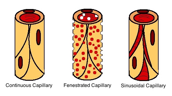
The capillary lamina of choroid or choriocapillaris is a part of the choroid of the eye. It is a layer of capillaries immediately adjacent to Bruch's membrane of the choroid. The choriocapillaris consists of a dense network of freely anastomosing highly permeable fenestrated large-calibre capillaries. It nourishes the outer avascular layers of the retina. Structure Microstructure In the capillaries that compose the choriocapillaris, the fenestrations are densest at the aspect of the capillaries that faces retina, whereas pericytes are situated at the obverse aspect. The choroidal blood vessels can be divided into two categories: the choriocapillaris, and the larger caliber arteries and veins that lie just posterior to the choriocapillaris (these can easily be seen in an albino fundus because there is minimal pigment obscuring the vessels). The choriocapillaris forms a single layer of anastomosing, fenestrated capillaries having wide lumina with most of the fenestrations fac ... [...More Info...] [...Related Items...] OR: [Wikipedia] [Google] [Baidu] |
Choroid
The choroid, also known as the choroidea or choroid coat, is a part of the uvea, the vascular layer of the eye. It contains connective tissues, and lies between the retina and the sclera. The human choroid is thickest at the far extreme rear of the eye (at 0.2 mm), while in the outlying areas it narrows to 0.1 mm. The choroid provides oxygen and nourishment to the outer layers of the retina. Along with the ciliary body and iris, the choroid forms the uveal tract. The structure of the choroid is generally divided into four layers (classified in order of furthest away from the retina to closest): *Haller's layer – outermost layer of the choroid consisting of larger diameter blood vessels; * Sattler's layer – layer of medium diameter blood vessels; * Choriocapillaris – layer of capillaries; and * Bruch's membrane (synonyms: Lamina basalis, Complexus basalis, Lamina vitra) – innermost layer of the choroid. Blood supply There are two circulations of the eye: ... [...More Info...] [...Related Items...] OR: [Wikipedia] [Google] [Baidu] |
Capillary
A capillary is a small blood vessel, from 5 to 10 micrometres in diameter, and is part of the microcirculation system. Capillaries are microvessels and the smallest blood vessels in the body. They are composed of only the tunica intima (the innermost layer of an artery or vein), consisting of a thin wall of simple squamous endothelial cells. They are the site of the exchange of many substances from the surrounding interstitial fluid, and they convey blood from the smallest branches of the arteries (arterioles) to those of the veins (venules). Other substances which cross capillaries include water, oxygen, carbon dioxide, urea, glucose, uric acid, lactic acid and creatinine. Lymph capillaries connect with larger lymph vessels to drain lymphatic fluid collected in microcirculation. Etymology ''Capillary'' comes from the Latin word , meaning "of or resembling hair", with use in English beginning in the mid-17th century. The meaning stems from the tiny, hairlike diameter of a capi ... [...More Info...] [...Related Items...] OR: [Wikipedia] [Google] [Baidu] |
Bruch's Membrane
Bruch's membrane or lamina vitrea is the innermost layer of the choroid of the eye. It is also called the ''vitreous lamina'' or ''Membrane vitriae'', because of its glassy microscopic appearance. It is 2–4 μm thick. Anatomy Structure Bruch's membrane consists of five layers (from inside to outside): #the basement membrane of the retinal pigment epithelium #the inner collagenous zone #a central band of elastic fibers #the outer collagenous zone #the basement membrane of the choriocapillaris Development The membrane grows thicker with age. With age, lipid-containing extracellular deposits may accumulate between the membrane and the basal lamina of the retinal pigmental epithelium, impairing exchange of solutes and contributing to age-related pathology. Embryology Bruch's membrane is present by midterm in fetal development as an elastic sheet. Function The membrane is involved in the regulation of fluid and solute passage from the choroid to the retina. Pathology ... [...More Info...] [...Related Items...] OR: [Wikipedia] [Google] [Baidu] |
Pericyte
Pericytes (formerly called Rouget cells) are multi-functional mural cells of the microcirculation that wrap around the endothelial cells that line the capillaries throughout the body. Pericytes are embedded in the basement membrane of blood capillaries, where they communicate with endothelial cells by means of both direct physical contact and paracrine signaling. The morphology, distribution, density and molecular fingerprints of pericytes vary between organs and vascular beds. Pericytes help in the maintainenance of homeostatic and hemostatic functions in the brain, where one of the organs is characterized with a higher pericyte coverage, and also sustain the blood–brain barrier. These cells are also a key component of the neurovascular unit, which includes endothelial cells, astrocytes, and neurons. Pericytes have been postulated to regulate capillary blood flow and the clearance and phagocytosis of cellular debris ''in vitro.'' Pericytes stabilize and monitor the ma ... [...More Info...] [...Related Items...] OR: [Wikipedia] [Google] [Baidu] |
Photoreceptor Cell
A photoreceptor cell is a specialized type of neuroepithelial cell found in the retina that is capable of visual phototransduction. The great biological importance of photoreceptors is that they convert light (visible electromagnetic radiation) into signals that can stimulate biological processes. To be more specific, photoreceptor proteins in the cell absorb photons, triggering a change in the cell's membrane potential. There are currently three known types of photoreceptor cells in mammalian eyes: rod cell, rods, cone cell, cones, and intrinsically photosensitive retinal ganglion cells. The two classic photoreceptor cells are rods and cones, each contributing information used by the visual system to form an image of the environment, Visual perception, sight. Rods primarily mediate scotopic vision (dim conditions) whereas cones primarily mediate photopic vision (bright conditions), but the processes in each that supports phototransduction is similar. The intrinsically photosen ... [...More Info...] [...Related Items...] OR: [Wikipedia] [Google] [Baidu] |
Macula Lutea
The macula (/ˈmakjʊlə/) or macula lutea is an oval-shaped pigmented area in the center of the retina of the human eye and in other animals. The macula in humans has a diameter of around and is subdivided into the umbo, foveola, foveal avascular zone, fovea, parafovea, and perifovea areas. The anatomical macula at a size of is much larger than the clinical macula which, at a size of , corresponds to the anatomical fovea. The macula is responsible for the central, high-resolution, color vision that is possible in good light. This kind of vision is impaired if the macula is damaged, as in macular degeneration. The clinical macula is seen when viewed from the pupil, as in ophthalmoscopy or retinal photography. The term macula lutea comes from Latin ''macula'', "spot", and ''lutea'', "yellow". Structure The macula is an oval-shaped pigmented area in the center of the retina of the human eye and other animal eyes. Its center is shifted slightly away from the optical ... [...More Info...] [...Related Items...] OR: [Wikipedia] [Google] [Baidu] |
Vitamin A
Vitamin A is a fat-soluble vitamin that is an essential nutrient. The term "vitamin A" encompasses a group of chemically related organic compounds that includes retinol, retinyl esters, and several provitamin (precursor) carotenoids, most notably Β-Carotene, β-carotene (''beta''-carotene). Vitamin A has multiple functions: growth during embryo development, maintaining the immune system, and healthy vision. For aiding vision specifically, it combines with the protein opsin to form rhodopsin, the light-absorbing molecule necessary for both low-light (scotopic vision) and color vision. Vitamin A occurs as two principal forms in foods: A) retinoids, found in Animal source foods, animal-sourced foods, either as retinol or bound to a fatty acid to become a retinyl ester, and B) the carotenoids Α-Carotene, α-carotene (''alpha''-carotene), β-carotene, Γ-Carotene, γ-carotene (''gamma''-carotene), and the xanthophyll beta-cryptoxanthin (all of which contain β-ionone rings) that ... [...More Info...] [...Related Items...] OR: [Wikipedia] [Google] [Baidu] |
Retinal Pigment Epithelium
The pigmented layer of retina or retinal pigment epithelium (RPE) is the pigment A pigment is a powder used to add or alter color or change visual appearance. Pigments are completely or nearly solubility, insoluble and reactivity (chemistry), chemically unreactive in water or another medium; in contrast, dyes are colored sub ...ed cell layer just outside the neurosensory retina that nourishes retinal visual cells, and is firmly attached to the underlying choroid and overlying retinal visual cells. History The RPE was known in the 18th and 19th centuries as the pigmentum nigrum, referring to the observation that the RPE is dark (black in many animals, brown in humans); and as the tapetum nigrum, referring to the observation that in animals with a tapetum lucidum, in the region of the tapetum lucidum the RPE is not pigmented. Anatomy The RPE is composed of a single layer of hexagonal cells that are densely packed with pigment granules. When viewed from the outer surface, ... [...More Info...] [...Related Items...] OR: [Wikipedia] [Google] [Baidu] |
Eschricht
Daniel Frederik Eschricht (18 March 1798 – 22 February 1863) was a Danish zoologist, physiologist, and anatomist known as an authority on whales. He was born in Copenhagen, and studied medicine at Frederiks Hospital, graduating in 1822. He was a student of François Magendie in Paris from 1824-1825, composing a thesis on cranial nerves, after which he studied with prominent European naturalists and anatomists, including Georges Cuvier. He joined the University of Copenhagen in 1829, becoming Professor of Anatomy and Physiology in 1836. The gray whale genus ''Eschrichtius'' was named for him a year after his death. In 1861, Eschricht dissected an orca and found thirteen common porpoises and fourteen seals inside. Jules Verne referred to the incident in the Sargasso chapter of his 1870 novel ''Twenty Thousand Leagues Under the Seas''. He was elected as a member of the American Philosophical Society The American Philosophical Society (APS) is an American scholarly organization ... [...More Info...] [...Related Items...] OR: [Wikipedia] [Google] [Baidu] |
Stewart Duke-Elder
Sir William Stewart Duke-Elder (22 April 1898 – 27 March 1978) was a Scottish ophthalmologist, a dominant force in his field for more than a quarter of a century. Life Duke-Elder was born in the manse in Tealing near Dundee. His father, Rev Neil Stewart Elder, was the village minister of the Free Church of Scotland. His mother was Isabelle Duke, daughter of Rev John Duke of the Free Church in Campsie, Stirlingshire. Duke-Elder was educated at Morgan Academy in Dundee, and was school dux for 1914–1915. Duke-Elder entered the University of St Andrews in 1915 on scholarship, and graduated in 1919 with a BSc in Physiology and MA (Hons) in Natural Sciences. He graduated from the University of St Andrews School of Medicine in 1923 with an MB ChB. In 1925, he earned an MD from St Andrews for his dissertation on 'Reaction of the eye to changes in osmotic pressure of the blood'. In 1927, Duke-Elder earned a DSc from St Andrews for his thesis on "The nature of the intrao ... [...More Info...] [...Related Items...] OR: [Wikipedia] [Google] [Baidu] |
Angiology
Angiology (from Greek , ''angeīon'', "vessel"; and , ''-logia'') is the medical specialty dedicated to studying the circulatory system and of the lymphatic system, i.e., arteries, veins and lymphatic vessels. In the UK, this field is more often termed ''angiology'', and in the United States the term vascular medicine is more frequent. The field of vascular medicine (angiology) is the field that deals with preventing, diagnosing, and treating lymphatic and blood vessel related diseases. Overview Arterial diseases include the aorta ( aneurysms/dissection) and arteries supplying the legs, hands, kidneys, brain, intestines. It also covers arterial thrombosis and embolism; vasculitides; and vasospastic disorders. Naturally, it deals with preventing cardiovascular diseases such as heart attack and stroke. Venous diseases include venous thrombosis, chronic venous insufficiency, and varicose veins. Lymphatic diseases include primary and secondary forms of lymphedema. It ... [...More Info...] [...Related Items...] OR: [Wikipedia] [Google] [Baidu] |


