|
CIP2A
Protein CIP2A also known as cancerous inhibitor of PP2A (CIP2A) is a protein that in humans is encoded by the ''KIAA1524'' gene. CIP2A is a regulatory protein involved in the inhibition of the serine-threonine phosphatase activity of Protein phosphatase 2A (PP2A). PP2A is a trimeric enzyme composed of a catalytic C-subunit, a scaffolding A-subunit, and various regulatory B-subunits, which collectively dephosphorylate a vast majority of cellular serine/threonine phosphorylated proteins, including many involved in cancer progression. CIP2A has been shown to regulate phosphorylation and activity of numerous oncoproteins, promoting malignant cell growth and tumorigenesis in various human cancers. High expression levels of CIP2A have been observed in multiple cancer types, such as head and neck cancer, colon cancer, gastric cancer, breast cancer, prostate cancer, and lung cancer, correlating with poor patient prognosis and disease aggressivity. Additionally, CIP2A is implicated in resi ... [...More Info...] [...Related Items...] OR: [Wikipedia] [Google] [Baidu] |
TOPBP1
DNA topoisomerase 2-binding protein 1 (TOPBP1) is a scaffold protein that in humans is encoded by the ''TOPBP1'' gene. TOPBP1 was first identified as a protein binding partner of DNA TOP2B, topoisomerase-IIβ by a Two-hybrid screening, yeast 2-hybrid screen, giving it its name. TOPBP1 is involved in a variety of nuclear specific events. These include DNA repair, DNA damage repair, Eukaryotic DNA replication, DNA replication, transcriptional regulation, and Cell cycle checkpoint, cell cycle checkpoint activation. TOPBP1 primarily regulates the DNA damage repair response through its ability to activate the damage response kinase, Ataxia telangiectasia and Rad3 related, ataxia-telangiectasia mutated and RAD3-related (ATR). It also plays a critical role in DNA replication initiation and regulation of the cell cycle. Changes in TOPBP1 gene expression are associated with pulmonary hypertension, breast cancer, glioblastoma, Non-small-cell lung carcinoma, non-small cell lung cancer, and ... [...More Info...] [...Related Items...] OR: [Wikipedia] [Google] [Baidu] |
MDC1
Mediator of DNA damage checkpoint protein 1 is a 2080 amino acid long protein that in humans is encoded by the ''MDC1'' gene located on the short arm (p) of chromosome 6. MDC1 protein is a regulator of the Intra-S phase and the G2/M cell cycle checkpoints and recruits repair proteins to the site of DNA damage. It is involved in determining cell survival fate in association with tumor suppressor protein p53. This protein also goes by the name Nuclear Factor with BRCT Domain 1 (NFBD1). Function Role in DNA damage response The ''MDC1'' gene encodes the MDC1 nuclear protein which is part of the DNA damage response (DDR) pathway, the mechanism through which eukaryotic cells respond to damaged DNA, specifically DNA double-strand breaks (DSB) that are caused by ionizing radiation or chemical clastogens. The DDR of mammalian cells is made up of kinases, and mediator/adaptors factors. In mammalian cells the DDR is a network of pathways made up of proteins that function as either ... [...More Info...] [...Related Items...] OR: [Wikipedia] [Google] [Baidu] |
Protein
Proteins are large biomolecules and macromolecules that comprise one or more long chains of amino acid residue (biochemistry), residues. Proteins perform a vast array of functions within organisms, including Enzyme catalysis, catalysing metabolic reactions, DNA replication, Cell signaling, responding to stimuli, providing Cytoskeleton, structure to cells and Fibrous protein, organisms, and Intracellular transport, transporting molecules from one location to another. Proteins differ from one another primarily in their sequence of amino acids, which is dictated by the Nucleic acid sequence, nucleotide sequence of their genes, and which usually results in protein folding into a specific Protein structure, 3D structure that determines its activity. A linear chain of amino acid residues is called a polypeptide. A protein contains at least one long polypeptide. Short polypeptides, containing less than 20–30 residues, are rarely considered to be proteins and are commonly called pep ... [...More Info...] [...Related Items...] OR: [Wikipedia] [Google] [Baidu] |
TP53
p53, also known as tumor protein p53, cellular tumor antigen p53 (UniProt name), or transformation-related protein 53 (TRP53) is a regulatory transcription factor protein that is often mutated in human cancers. The p53 proteins (originally thought to be, and often spoken of as, a single protein) are crucial in vertebrates, where they prevent cancer formation. As such, p53 has been described as "the guardian of the genome" because of its role in conserving stability by preventing genome mutation. Hence ''TP53''Gene nomenclature#Vertebrate gene and protein symbol conventions, ''italics'' are used to denote the ''TP53'' gene name and distinguish it from the protein it encodes is classified as a tumor suppressor gene. The ''TP53'' gene is the most frequently mutated gene (>50%) in human cancer, indicating that the ''TP53'' gene plays a crucial role in preventing cancer formation. ''TP53'' gene encodes proteins that bind to DNA and regulate gene expression to prevent mutations of the ... [...More Info...] [...Related Items...] OR: [Wikipedia] [Google] [Baidu] |
Chromosomes
A chromosome is a package of DNA containing part or all of the genetic material of an organism. In most chromosomes, the very long thin DNA fibers are coated with nucleosome-forming packaging proteins; in eukaryotic cells, the most important of these proteins are the histones. Aided by chaperone proteins, the histones bind to and condense the DNA molecule to maintain its integrity. These eukaryotic chromosomes display a complex three-dimensional structure that has a significant role in transcriptional regulation. Normally, chromosomes are visible under a light microscope only during the metaphase of cell division, where all chromosomes are aligned in the center of the cell in their condensed form. Before this stage occurs, each chromosome is duplicated ( S phase), and the two copies are joined by a centromere—resulting in either an X-shaped structure if the centromere is located equatorially, or a two-armed structure if the centromere is located distally; the joined ... [...More Info...] [...Related Items...] OR: [Wikipedia] [Google] [Baidu] |
Spindle Pole Body
The spindle pole body (SPB) is the microtubule organizing center in yeast cells, functionally equivalent to the centrosome. Unlike the centrosome the SPB does not contain centrioles. The SPB organises the microtubule cytoskeleton which plays many roles in the cell. It is important for organising the spindle and thus in cell division. SPB structure in ''Saccharomyces cerevisiae'' The molecular mass of a diploid SPB, including microtubules and microtubule associated proteins, is estimated to be 1–1.5 GDa whereas a core SPB is 0.3–0.5 GDa. The SPB is a cylindrical multilayer organelle. These layers are: an outer plaque (OP), which connects to the cytoplasmic microtubules (cMT); a first intermediate layer (IL1) and an electrondense second intermediate layer (IL2); an electrondense central plaque (CP), which is at the level of the nuclear envelope and is connected to it by hook-like structures, an ill-defined inner plaque (IP); and a layer of the inner plaque that contains capped nuc ... [...More Info...] [...Related Items...] OR: [Wikipedia] [Google] [Baidu] |
Microtubule
Microtubules are polymers of tubulin that form part of the cytoskeleton and provide structure and shape to eukaryotic cells. Microtubules can be as long as 50 micrometres, as wide as 23 to 27 nanometer, nm and have an inner diameter between 11 and 15 nm. They are formed by the polymerization of a Protein dimer, dimer of two globular proteins, Tubulin#Eukaryotic, alpha and beta tubulin into #Structure, protofilaments that can then associate laterally to form a hollow tube, the microtubule. The most common form of a microtubule consists of 13 protofilaments in the tubular arrangement. Microtubules play an important role in a number of cellular processes. They are involved in maintaining the structure of the cell and, together with microfilaments and intermediate filaments, they form the cytoskeleton. They also make up the internal structure of cilia and flagella. They provide platforms for intracellular transport and are involved in a variety of cellular processes, in ... [...More Info...] [...Related Items...] OR: [Wikipedia] [Google] [Baidu] |
DNA Damage (naturally Occurring)
Natural DNA damage is an alteration in the chemical structure of DNA, such as a break in a strand of DNA, a nucleobase missing from the backbone of DNA, or a chemically changed base such as 8-OHdG. DNA damage can occur naturally or via environmental factors, but is distinctly different from mutation, although both are types of error in DNA. DNA damage is an abnormal chemical structure in DNA, while a mutation is a change in the sequence of base pairs. DNA damages cause changes in the structure of the genetic material and prevents the replication mechanism from functioning and performing properly. The DNA damage response (DDR) is a complex signal transduction pathway which recognizes when DNA is damaged and initiates the cellular response to the damage. DNA damage and mutation have different biological consequences. While most DNA damages can undergo DNA repair, such repair is not 100% efficient. Un-repaired DNA damages accumulate in non-replicating cells, such as cells in the brai ... [...More Info...] [...Related Items...] OR: [Wikipedia] [Google] [Baidu] |
Oocyte
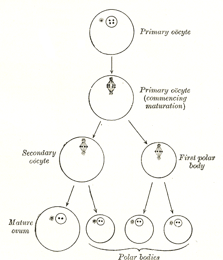
An oocyte (, oöcyte, or ovocyte) is a female gametocyte or germ cell involved in reproduction. In other words, it is an immature ovum, or egg cell. An oocyte is produced in a female fetus in the ovary during female gametogenesis. The female germ cells produce a primordial germ cell (PGC), which then undergoes mitosis, forming oogonia. During oogenesis, the oogonia become primary oocytes. An oocyte is a form of genetic material that can be collected for cryoconservation. Formation The formation of an oocyte is called oocytogenesis, which is a part of oogenesis. Oogenesis results in the formation of both primary oocytes during fetal period, and of secondary oocytes after it as part of ovulation. Characteristics Cytoplasm Oocytes are rich in cytoplasm, which contains yolk granules to nourish the cell early in development. Nucleus During the primary oocyte stage of oogenesis, the nucleus is called a germinal vesicle. The only normal human type of secondary oocyte has the ... [...More Info...] [...Related Items...] OR: [Wikipedia] [Google] [Baidu] |
Prophase
Prophase () is the first stage of cell division in both mitosis and meiosis. Beginning after interphase, DNA has already been replicated when the cell enters prophase. The main occurrences in prophase are the condensation of the chromatin reticulum and the disappearance of the nucleolus. Staining and microscopy Microscopy can be used to visualize condensed chromosomes as they move through meiosis and mitosis. Various DNA stains are used to treat cells such that condensing chromosomes can be visualized as the move through prophase. The giemsa G-banding technique is commonly used to identify mammalian chromosomes, but utilizing the technology on plant cells was originally difficult due to the high degree of chromosome compaction in plant cells. G-banding was fully realized for plant chromosomes in 1990. During both meiotic and mitotic prophase, giemsa staining can be applied to cells to elicit G-banding in chromosomes. Silver staining, a more modern technology, in conj ... [...More Info...] [...Related Items...] OR: [Wikipedia] [Google] [Baidu] |
Meiosis
Meiosis () is a special type of cell division of germ cells in sexually-reproducing organisms that produces the gametes, the sperm or egg cells. It involves two rounds of division that ultimately result in four cells, each with only one copy of each chromosome (haploid). Additionally, prior to the division, genetic material from the paternal and maternal copies of each chromosome is crossed over, creating new combinations of code on each chromosome. Later on, during fertilisation, the haploid cells produced by meiosis from a male and a female will fuse to create a zygote, a cell with two copies of each chromosome. Errors in meiosis resulting in aneuploidy (an abnormal number of chromosomes) are the leading known cause of miscarriage and the most frequent genetic cause of developmental disabilities. In meiosis, DNA replication is followed by two rounds of cell division to produce four daughter cells, each with half the number of chromosomes as the original parent cell. ... [...More Info...] [...Related Items...] OR: [Wikipedia] [Google] [Baidu] |
SET (gene)
Protein SET, also known as Protein SET 1, is a protein that in humans is encoded by the ''SET'' gene. Interactions Protein SET has been shown to interact with: * Acidic leucine-rich nuclear phosphoprotein 32 family member A, * CDK5R1, * KLF5, * NME1 Nucleoside diphosphate kinase A is an enzyme that in humans is encoded by the ''NME1'' gene. It is thought to be a metastasis suppressor. Function This gene (NME1) was identified because of its reduced mRNA transcript levels in highly metast ..., and * TAF1A. References Further reading * * * * * * * * * * * * * * * * * Histone Acetyltransferase Inhibitor {{protein-stub ... [...More Info...] [...Related Items...] OR: [Wikipedia] [Google] [Baidu] |


