|
Birth Trauma (physical)
Birth trauma refers to damage of the tissues and organs of a newly delivered child, often as a result of physical pressure or trauma during childbirth. It encompasses the long term consequences, often of cognitive nature, of damage to the brain or cranium. Medical study of birth trauma dates to the 16th century, and the morphological consequences of mishandled delivery are described in Renaissance-era medical literature. Birth injury occupies a unique area of concern and study in the medical canon. In ICD-10 "birth trauma" occupied 49 individual codes (P10–Р15). However, there are often clear distinctions to be made between brain damage caused by birth trauma and that induced by intrauterine asphyxia. It is also crucial to distinguish between "birth trauma" and "birth injury". Birth injuries encompass any systemic damages incurred during delivery (Intrauterine hypoxia, hypoxic, toxic, biochemical, infection factors, etc.), but "birth trauma" focuses largely on mechanical damage. ... [...More Info...] [...Related Items...] OR: [Wikipedia] [Google] [Baidu] |
Obstetrics
Obstetrics is the field of study concentrated on pregnancy, childbirth and the postpartum period. As a medical specialty, obstetrics is combined with gynecology under the discipline known as obstetrics and gynecology (OB/GYN), which is a surgical field. Main areas Prenatal care Prenatal care is important in screening for various complications of pregnancy. This includes routine office visits with physical exams and routine lab tests along with telehealth care for women with low-risk pregnancies: Image:Ultrasound_image_of_a_fetus.jpg, 3D ultrasound of fetus (about 14 weeks gestational age) Image:Sucking his thumb and waving.jpg, Fetus at 17 weeks Image:3dultrasound 20 weeks.jpg, Fetus at 20 weeks First trimester Routine tests in the first trimester of pregnancy generally include: * Complete blood count * Blood type ** Rh-negative antenatal patients should receive RhoGAM at 28 weeks to prevent Rh disease. * Indirect Coombs test (AGT) to assess risk of hem ... [...More Info...] [...Related Items...] OR: [Wikipedia] [Google] [Baidu] |
Intracranial Hemorrhage
Intracranial hemorrhage (ICH) refers to any form of Hemorrhage, bleeding Internal bleeding, within the Human skull, skull. It can result from trauma, vascular abnormalities, hypertension, or other medical conditions. ICH is broadly categorized into several subtypes based on the location of the bleed: intracerebral hemorrhage (including Intraparenchymal hemorrhage, intraparenchymal and Intraventricular hemorrhage, intraventricular hemorrhages), subarachnoid hemorrhage, Epidural hematoma, epidural hemorrhage, and Subdural hematoma, subdural hematoma. Each subtype has distinct causes, clinical features, and treatment approaches. Epidemiology Acute, spontaneous intracranial hemorrhage (ICH) is the second most common form of stroke, affecting approximately 2 million people worldwide each year. In the United States, intracranial hemorrhage accounts for about 20% of all cerebrovascular accidents, with an incidence of approximately 20 cases per 100,000 people annually. Intracranial h ... [...More Info...] [...Related Items...] OR: [Wikipedia] [Google] [Baidu] |
Subarachnoid Hemorrhage
Subarachnoid hemorrhage (SAH) is bleeding into the subarachnoid space—the area between the arachnoid (brain), arachnoid membrane and the pia mater surrounding the human brain, brain. Symptoms may include a thunderclap headache, severe headache of rapid onset, vomiting, decreased level of consciousness, fever, weakness, numbness, and sometimes seizures. Neck stiffness or neck pain are also relatively common. In about a quarter of people a small bleed with resolving symptoms occurs within a month of a larger bleed. SAH may occur as a result of a head injury or spontaneously, usually from a ruptured cerebral aneurysm. Risk factors for spontaneous cases include high blood pressure, smoking, family history, alcoholism, and cocaine use. Generally, the diagnosis can be determined by a computed tomography, CT scan of the head if done within six hours of symptom onset. Occasionally, a lumbar puncture is also required. After confirmation further tests are usually performed to determi ... [...More Info...] [...Related Items...] OR: [Wikipedia] [Google] [Baidu] |
Subdural Hemorrhage
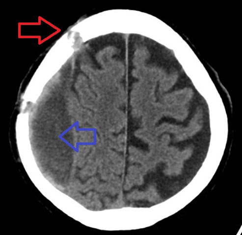
A subdural hematoma (SDH) is a type of bleeding in which a collection of blood—usually but not always associated with a traumatic brain injury—gathers between the inner layer of the dura mater and the arachnoid mater of the meninges surrounding the brain. It usually results from rips in bridging veins that cross the subdural space. Subdural hematomas may cause an increase in the pressure inside the skull, which in turn can cause compression of and damage to delicate brain tissue. Acute subdural hematomas are often life-threatening. Chronic subdural hematomas have a better prognosis if properly managed. In contrast, epidural hematomas are usually caused by rips in arteries, resulting in a build-up of blood between the dura mater and the skull. The third type of brain hemorrhage, known as a subarachnoid hemorrhage (SAH), causes bleeding into the subarachnoid space between the arachnoid mater and the pia mater. SAH are often seen in trauma settings, or after rupture of int ... [...More Info...] [...Related Items...] OR: [Wikipedia] [Google] [Baidu] |
Subgaleal Hemorrhage
Subgaleal hemorrhage, also known as subgaleal hematoma, is bleeding in the potential space between the skull periosteum and the scalp galea aponeurosis (dense fibrous tissue surrounding the skull). Symptoms The diagnosis is generally clinical, with a fluctuant boggy mass developing over the scalp (especially over the occiput) with superficial skin bruising. The swelling develops gradually 12–72 hours after delivery, although it may be noted immediately after delivery in severe cases. Subgaleal hematoma growth is insidious, as it spreads across the whole calvaria and may not be recognized for hours to days. If enough blood accumulates, a visible fluid wave may be seen. Patients may develop periorbital ecchymosis (" raccoon eyes"). Patients with subgaleal hematoma may present with hemorrhagic shock given the volume of blood that can be lost into the potential space between the skull periosteum and the scalp galea aponeurosis, which has been found to be as high as 20-40% of ... [...More Info...] [...Related Items...] OR: [Wikipedia] [Google] [Baidu] |
Cephalohematoma
A cephalohematoma (American English), also spelled cephalohaematoma (British English), is a hemorrhage of blood between the skull and the periosteum at any age, including a newborn baby secondary to rupture of blood vessels crossing the periosteum. Because the swelling is subperiosteal, its boundaries are limited by the individual bones, in contrast to a caput succedaneum. Symptoms and signs Swelling appears 2-3 days after birth. If severe the child may develop jaundice, anemia or hypotension. In some cases it may be an indication of a linear skull fracture or be at risk of an infection leading to osteomyelitis or meningitis. The swelling of a cephalohematoma takes weeks to resolve as the blood clot is slowly absorbed from the periphery towards the centre. In time the swelling hardens (calcification) leaving a relatively softer centre so that it appears as a 'depressed fracture'. Cephalohematoma should be distinguished from another scalp bleeding called subgaleal hemorrhage ... [...More Info...] [...Related Items...] OR: [Wikipedia] [Google] [Baidu] |
Brachial Plexus Injury
A brachial plexus injury (BPI), also known as brachial plexus lesion, is an injury to the brachial plexus, the network of nerves that conducts signals from the spinal cord to the shoulder, arm and hand. These nerves originate in the fifth, sixth, seventh and eighth cervical (C5–C8), and first thoracic (T1) spinal nerves, and innervate the muscles and skin of the chest, shoulder, arm and hand. Brachial plexus injuries can occur as a result of shoulder trauma (e.g. dislocation), tumours, or inflammation, or obstetric. Obstetric injuries may occur from mechanical injury involving shoulder dystocia during difficult Childbirth#Mechanical fetal injury, childbirth, with a prevalence of 1 in 1000 births. "The brachial plexus may be injured by falls from a height on to the side of the head and shoulder, whereby the nerves of the plexus are violently stretched. The brachial plexus may also be injured by direct violence or gunshot wounds, by violent traction on the arm, or by efforts at red ... [...More Info...] [...Related Items...] OR: [Wikipedia] [Google] [Baidu] |
Vacuum Extraction
Vacuum extraction (VE), also known as ventouse, is a method to assist delivery of a baby using a vacuum device. It is used in the second stage of labor if it has not progressed adequately. It may be an alternative to a forceps delivery and caesarean section. It cannot be used when the baby is in the breech position or for premature births. The use of VE is generally safe, but it can occasionally have negative effects on either the mother or the child. The term ''ventouse'' comes from the French word for "suction cup". Medical uses There are several indications to use a vacuum extraction to aid delivery: * Maternal exhaustion * Prolonged second stage of labor * Foetal distress in the second stage of labor, generally indicated by changes in the foetal heart-rate (usually measured on a CTG) * Maternal illness where prolonged "bearing down" or pushing efforts would be risky (e.g. cardiac conditions, blood pressure, aneurysm, glaucoma). If these conditions are known about befor ... [...More Info...] [...Related Items...] OR: [Wikipedia] [Google] [Baidu] |
Obstetrical Forceps
Obstetrical forceps are a medical instrument used in childbirth. Their use can serve as an alternative to the ventouse (vacuum extraction) method. Medical uses Forceps births, like all assisted births, should be undertaken only to help promote the health of the mother or baby. In general, a forceps birth is likely to be safer for both the mother and baby than the alternatives – either a ventouse birth or a caesarean section – although caveats such as operator skill apply. Advantages of forceps use include avoidance of caesarean section (and the short and long-term complications that accompany this), reduction of delivery time, and general applicability with cephalic presentation (head presentation). Common complications include the possibility of bruising the baby and causing more severe vaginal tears (perineal laceration) than would otherwise be the case. Severe and rare complications (occurring less frequently than 1 in 200) include nerve damage, Descemet's membran ... [...More Info...] [...Related Items...] OR: [Wikipedia] [Google] [Baidu] |
Breech Presentation
A breech birth is when a baby is born bottom first instead of Cephalic presentation, head first, as is normal. Around 3–5% of pregnant women at term (37–40 weeks pregnant) have a breech baby. Due to their higher than average rate of possible complications for the baby, breech births are generally considered higher risk. Breech births also occur in many other mammals such as dogs and horses, see veterinary obstetrics. Most babies in the breech position are delivered via caesarean section because it is seen as safer than being Vaginal birth, born vaginally. Doctors and Midwife, midwives in the developing world often lack many of the skills required to safely assist women giving birth to a breech baby vaginally. Also, delivering all breech babies by caesarean section in developing countries is difficult to implement as there are not always resources available to provide this service. Cause With regard to the fetal presentation during pregnancy, three periods have been distingu ... [...More Info...] [...Related Items...] OR: [Wikipedia] [Google] [Baidu] |
Asynclitic Birth
In obstetrics, asynclitic birth, or asynclitism, refers to the malposition of the fetal head in the uterus relative to the birth canal. Many babies enter the pelvis in an asynclitic presentation, but in most cases, the issue is corrected during labor. Asynclitic presentation is not the same as shoulder presentation, where the shoulder enters first. Fetal head asynclitism may affect the progression of labor, increase the need for obstetrical intervention, and be associated with difficult instrumental delivery. The prevalence of asynclitism at transperineal ultrasound was common in nulliparous women (those who have never given birth) at labor stage two and seemed more commonly associated with non occiput anterior position, suggesting an autocorrection typically occurs. When self-correction does not occur, obstetrical intervention is necessary. Persistent asynclitism can cause problems with dystocia, and has often been associated with cesarean births. However, a skilled midwife ... [...More Info...] [...Related Items...] OR: [Wikipedia] [Google] [Baidu] |
Cephalopelvic Disproportion
Cephalopelvic disproportion (CPD) exists when the capacity of the pelvis is inadequate to allow the fetus to negotiate the birth canal. This may be due to a small pelvis, a nongynecoid pelvic formation, a large fetus, an unfavorable orientation of the fetus, or a combination of these factors. Certain medical conditions may distort pelvic bones, such as rickets or a pelvic fracture, and lead to CPD. Transverse diagonal measurement has been proposed as a predictive method. Signs and symptoms 1. Prolonged Labor: Labor that does not progress as expected, particularly during the active phase. 2. Failure to Progress: Lack of dilation or descent of the baby despite strong contractions. 3. Severe Pain: Intense pain that is disproportionate to normal labor pain. 4. Fetal Distress: Signs like abnormal heart rate patterns detected via fetal monitoring. 5. Maternal Exhaustion: Extreme fatigue in the mother due to prolonged labor. 6. High Station: The baby’s head remains high in the ... [...More Info...] [...Related Items...] OR: [Wikipedia] [Google] [Baidu] |


