|
Biot's Respiration
Ataxic respirations, also known as Biot's respirations or Biot's breathing, is an abnormal pattern of breathing characterized by variable tidal volume, random apneas, and no regularity. It is named for Camille Biot, who characterized it in 1876. Biot MC. Contribution a l'étude du phénomène respiratoire de Cheyne-Stokes. Lyon Med. 1876;23:517-528, 561-567. Biot's respiration is caused by damage to the medulla oblongata and pons due to trauma, stroke, opioid use, and increased intracranial pressure due to uncal or tentorial herniation. Often this condition is also associated with meningitis. In common medical practice, Biot's respiration is often mistaken for Cheyne–Stokes respiration, part of which may have been caused by them both being described by the same person and subtle differences between the types of breathing. Ataxic respirations were discovered by Dr. Camille Biot in the late 19th century as he wrote multiple papers analyzing subtle differences in Cheyne-Stokes ... [...More Info...] [...Related Items...] OR: [Wikipedia] [Google] [Baidu] |
Camille Biot
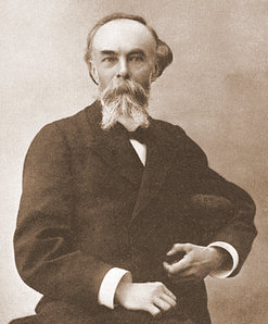
Camille Biot (19 December 1850, Châtenoy-le-Royal - 1918, Mâcon) was a French physician who is remembered for describing Biot's respiration. Biography Camille Biot was born in Chatenoy-le-Royal, Saône-et-Loire Saône-et-Loire (; Arpitan: ''Sona-et-Lêre'') is a department in the Bourgogne-Franche-Comté region in France. It is named after the rivers Saône and Loire, between which it lies, in the country's central-eastern part. Saône-et-Loire is B ..., France in 1850. He made observations about breathing patterns while working as an intern at the Hôtel Dieu Hospital in Lyon, which were published in 1876. After 1875 he practised medicine in Mâcon. References 19th-century French physicians 1850 births 1918 deaths 20th-century French physicians {{france-med-bio-stub ... [...More Info...] [...Related Items...] OR: [Wikipedia] [Google] [Baidu] |
Medulla Oblongata
The medulla oblongata or simply medulla is a long stem-like structure which makes up the lower part of the brainstem. It is anterior and partially inferior to the cerebellum. It is a cone-shaped neuronal mass responsible for autonomic (involuntary) functions, ranging from vomiting to sneezing. The medulla contains the cardiovascular center, the respiratory center, vomiting and vasomotor centers, responsible for the autonomic functions of breathing, heart rate and blood pressure as well as the sleep–wake cycle. "Medulla" is from Latin, ‘pith or marrow’. And "oblongata" is from Latin, ‘lengthened or longish or elongated'. During embryonic development, the medulla oblongata develops from the myelencephalon. The myelencephalon is a secondary brain vesicle which forms during the maturation of the rhombencephalon, also referred to as the hindbrain. The bulb is an archaic term for the medulla oblongata. In modern clinical usage, the word bulbar (as in bulbar palsy) is r ... [...More Info...] [...Related Items...] OR: [Wikipedia] [Google] [Baidu] |
Pons
The pons (from Latin , "bridge") is part of the brainstem that in humans and other mammals, lies inferior to the midbrain, superior to the medulla oblongata and anterior to the cerebellum. The pons is also called the pons Varolii ("bridge of Varolius"), after the Italian anatomist and surgeon Costanzo Varolio (1543–75). This region of the brainstem includes neural pathways and tracts that conduct signals from the brain down to the cerebellum and medulla, and tracts that carry the sensory signals up into the thalamus. Structure The pons in humans measures about in length. It is the part of the brainstem situated between the midbrain and the medulla oblongata. The horizontal ''medullopontine sulcus'' demarcates the boundary between the pons and medulla oblongata on the ventral aspect of the brainstem, and the roots of cranial nerves VI/VII/VIII emerge from the brainstem along this groove. The junction of pons, medulla oblongata, and cerebellum forms the cerebellopontine ... [...More Info...] [...Related Items...] OR: [Wikipedia] [Google] [Baidu] |
Uncal Herniation
Brain herniation is a potentially deadly side effect of very high pressure within the skull that occurs when a part of the brain is squeezed across structures within the skull. The brain can shift across such structures as the falx cerebri, the tentorium cerebelli, and even through the foramen magnum (the hole in the base of the skull through which the spinal cord connects with the brain). Herniation can be caused by a number of factors that cause a mass effect and increase intracranial pressure (ICP): these include traumatic brain injury, intracranial hemorrhage, or brain tumor. Herniation can also occur in the absence of high ICP when mass lesions such as hematomas occur at the borders of brain compartments. In such cases local pressure is increased at the place where the herniation occurs, but this pressure is not transmitted to the rest of the brain, and therefore does not register as an increase in ICP. Because herniation puts extreme pressure on parts of the brain and the ... [...More Info...] [...Related Items...] OR: [Wikipedia] [Google] [Baidu] |
Tentorium Cerebelli
The cerebellar tentorium or tentorium cerebelli (Latin for "tent of the cerebellum") is one of four dural folds that separate the cranial cavity into four (incomplete) compartments. The cerebellar tentorium separates the cerebellum from the cerebrum forming a supratentorial and an infratentorial region; the cerebrum is supratentorial and the cerebellum infratentorial. The free border of the tentorium gives passage to the midbrain (the upper-most part of the brainstem). Structure Free border The free border of the tentorium is U-shaped; it forms an aperture - the tentorial notch (tentorial incisure) - which gives passage to the midbrain. The free border of each side extends anteriorly beyond the medial end of the superior petrosal sinus (i.e. the apex of the petrous part of the temporal bone) to overlap the attached margin, thenceforth forming a ridge of dura matter upon the roof of the cavernous sinus, terminating anteriorly by attaching at the anterior clinoid process. ... [...More Info...] [...Related Items...] OR: [Wikipedia] [Google] [Baidu] |
Cheyne–Stokes Respiration
Cheyne–Stokes respiration is an abnormal pattern of breathing characterized by progressively deeper, and sometimes faster, breathing followed by a gradual decrease that results in a temporary stop in breathing called an apnea. The pattern repeats, with each cycle usually taking 30 seconds to 2 minutes. It is an oscillation of ventilation between apnea and hyperpnea with a crescendo-diminuendo pattern, and is associated with changing serum partial pressures of oxygen and carbon dioxide. Cheyne–Stokes respiration and periodic breathing are the two regions on a spectrum of severity of oscillatory tidal volume. The distinction lies in what is observed at the trough of ventilation: Cheyne–Stokes respiration involves apnea (since apnea is a prominent feature in their original description) while periodic breathing involves hypopnea (abnormally small but not absent breaths). These phenomena can occur during wakefulness or during sleep, where they are called the central sleep ap ... [...More Info...] [...Related Items...] OR: [Wikipedia] [Google] [Baidu] |
Brainstem
The brainstem (or brain stem) is the posterior stalk-like part of the brain that connects the cerebrum with the spinal cord. In the human brain the brainstem is composed of the midbrain, the pons, and the medulla oblongata. The midbrain is continuous with the thalamus of the diencephalon through the tentorial notch, and sometimes the diencephalon is included in the brainstem. The brainstem is very small, making up around only 2.6 percent of the brain's total weight. It has the critical roles of regulating heart and respiratory system, respiratory function, helping to control heart rate and breathing rate. It also provides the main motor and sensory nerve supply to the face and neck via the cranial nerves. Ten pairs of cranial nerves come from the brainstem. Other roles include the regulation of the central nervous system and the body's sleep cycle. It is also of prime importance in the conveyance of motor and sensory pathways from the rest of the brain to the body, and from the b ... [...More Info...] [...Related Items...] OR: [Wikipedia] [Google] [Baidu] |
Apnea
Apnea (also spelled apnoea in British English) is the temporary cessation of breathing. During apnea, there is no movement of the muscles of inhalation, and the volume of the lungs initially remains unchanged. Depending on how blocked the airways are ( patency), there may or may not be a flow of gas between the lungs and the environment. If there is sufficient flow, gas exchange within the lungs and cellular respiration would not be severely affected. Voluntarily doing this is called holding one's breath. Apnea may first be diagnosed in childhood, and it is recommended to consult an ENT specialist, allergist or sleep physician to discuss symptoms when noticed; malformation and/or malfunctioning of the upper airways may be observed by an orthodontist. Cause Apnea can be involuntary—for example, drug-induced (such as by opiate toxicity), mechanically / physiologically induced (for example, by strangulation or choking), or a consequence of neurological disease or tra ... [...More Info...] [...Related Items...] OR: [Wikipedia] [Google] [Baidu] |
Mechanical Ventilation
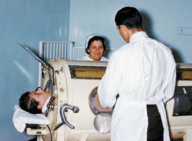
Mechanical ventilation or assisted ventilation is the Medicine, medical term for using a ventilator, ventilator machine to fully or partially provide artificial ventilation. Mechanical ventilation helps move air into and out of the lungs, with the main goal of helping the delivery of oxygen and removal of carbon dioxide. Mechanical ventilation is used for many reasons, including to protect the airway due to mechanical or neurologic cause, to ensure adequate oxygenation, or to remove excess carbon dioxide from the lungs. Various healthcare providers are involved with the use of mechanical ventilation and people who require ventilators are typically monitored in an intensive care unit. Mechanical ventilation is termed invasive if it involves an instrument to create an airway that is placed inside the trachea. This is done through an endotracheal tube or nasotracheal tube. For non-invasive ventilation in people who are conscious, face or nasal masks are used. The two main types o ... [...More Info...] [...Related Items...] OR: [Wikipedia] [Google] [Baidu] |
Breathing Abnormalities
Breathing (spiration or ventilation) is the neuroscience of rhythm, rhythmical process of moving air into (inhalation) and out of (exhalation) the lungs to facilitate gas exchange with the Milieu intérieur, internal environment, mostly to flush out carbon dioxide and bring in oxygen. All Aerobic respiration, aerobic creatures need oxygen for cellular respiration, which extracts energy from the reaction of oxygen with molecules derived from food and produces carbon dioxide as a Waste, waste product. Breathing, or external respiration, brings Atmosphere of Earth, air into the lungs where gas exchange takes place in the Pulmonary alveolus, alveoli through diffusion. The body's circulatory system transports these gases to and from the cells, where cellular Respiration (physiology), respiration takes place. The breathing of all vertebrates with lungs consists of repetitive cycles of inhalation and exhalation through a highly branched system of tubes or Respiratory tract, airways w ... [...More Info...] [...Related Items...] OR: [Wikipedia] [Google] [Baidu] |


