|
Articular Cartilage
Hyaline cartilage is the glass-like (hyaline) and translucent cartilage found on many joint surfaces. It is also most commonly found in the ribs, nose, larynx, and trachea. Hyaline cartilage is pearl-gray in color, with a firm consistency and has a considerable amount of collagen. It contains no nerves or blood vessels, and its structure is relatively simple. Structure Hyaline cartilage is the most common kind of cartilage in the human body. It is primarily composed of type II collagen and proteoglycans. Hyaline cartilage is located in the trachea, nose, epiphyseal plate, sternum, and ribs. Hyaline cartilage is covered externally by a fibrous membrane known as the perichondrium. The primary cells of cartilage are chondrocytes, which are in a Matrix (biology), matrix of fibrous tissue, proteoglycans and glycosaminoglycans. As cartilage does not have lymph glands or blood vessels, the movements of solutes, including nutrients, occur via diffusion within the fluid compartments con ... [...More Info...] [...Related Items...] OR: [Wikipedia] [Google] [Baidu] |
Light Micrograph
A micrograph is an image, captured photographically or digitally, taken through a microscope or similar device to show a magnify, magnified image of an object. This is opposed to a macrograph or photomacrograph, an image which is also taken on a microscope but is only slightly magnified, usually less than 10 times. Micrography is the practice or art of using microscopes to make photographs. A photographic micrograph is a photomicrograph, and one taken with an electron microscope is an electron micrograph. A micrograph contains extensive details of microstructure. A wealth of information can be obtained from a simple micrograph like behavior of the material under different conditions, the phases found in the system, failure analysis, grain size estimation, elemental analysis and so on. Micrographs are widely used in all fields of microscopy. Types Photomicrograph A light micrograph or photomicrograph is a micrograph prepared using an optical microscope, a process referred to ... [...More Info...] [...Related Items...] OR: [Wikipedia] [Google] [Baidu] |
Matrix (biology)
In biology, matrix (: matrices) is the material (or tissue) in between a eukaryotic organism's cells. The structure of connective tissues is an extracellular matrix. Fingernails and toenails grow from matrices. It is found in various connective tissues. It serves as a jelly-like structure instead of cytoplasm in connective tissue. Tissue matrices Extracellular matrix (ECM) The main ingredients of the extracellular matrix are glycoproteins secreted by the cells. The most abundant glycoprotein in the ECM of most animal cells is collagen, which forms strong fibers outside the cells. In fact, collagen accounts for about 40% of the total protein in the human body. The collagen fibers are embedded in a network woven from proteoglycans. A proteoglycan molecule consists of a small core protein with many carbohydrate chains covalently attached, so that it may be up to 95% carbohydrate. Large proteoglycan complexes can form when hundreds of proteoglycans become noncovalently attached ... [...More Info...] [...Related Items...] OR: [Wikipedia] [Google] [Baidu] |
Histology Of Articular Cartilage Zones
Histology, also known as microscopic anatomy or microanatomy, is the branch of biology that studies the microscopic anatomy of biological tissues. Histology is the microscopic counterpart to gross anatomy, which looks at larger structures visible without a microscope. Although one may divide microscopic anatomy into ''organology'', the study of organs, ''histology'', the study of tissues, and ''cytology'', the study of cells, modern usage places all of these topics under the field of histology. In medicine, histopathology is the branch of histology that includes the microscopic identification and study of diseased tissue. In the field of paleontology, the term paleohistology refers to the histology of fossil organisms. Biological tissues Animal tissue classification There are four basic types of animal tissues: muscle tissue, nervous tissue, connective tissue, and epithelial tissue. All animal tissues are considered to be subtypes of these four principal tissue types (for ... [...More Info...] [...Related Items...] OR: [Wikipedia] [Google] [Baidu] |
Mitosis
Mitosis () is a part of the cell cycle in eukaryote, eukaryotic cells in which replicated chromosomes are separated into two new Cell nucleus, nuclei. Cell division by mitosis is an equational division which gives rise to genetically identical cells in which the total number of chromosomes is maintained. Mitosis is preceded by the S phase of interphase (during which DNA replication occurs) and is followed by telophase and cytokinesis, which divide the cytoplasm, organelles, and cell membrane of one cell into two new cell (biology), cells containing roughly equal shares of these cellular components. The different stages of mitosis altogether define the mitotic phase (M phase) of a cell cycle—the cell division, division of the mother cell into two daughter cells genetically identical to each other. The process of mitosis is divided into stages corresponding to the completion of one set of activities and the start of the next. These stages are preprophase (specific to plant ce ... [...More Info...] [...Related Items...] OR: [Wikipedia] [Google] [Baidu] |
Isogenous Group
An isogenous group (lat. "equal origin") is a cluster of up to eight chondrocytes found in hyaline cartilage, hyaline and elastic cartilage.Wheater's Functional Histology, 6th ed. Young, O'Dowd and Woodford. Formation Chondrocytes develop in the embryo from Mesenchymal stem cell, mesenchymal progenitor cells through a process known as chondrogenesis. A chondrocyte can then undergo mitosis to form an isogenous group within its lacuna. Function Isogenous groups differentiate into individual chondrocytes where they continue to produce and deposit extracellular matrix (ECM), lengthening the cartilage and increasing its diameter. This is termed interstitial growth and is one of only two ways cartilage can grow. See also *Endochondral ossification * Hyaline References Connective tissue cells [...More Info...] [...Related Items...] OR: [Wikipedia] [Google] [Baidu] |
Cartilage Lacunae
In histology, a lacuna is a small space, containing an osteocyte in bone, or chondrocyte in cartilage. Bone The lacuna are situated between the lamellae, and consist of a number of oblong spaces. In an ordinary microscopic section, viewed by transmitted light, they appear as fusiform opaque spots. Each lacuna is occupied during life by a branched cell, termed an osteocyte, bone-cell or bone-corpuscle. Lacunae are connected to one another by small canals called canaliculi. A lacuna never contains more than one osteocyte. Sinuses are an example of lacuna. Cartilage The cartilage cells or chondrocytes are contained in cavities in the matrix, called cartilage lacunae; around these, the matrix is arranged in concentric lines as if it had been formed in successive portions around the cartilage cells. This constitutes the so-called capsule of the space. Each lacuna is generally occupied by a single cell, but during the division of the cells, it may contain two, four, or eight cells. Lac ... [...More Info...] [...Related Items...] OR: [Wikipedia] [Google] [Baidu] |
Cell Nucleus
The cell nucleus (; : nuclei) is a membrane-bound organelle found in eukaryote, eukaryotic cell (biology), cells. Eukaryotic cells usually have a single nucleus, but a few cell types, such as mammalian red blood cells, have #Anucleated_cells, no nuclei, and a few others including osteoclasts have Multinucleate, many. The main structures making up the nucleus are the nuclear envelope, a double membrane that encloses the entire organelle and isolates its contents from the cellular cytoplasm; and the nuclear matrix, a network within the nucleus that adds mechanical support. The cell nucleus contains nearly all of the cell's genome. Nuclear DNA is often organized into multiple chromosomes – long strands of DNA dotted with various proteins, such as histones, that protect and organize the DNA. The genes within these chromosomes are Nuclear organization, structured in such a way to promote cell function. The nucleus maintains the integrity of genes and controls the activities of the ... [...More Info...] [...Related Items...] OR: [Wikipedia] [Google] [Baidu] |
Protoplasm
Protoplasm (; ) is the part of a cell that is surrounded by a plasma membrane. It is a mixture of small molecules such as ions, monosaccharides, amino acids, and macromolecules such as proteins, polysaccharides, lipids, etc. In some definitions, it is a general term for the cytoplasm (e.g., Mohl, 1846), but for others, it also includes the nucleoplasm (e.g., Strasburger, 1882). For Sharp (1921), "According to the older usage the extra-nuclear portion of the protoplast 'the entire cell, excluding the cell wall''was called "protoplasm," but the nucleus also is composed of protoplasm, or living substance in its broader sense. The current consensus is to avoid this ambiguity by employing Strasburger's (1882) terms cytoplasm Kölliker (1863), originally as synonym for protoplasm''and nucleoplasm van Beneden (1875), or karyoplasm, used by Walther Flemming">Flemming (1878)''">karyoplasm">'term coined by Edouard Van Beneden">van Beneden (1875), or karyoplasm, used by Walther Flemming ... [...More Info...] [...Related Items...] OR: [Wikipedia] [Google] [Baidu] |
Homogeneous
Homogeneity and heterogeneity are concepts relating to the uniformity of a substance, process or image. A homogeneous feature is uniform in composition or character (i.e., color, shape, size, weight, height, distribution, texture, language, income, disease, temperature, radioactivity, architectural design, etc.); one that is heterogeneous is distinctly nonuniform in at least one of these qualities. Etymology and spelling The words ''homogeneous'' and ''heterogeneous'' come from Medieval Latin ''homogeneus'' and ''heterogeneus'', from Ancient Greek ὁμογενής (''homogenēs'') and ἑτερογενής (''heterogenēs''), from ὁμός (''homos'', "same") and ἕτερος (''heteros'', "other, another, different") respectively, followed by γένος (''genos'', "kind"); -ous is an adjectival suffix. Alternate spellings omitting the last ''-e-'' (and the associated pronunciations) are common, but mistaken: ''homogenous'' is strictly a biological/pathological term whic ... [...More Info...] [...Related Items...] OR: [Wikipedia] [Google] [Baidu] |
Microscope
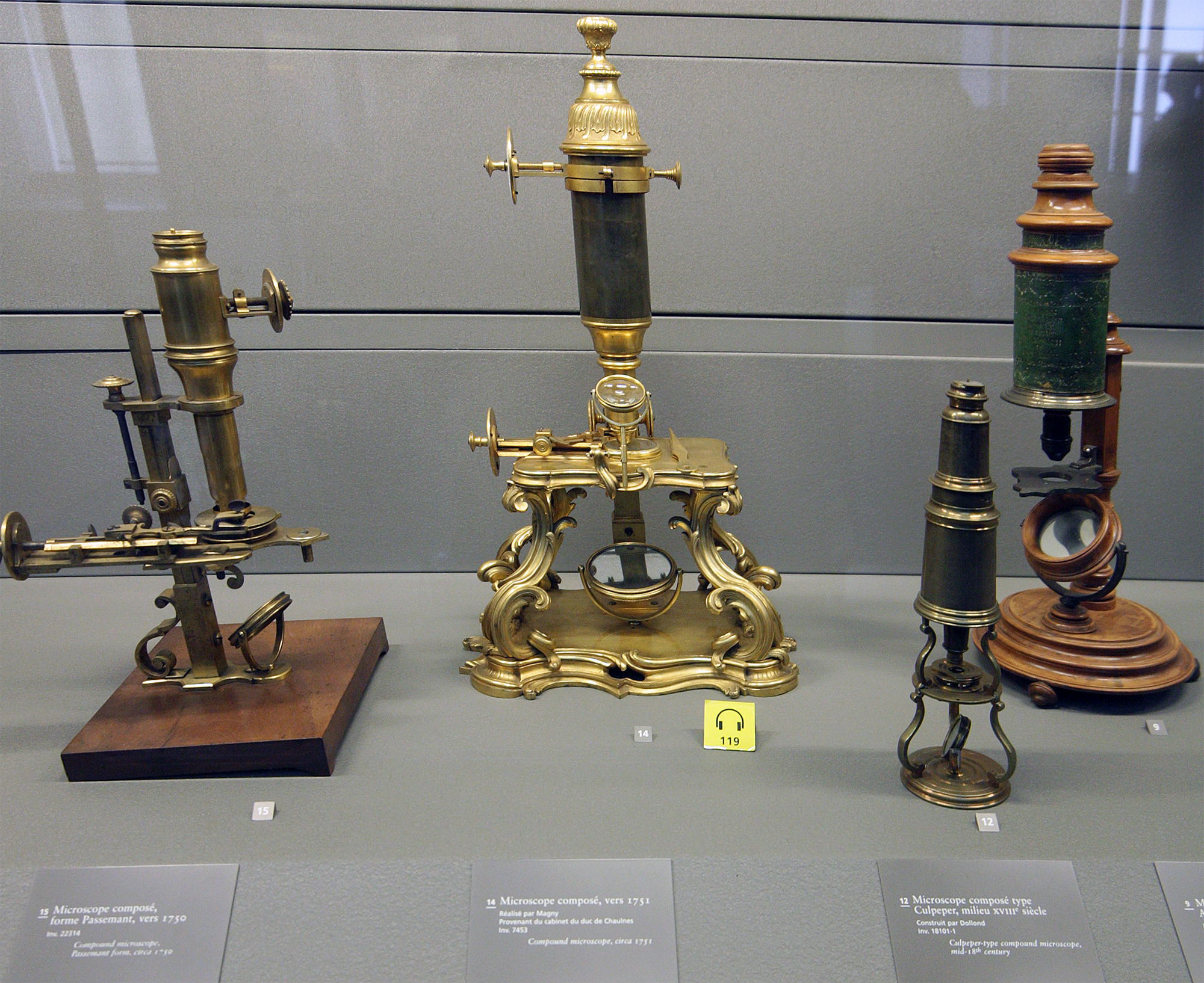
A microscope () is a laboratory equipment, laboratory instrument used to examine objects that are too small to be seen by the naked eye. Microscopy is the science of investigating small objects and structures using a microscope. Microscopic means being invisible to the eye unless aided by a microscope. There are many types of microscopes, and they may be grouped in different ways. One way is to describe the method an instrument uses to interact with a sample and produce images, either by sending a beam of light or electrons through a sample in its optical path, by detecting fluorescence, photon emissions from a sample, or by scanning across and a short distance from the surface of a sample using a probe. The most common microscope (and the first to be invented) is the optical microscope, which uses lenses to refract visible light that passed through a microtome, thinly sectioned sample to produce an observable image. Other major types of microscopes are the fluorescence micro ... [...More Info...] [...Related Items...] OR: [Wikipedia] [Google] [Baidu] |
Elasmobranch
Elasmobranchii () is a subclass of Chondrichthyes or cartilaginous fish, including modern sharks ( division Selachii), and batomorphs (division Batomorphi, including rays, skates, and sawfish). Members of this subclass are characterised by having five to seven pairs of gill slits opening individually to the exterior, rigid dorsal fins and small placoid scales on the skin. The teeth are in several series; the upper jaw is not fused to the cranium, and the lower jaw is articulated with the upper. The details of this jaw anatomy vary between species, and help distinguish the different elasmobranch clades. The pelvic fins in males are modified to create claspers for the transfer of sperm. There is no swim bladder; instead, these fish maintain buoyancy with large livers rich in oil. The definition of the clade is unclear with respect to fossil chondrichthyans. Some authors consider it as equivalent to Neoselachii (the crown group clade including modern sharks, rays, and all o ... [...More Info...] [...Related Items...] OR: [Wikipedia] [Google] [Baidu] |
Fluid Compartments
The human body and even its individual body fluids may be conceptually divided into various fluid compartments, which, although not literally anatomic compartments, do represent a real division in terms of how portions of the body's water, solutes, and suspended elements are segregated. The two main fluid compartments are the intracellular and extracellular compartments. The intracellular compartment is the space within the organism's cells; it is separated from the extracellular compartment by cell membranes. About two-thirds of the total body water of humans is held in the cells, mostly in the cytosol, and the remainder is found in the extracellular compartment. The extracellular fluids may be divided into three types: interstitial fluid in the "interstitial compartment" (surrounding tissue cells and bathing them in a solution of nutrients and other chemicals), blood plasma and lymph in the "intravascular compartment" (inside the blood vessels and lymphatic vessels), and sm ... [...More Info...] [...Related Items...] OR: [Wikipedia] [Google] [Baidu] |


