|
Aorta
The aorta ( ; : aortas or aortae) is the main and largest artery in the human body, originating from the Ventricle (heart), left ventricle of the heart, branching upwards immediately after, and extending down to the abdomen, where it splits at the aortic bifurcation into two smaller arteries (the common iliac artery, common iliac arteries). The aorta distributes Oxygen saturation (medicine), oxygenated blood to all parts of the body through the systemic circulation. Structure Sections In anatomical sources, the aorta is usually divided into sections. One way of classifying a part of the aorta is by anatomical compartment, where the thoracic aorta (or thoracic portion of the aorta) runs from the heart to the thoracic diaphragm, diaphragm. The aorta then continues downward as the abdominal aorta (or abdominal portion of the aorta) from the diaphragm to the aortic bifurcation. Another system divides the aorta with respect to its course and the direction of blood flow. In this s ... [...More Info...] [...Related Items...] OR: [Wikipedia] [Google] [Baidu] |
Systemic Circulation
In vertebrates, the circulatory system is a organ system, system of organs that includes the heart, blood vessels, and blood which is circulated throughout the body. It includes the cardiovascular system, or vascular system, that consists of the heart and blood vessels (from Greek meaning ''heart'', and Latin meaning ''vessels''). The circulatory system has two divisions, a systemic circulation, systemic circulation or circuit, and a pulmonary circulation, pulmonary circulation or circuit. Some sources use the terms ''cardiovascular system'' and ''vascular system'' interchangeably with ''circulatory system''. The network of blood vessels are the great vessels of the heart including large elastic arteries, and large veins; other arteries, smaller arterioles, capillaries that join with venules (small veins), and other veins. The Closed circulatory system, circulatory system is closed in vertebrates, which means that the blood never leaves the network of blood vessels. Many in ... [...More Info...] [...Related Items...] OR: [Wikipedia] [Google] [Baidu] |
Dorsal Aorta
The dorsal aortae are paired (left and right) embryological vessels which progress to form the descending aorta. The paired dorsal aortae arise from aortic arches that in turn arise from the aortic sac. The primary dorsal aorta is located deep to the lateral plate of mesoderm and move from lateral to medial position with development and eventually will fuse with the other dorsal aorta to form the descending aorta. Each primitive aorta anteriorly receives the vitelline vein from the yolk-sac, and is prolonged backward on the lateral aspect of the notochord under the name of the dorsal aorta. The dorsal aortae give branches to the yolk-sac, and are continued backward through the body-stalk as the umbilical arteries The umbilical artery is a paired artery (with one for each half of the body) that is found in the abdominal and pelvic regions. In the fetus, it extends into the umbilical cord. Structure Development The umbilical arteries supply systemic arte ... to the v ... [...More Info...] [...Related Items...] OR: [Wikipedia] [Google] [Baidu] |
Brachiocephalic Artery
The brachiocephalic artery, brachiocephalic trunk, or innominate artery is an artery of the mediastinum that supplies blood to the right arm, head, and neck. It is the first branch of the aortic arch. Soon after it emerges, the brachiocephalic artery divides into the right common carotid artery and the right subclavian artery. There is no brachiocephalic artery for the left side of the body. The left common carotid artery and the left subclavian artery come directly off the aortic arch. Despite this, there are two brachiocephalic veins. Structure The brachiocephalic artery arises on a level with the upper border of the second right costal cartilage from the start of the aortic arch on a plane anterior to the origin of the left carotid artery. It ascends obliquely upward, backward, and to the right to the level of the upper border of the right sternoclavicular articulation, where it divides into the right common carotid artery and right subclavian arteries. The artery then ... [...More Info...] [...Related Items...] OR: [Wikipedia] [Google] [Baidu] |
Subclavian Artery
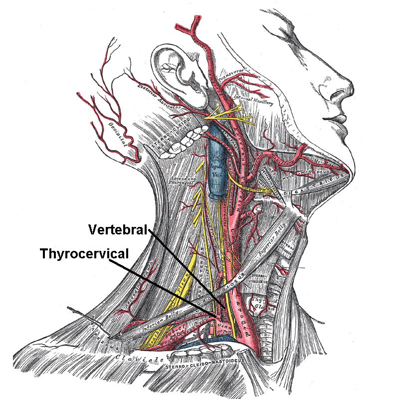
In human anatomy, the subclavian arteries are paired major arteries of the upper thorax, below the clavicle. They receive blood from the aortic arch. The left subclavian artery supplies blood to the left arm and the right subclavian artery supplies blood to the right arm, with some branches supplying the head and thorax. On the left side of the body, the subclavian comes directly off the aortic arch, while on the right side it arises from the relatively short brachiocephalic artery when it bifurcates into the subclavian and the right common carotid artery. The usual branches of the subclavian on both sides of the body are the vertebral artery, the internal thoracic artery, the thyrocervical trunk, the costocervical trunk and the dorsal scapular artery, which may branch off the transverse cervical artery, which is a branch of the thyrocervical trunk. The subclavian becomes the axillary artery at the lateral border of the first rib. Structure From its origin, the subclavian art ... [...More Info...] [...Related Items...] OR: [Wikipedia] [Google] [Baidu] |
Celiac Trunk
The celiac () artery (also spelled coeliac in British English), also known as the celiac trunk or truncus coeliacus, is the first major branch of the abdominal aorta. It is about 1.25 cm in length. Branching from the aorta at thoracic vertebra 12 (T12) in humans, it is one of three anterior/ midline branches of the abdominal aorta (the others are the superior and inferior mesenteric arteries). Structure The celiac artery is the first major branch of the descending abdominal aorta, branching at a 90° angle. This occurs just below the crus of the diaphragm. This is around the first lumbar vertebra. There are three main divisions of the celiac artery, and each in turn has its own named branches: The celiac artery may also give rise to the inferior phrenic arteries. Function The celiac artery supplies oxygenated blood to the liver, stomach, abdominal esophagus, spleen, and the superior half of both the duodenum and the pancreas. These structures correspond to the em ... [...More Info...] [...Related Items...] OR: [Wikipedia] [Google] [Baidu] |
Ventricle (heart)
A ventricle is one of two large chambers located toward the bottom of the heart that collect and expel blood towards the peripheral beds within the body and lungs. The blood pumped by a ventricle is supplied by an atrium, an adjacent chamber in the upper heart that is smaller than a ventricle. Interventricular means between the ventricles (for example the interventricular septum), while intraventricular means within one ventricle (for example an intraventricular block). In a four-chambered heart, such as that in humans, there are two ventricles that operate in a double circulatory system: the right ventricle pumps blood into the pulmonary circulation to the lungs, and the left ventricle pumps blood into the systemic circulation through the aorta. Structure Ventricles have thicker walls than atria and generate higher blood pressures. The physiological load on the ventricles requiring pumping of blood throughout the body and lungs is much greater than the pressure generated by ... [...More Info...] [...Related Items...] OR: [Wikipedia] [Google] [Baidu] |
Bronchial Arteries
In human anatomy, the bronchial arteries supply the lungs with oxygenated blood, and nutrition. Although there is much variation, there are usually two bronchial arteries that run to the left lung, and one to the right lung, and are a vital part of the respiratory system. Structure There are typically two left and one right bronchial arteries. The ''left bronchial arteries'' (superior and inferior) usually arise directly from the thoracic aorta. The single ''right bronchial artery'' may arise from one of the following: * 1) in typical anatomy, the thoracic aorta at a common trunk with the right 3rd posterior intercostal artery (known as the ''intercostobronchial trunk''). * 2) the superior bronchial artery on the left side. * 3) any number of the right intercostal arteries mostly the third right posterior. Function The bronchial arteries supply blood to the bronchi and connective tissue of the lungs. They travel with and branch with the bronchi, ending about at the le ... [...More Info...] [...Related Items...] OR: [Wikipedia] [Google] [Baidu] |
Esophageal Branches Of Thoracic Part Of Aorta
The esophageal arteries four or five in number, arise from the front of the aorta, and pass obliquely downward to the esophagus, forming a chain of anastomoses along that tube, anastomosing with the esophageal branches of the inferior thyroid arteries above, and with ascending branches from the left inferior phrenic and left gastric arteries below. These arteries supply the middle third of the esophagus. References External links * - "Branches of the ascending aorta, arch of the aorta, and the descending aorta In human anatomy, the descending aorta is part of the aorta, the largest artery in the body. The descending aorta begins at the aortic arch and runs down through the chest and abdomen. The descending aorta anatomically consists of two portions o ...." Arteries of the thorax {{circulatory-stub ... [...More Info...] [...Related Items...] OR: [Wikipedia] [Google] [Baidu] |
Aortic Arches
The aortic arches or pharyngeal arch arteries (previously referred to as branchial arches in human embryos) are a series of six paired embryological vascular structures which give rise to the great arteries of the neck and head. They are ventral to the dorsal aorta and arise from the aortic sac. The aortic arches are formed sequentially within the pharyngeal arches and initially appear symmetrical on both sides of the embryo, but then undergo a significant remodelling to form the final asymmetrical structure of the great arteries. Structure Arches 1 and 2 The ''first'' and ''second arches'' disappear early. A remnant of the 1st arch forms part of the maxillary artery, a branch of the external carotid artery. The ventral end of the second develops into the ascending pharyngeal artery, and its dorsal end gives origin to the stapedial artery, a vessel which typically atrophies in humans but persists in some mammals. The stapedial artery passes through the ring of the stapes an ... [...More Info...] [...Related Items...] OR: [Wikipedia] [Google] [Baidu] |
Posterior Intercostal Arteries
The intercostal arteries are a group of arteries passing within an intercostal space (the space between two adjacent ribs). There are 9 anterior and 11 posterior intercostal arteries on each side of the body. The anterior intercostal arteries are branches of the internal thoracic artery and its terminal branchthe musculophrenic artery. The posterior intercostal arteries are branches of the supreme intercostal artery and thoracic aorta. Each anterior intercostal artery anastomoses with the corresponding posterior intercostal artery arising from the thoracic aorta. Anterior intercostal arteries Origin The upper six anterior intercostal arteries are branches of the internal thoracic artery (anterior intercostal branches of internal thoracic artery). The internal thoracic artery then divides into its two terminal branches, one of which - the musculophrenic artery - proceeds to issue anterior intercostal arteries to the remaining 7th, 8th, and 9th intercostal spaces; these dimi ... [...More Info...] [...Related Items...] OR: [Wikipedia] [Google] [Baidu] |
Renal Arteries
The renal arteries are paired arteries that supply the kidneys with blood. Each is directed across the crus of the diaphragm, so as to form nearly a right angle. The renal arteries carry a large portion of total blood flow to the kidneys. Up to a third of total cardiac output can pass through the renal arteries to be filtered by the kidneys. Structure The renal arteries normally arise at a 90° angle off of the left interior side of the abdominal aorta, immediately below the superior mesenteric artery. They have a radius of approximately 0.25 cm, 0.26 cm at the root. The measured mean diameter can differ depending on the imaging method used. For example, the diameter was found to be 5.04 ± 0.74 mm using ultrasound but 5.68 ± 1.19 mm using angiography. Due to the anatomical position of the aorta, the inferior vena cava, and the kidneys, the right renal artery is normally longer than the left renal artery. * The right passes behind the inferior vena cava, th ... [...More Info...] [...Related Items...] OR: [Wikipedia] [Google] [Baidu] |
Truncus Arteriosus (embryology)
The truncus arteriosus is a structure that is present during embryonic development. It is an arterial trunk that originates from both ventricles of the heart that later divides into the aorta and the pulmonary trunk. Structure The truncus arteriosus and bulbus cordis are divided by the aorticopulmonary septum. The truncus arteriosus gives rise to the ascending aorta and the pulmonary trunk. The caudal end of the bulbus cordis gives rise to the smooth parts (outflow tract) of the left and right ventricles (aortic vestibule & conus arteriosus respectively). The cranial end of the bulbus cordis (also known as the conus cordis) gives rise to the aorta and pulmonary trunk with the truncus arteriosus. This makes its appearance in three portions. # Two distal ridge-like thickenings project into the lumen of the tube: the truncal and bulbar ridges. These increase in size, and ultimately meet and fuse to form a septum ( aorticopulmonary septum), which takes a spiral course toward the ... [...More Info...] [...Related Items...] OR: [Wikipedia] [Google] [Baidu] |


