|
Anterior Atlantoaxial Ligament
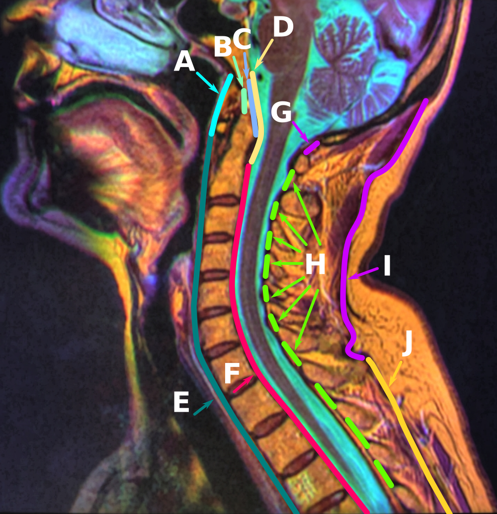
The anterior atlantoaxial ligament is a strong membrane, fixed above the lower border of the anterior arch of the atlas; below, to the front of the body of the axis. It is strengthened in the middle line by a rounded cord, which connects the tubercle on the anterior arch of the atlas to the body of the axis. It is a continuation upward of the anterior longitudinal ligament. Structure Anatomical relations The anterior atlantoaxial ligament is situated anterior to the longus capitis muscle The longus capitis muscle (Latin for ''long muscle of the head'', alternatively rectus capitis anticus major) is broad and thick above, narrow below, and arises by four tendinous slips, from the anterior tubercles of the transverse processes of t .... See also * Atlanto-axial joint References External links Description at spineuniverse.com Ligaments of the head and neck Bones of the vertebral column {{Portal bar, Anatomy ... [...More Info...] [...Related Items...] OR: [Wikipedia] [Google] [Baidu] |
Sagittal Section
The sagittal plane (; also known as the longitudinal plane) is an anatomical plane that divides the body into right and left sections. It is perpendicular to the transverse and coronal planes. The plane may be in the center of the body and divide it into two equal parts ( mid-sagittal), or away from the midline and divide it into unequal parts (para-sagittal). The term ''sagittal'' was coined by Gerard of Cremona. Variations in terminology Examples of sagittal planes include: * The terms '' median plane'' or ''mid-sagittal plane'' are sometimes used to describe the sagittal plane running through the midline. This plane cuts the body into halves (assuming bilateral symmetry), passing through midline structures such as the navel and spine. It is one of the planes which, combined with the umbilical plane, defines the four quadrants of the human abdomen. * The term ''parasagittal'' is used to describe any plane parallel or adjacent to a given sagittal plane. Specific named par ... [...More Info...] [...Related Items...] OR: [Wikipedia] [Google] [Baidu] |
Occipital Bone
The occipital bone () is a neurocranium, cranial dermal bone and the main bone of the occiput (back and lower part of the skull). It is trapezoidal in shape and curved on itself like a shallow dish. The occipital bone lies over the occipital lobes of the cerebrum. At the base of the skull in the occipital bone, there is a large oval opening called the foramen magnum, which allows the passage of the spinal cord. Like the other cranial bones, it is classed as a flat bone. Due to its many attachments and features, the occipital bone is described in terms of separate parts. From its front to the back is the basilar part of occipital bone, basilar part, also called the basioccipital, at the sides of the foramen magnum are the lateral parts of occipital bone, lateral parts, also called the exoccipitals, and the back is named as the squamous part of occipital bone, squamous part. The basilar part is a thick, somewhat quadrilateral piece in front of the foramen magnum and directed toward ... [...More Info...] [...Related Items...] OR: [Wikipedia] [Google] [Baidu] |
Cervical Vertebrae
In tetrapods, cervical vertebrae (: vertebra) are the vertebrae of the neck, immediately below the skull. Truncal vertebrae (divided into thoracic and lumbar vertebrae in mammals) lie caudal (toward the tail) of cervical vertebrae. In sauropsid species, the cervical vertebrae bear cervical ribs. In lizards and saurischian dinosaurs, the cervical ribs are large; in birds, they are small and completely fused to the vertebrae. The vertebral transverse processes of mammals are homologous to the cervical ribs of other amniotes. Most mammals have seven cervical vertebrae, with the only three known exceptions being the manatee with six, the two-toed sloth with five or six, and the three-toed sloth with nine. In humans, cervical vertebrae are the smallest of the true vertebrae and can be readily distinguished from those of the thoracic or lumbar regions by the presence of a transverse foramen, an opening in each transverse process, through which the vertebral artery, verteb ... [...More Info...] [...Related Items...] OR: [Wikipedia] [Google] [Baidu] |
Anterior Atlantooccipital Membrane
The anterior atlantooccipital membrane (anterior atlantooccipital ligament) is a broad, dense membrane extending between the anterior margin of the foramen magnum (superiorly), and (the superior margin of) the anterior arch of atlas (inferiorly). The membrane helps limit excessive movement at the atlanto-occipital joints. Anatomy Structure It is composed of broad, densely woven fibers; especially towards the midline where the membrane is continuous medially with the anterior longitudinal ligament. It is innervated by the cervical spinal nerve 1. Relations Medially, it is continuous with the anterior longitudinal ligament. Laterally, it is blends with either articular capsule In anatomy, a joint capsule or articular capsule is an envelope surrounding a synovial joint. [...More Info...] [...Related Items...] OR: [Wikipedia] [Google] [Baidu] |
Axis (anatomy)
In anatomy, the axis (from Latin ''axis'', "axle") is the second cervical vertebra (C2) of the spine, immediately inferior to the atlas, upon which the head rests. The spinal cord passes through the axis. The defining feature of the axis is its strong bony protrusion known as the dens, which rises from the superior aspect of the bone. Structure The body is deeper in front or in the back and is prolonged downward anteriorly to overlap the upper and front part of the third vertebra. It presents a median longitudinal ridge in front, separating two lateral depressions for the attachment of the longus colli muscles. Dens The dens, also called the odontoid process, or the peg, is the most pronounced projecting feature of the axis. The dens exhibits a slight constriction where it joins the main body of the vertebra. The condition where the dens is separated from the body of the axis is called ''os odontoideum'' and may cause nerve and circulation compression syndrome. On its an ... [...More Info...] [...Related Items...] OR: [Wikipedia] [Google] [Baidu] |
Anterior Longitudinal Ligament
The anterior longitudinal ligament is a ligament that extends across the anterior/ventral aspect of the vertebral bodies and intervertebral discs the spine. It may be partially cut to treat certain abnormal curvatures in the vertebral column, such as kyphosis. Anatomy The anterior longitudinal ligament extends superoinferiorly between the basiocciput of the skull and the anterior tubercle of the atlas (cervical vertebra C1) superiorly, and the superior part of the sacrum inferiorly; inferiorly, it ends at the sacral promontory. It broadens inferiorly. Inferiorly, it becomes continuous with the anterior sacrococcygeal ligament. Superiorly, between the skull and atlas, the ligament is continuous laterally with the anterior atlantooccipital membrane. The ligament is thick and slightly more narrow over the vertebral bodies and thinner but slightly wider over the intervertebral discs. It tends to be narrower and thicker around thoracic vertebrae, and wider and thinner around ... [...More Info...] [...Related Items...] OR: [Wikipedia] [Google] [Baidu] |
Longi Capitis
The longus capitis muscle (Latin for ''long muscle of the head'', alternatively rectus capitis anticus major) is broad and thick above, narrow below, and arises by four tendinous slips, from the anterior tubercles of the transverse processes of the third, fourth, fifth, and sixth cervical vertebræ, and ascends, converging toward its fellow of the opposite side, to be inserted into the inferior surface of the basilar part of the occipital bone The occipital bone () is a neurocranium, cranial dermal bone and the main bone of the occiput (back and lower part of the skull). It is trapezoidal in shape and curved on itself like a shallow dish. The occipital bone lies over the occipital lob .... It is innervated by a branch of cervical plexus. Longus capitis has several actions: acting unilaterally, to: *flex the head and neck laterally *rotate the head ipsilaterally acting bilaterally: *flex the head and neck Additional images File:Gray129.png, Occipital bone. Outer surface. ... [...More Info...] [...Related Items...] OR: [Wikipedia] [Google] [Baidu] |
Atlanto-axial Joint
The atlanto-axial joint is a joint in the upper part of the neck between the atlas bone and the axis bone, which are the first and second cervical vertebrae. It is a pivot joint, that can start from C2 To C7. Structure The atlanto-axial joint is a joint between the atlas bone and the axis bone, which are the first and second cervical vertebrae. It is a pivot joint that provides 40 to 70% of axial rotation of the head. There is a pivot articulation between the odontoid process of the axis and the ring formed by the anterior arch and the transverse ligament of the atlas. Lateral and median joints There are three atlanto-axial joints: one median and two lateral: * The median atlanto-axial joint is sometimes considered a triple joint: ** one between the posterior surface of the anterior arch of atlas and the front of the odontoid process ** one between the anterior surface of the ligament and the back of the odontoid process * The lateral atlantoaxial joint involves the ... [...More Info...] [...Related Items...] OR: [Wikipedia] [Google] [Baidu] |
Ligaments Of The Head And Neck
A ligament is a type of Connective tissue#Types, fibrous connective tissue in the body that connects bones to other bones. It also connects Flight_feather#Remiges, flight feathers to bones, in dinosaurs and birds. All 30,000 species of amniotes (land animals with internal bones) have ligaments. It is also known as ''articular ligament'', ''articular larua'', ''fibrous ligament'', or ''true ligament''. Comparative anatomy Ligaments are similar to tendons and fasciae as they are all made of connective tissue. The differences among them are in the connections that they make: ligaments connect one bone to another bone, tendons connect muscle to bone, and fasciae connect muscles to other muscles. These are all found in the skeletal system of the human body. Ligaments cannot usually be regenerated naturally; however, there are periodontal ligament stem cells located near the periodontal ligament which are involved in the adult regeneration of periodontist ligament. The study of li ... [...More Info...] [...Related Items...] OR: [Wikipedia] [Google] [Baidu] |


