|
Acrokeratoelastoidosis Of Costa
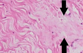
Acrokeratoelastoidosis of Costa or Acrokeratoelastoidosis is a hereditary form of marginal keratoderma, and can be defined as a palmoplantar keratoderma. It is distinguished by tiny, firm pearly or warty papules on the sides of the hands and, occasionally, the feet. It is less common than the hereditary type of marginal keratoderma, keratoelastoidosis marginalis. Costa described acrokeratoelastoidosis in 1953, as a result, it is also known as Costa acrokeratoelastoidosis. Acrokeratoelastoidosis is a form of punctate palmoplantar keratoderma type 3 characterized by keratin and elastic tissue abnormalities. There have been autosomal dominant and sporadic forms observed. Acrokeratoelastoidosis is not congenital; it develops gradually during puberty, or sometimes afterwards, and then stabilizes. In most cases, no treatment is required. Signs and symptoms Acrokeratoelastoidosis typically manifests in children and adolescents, though reports of adult onset exist. Acrokeratoelastoido ... [...More Info...] [...Related Items...] OR: [Wikipedia] [Google] [Baidu] |
Dermatology
Dermatology is the branch of medicine dealing with the Human skin, skin.''Random House Webster's Unabridged Dictionary.'' Random House, Inc. 2001. Page 537. . It is a speciality with both medical and surgical aspects. A List of dermatologists, dermatologist is a specialist medical doctor who manages diseases related to skin. Etymology Attested in English in 1819, the word "dermatology" derives from the Ancient Greek, Greek δέρματος (''dermatos''), genitive of δέρμα (''derma''), "skin" (itself from δέρω ''dero'', "to flay") and -λογία ''wikt:-logia, -logia''. Neo-Latin ''dermatologia'' was coined in 1630, an anatomical term with various French and German uses attested from the 1730s. History In 1708, the first great school of dermatology became a reality at the famous Hôpital Saint-Louis in Paris, and the first textbooks (Willan's, 1798–1808) and atlases (Jean-Louis-Marc Alibert, Alibert's, 1806–1816) appeared in print around the same time.Freedber ... [...More Info...] [...Related Items...] OR: [Wikipedia] [Google] [Baidu] |
Connective Tissue
Connective tissue is one of the four primary types of animal tissue, a group of cells that are similar in structure, along with epithelial tissue, muscle tissue, and nervous tissue. It develops mostly from the mesenchyme, derived from the mesoderm, the middle embryonic germ layer. Connective tissue is found in between other tissues everywhere in the body, including the nervous system. The three meninges, membranes that envelop the brain and spinal cord, are composed of connective tissue. Most types of connective tissue consists of three main components: elastic and collagen fibers, ground substance, and cells. Blood and lymph are classed as specialized fluid connective tissues that do not contain fiber. All are immersed in the body water. The cells of connective tissue include fibroblasts, adipocytes, macrophages, mast cells and leukocytes. The term "connective tissue" (in German, ) was introduced in 1830 by Johannes Peter Müller. The tissue was already recognized as ... [...More Info...] [...Related Items...] OR: [Wikipedia] [Google] [Baidu] |
Elastic Fiber
Elastic fibers (or yellow fibers) are an essential component of the extracellular matrix composed of bundles of proteins (elastin) which are produced by a number of different cell types including fibroblasts, endothelial, smooth muscle, and airway epithelial cells. These fibers are able to stretch many times their length, and snap back to their original length when relaxed without loss of energy. Elastic fibers include elastin, elaunin and oxytalan. Elastic fibers are formed via elastogenesis, a highly complex process involving several key proteins including fibulin-4, fibulin-5, latent transforming growth factor β binding protein 4, and microfibril associated protein 4. In this process tropoelastin, the soluble monomeric precursor to elastic fibers is produced by elastogenic cells and chaperoned to the cell surface. Following excretion from the cell, tropoelastin self associates into ~200 nm particles by coacervation, an entropically driven process involving interact ... [...More Info...] [...Related Items...] OR: [Wikipedia] [Google] [Baidu] |
Homogenization (biology)
Homogenization, in cell biology or molecular biology, is a process whereby different fractions of a biological sample become equal in composition. It can be a disease sign in histopathology, or an intentional process in research: A homogenized sample is equal in composition throughout, so that removing a fraction does not alter the overall molecular make-up of the sample remaining, and is identical to the fraction removed. Induced homogenization in biology is often followed by molecular extraction and various analytical techniques, including ELISA and western blot. Methods Homogenization of tissue in solution is often performed simultaneously with cell lysis. To prevent lysis however, the tissue (or collection of cells, e.g. from cell culture) can be kept at temperatures slightly above zero to prevent Autolysis (biology), autolysis, and in an Isotonicity, isotonic solution to prevent osmosis, osmotic damage. If freezing the tissue is possible, cryohomogenization can be performed un ... [...More Info...] [...Related Items...] OR: [Wikipedia] [Google] [Baidu] |
Acanthosis
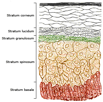
The epidermis is the outermost of the three layers that comprise the skin, the inner layers being the dermis and hypodermis. The epidermal layer provides a barrier to infection from environmental pathogens and regulates the amount of water released from the body into the atmosphere through transepidermal water loss. The epidermis is composed of multiple layers of flattened cells that overlie a base layer (stratum basale) composed of columnar cells arranged perpendicularly. The layers of cells develop from stem cells in the basal layer. The thickness of the epidermis varies from 31.2μm for the penis to 596.6μm for the sole of the foot with most being roughly 90μm. Thickness does not vary between the sexes but becomes thinner with age. The human epidermis is an example of epithelium, particularly a stratified squamous epithelium. The word epidermis is derived through Latin , itself and . Something related to or part of the epidermis is termed epidermal. Structure Cel ... [...More Info...] [...Related Items...] OR: [Wikipedia] [Google] [Baidu] |
Hypergranulosis
Hypergranulosis is an increased thickness of the stratum granulosum. It is seen in skin diseases with epidermal hyperplasia and orthokeratotic hyperkeratosis. See also * Skin lesion * Skin disease * List of skin diseases Many skin conditions affect the human integumentary system—the organ system covering the entire surface of the body and composed of skin, hair, nails, and related muscle and glands. The major function of this system is as a barrier against t ... References Dermatologic terminology {{Cutaneous-condition-stub} ... [...More Info...] [...Related Items...] OR: [Wikipedia] [Google] [Baidu] |
Hyperkeratosis
Hyperkeratosis is thickening of the stratum corneum (the outermost layer of the epidermis, or skin), often associated with the presence of an abnormal quantity of keratin,Kumar, Vinay; Fausto, Nelso; Abbas, Abul (2004) ''Robbins & Cotran Pathologic Basis of Disease'' (7th ed.). Saunders. Page 1230. . and is usually accompanied by an increase in the granular layer. As the corneum layer normally varies greatly in thickness in different sites, some experience is needed to assess minor degrees of hyperkeratosis. It can be caused by vitamin A deficiency or chronic exposure to arsenic. Hyperkeratosis can also be caused by B-Raf inhibitor drugs such as vemurafenib and dabrafenib.Niezgoda, Anna; Niezgoda, Piotr; Czajkowski, Rafal (2015) ''Novel Approaches to Treatment of Advanced Melanoma: A Review of Targeted Therapy and Immunotherapy'' BioMed Research International It can be treated with urea-containing creams, which dissolve the intercellular matrix of the cells of the strat ... [...More Info...] [...Related Items...] OR: [Wikipedia] [Google] [Baidu] |
Histopathology
Histopathology (compound of three Greek words: 'tissue', 'suffering', and '' -logia'' 'study of') is the microscopic examination of tissue in order to study the manifestations of disease. Specifically, in clinical medicine, histopathology refers to the examination of a biopsy or surgical specimen by a pathologist, after the specimen has been processed and histological sections have been placed onto glass slides. In contrast, cytopathology examines free cells or tissue micro-fragments (as "cell blocks "). Collection of tissues Histopathological examination of tissues starts with surgery, biopsy, or autopsy. The tissue is removed from the body or plant, and then, often following expert dissection in the fresh state, placed in a fixative which stabilizes the tissues to prevent decay. The most common fixative is 10% neutral buffered formalin (corresponding to 3.7% w/v formaldehyde in neutral buffered water, such as phosphate buffered saline). Preparation for h ... [...More Info...] [...Related Items...] OR: [Wikipedia] [Google] [Baidu] |
Dermatoscopy
Dermatoscopy, also known as dermoscopy or epiluminescence microscopy, is the examination of skin lesions with a dermatoscope. It is a tool similar to a camera to allow for inspection of skin lesions unobstructed by skin surface reflections. The dermatoscope consists of a magnifier, a light source (polarized or non-polarized), a transparent plate and sometimes a liquid medium between the instrument and the skin. The dermatoscope is often handheld, although there are stationary cameras allowing the capture of whole body images in a single shot. When the images or video clips are digitally captured or processed, the instrument can be referred to as a digital epiluminescence dermatoscope. The image is then analyzed automatically and given a score indicating how dangerous it is. This technique is useful to dermatologists and skin cancer practitioners in distinguishing benign from malignant (cancerous) lesions, especially in the diagnosis of melanoma. Types There are two main types o ... [...More Info...] [...Related Items...] OR: [Wikipedia] [Google] [Baidu] |
Elastic Fiber
Elastic fibers (or yellow fibers) are an essential component of the extracellular matrix composed of bundles of proteins (elastin) which are produced by a number of different cell types including fibroblasts, endothelial, smooth muscle, and airway epithelial cells. These fibers are able to stretch many times their length, and snap back to their original length when relaxed without loss of energy. Elastic fibers include elastin, elaunin and oxytalan. Elastic fibers are formed via elastogenesis, a highly complex process involving several key proteins including fibulin-4, fibulin-5, latent transforming growth factor β binding protein 4, and microfibril associated protein 4. In this process tropoelastin, the soluble monomeric precursor to elastic fibers is produced by elastogenic cells and chaperoned to the cell surface. Following excretion from the cell, tropoelastin self associates into ~200 nm particles by coacervation, an entropically driven process involving interact ... [...More Info...] [...Related Items...] OR: [Wikipedia] [Google] [Baidu] |
Dermal Fibroblast
Dermal fibroblasts are cells within the dermis layer of skin which are responsible for generating connective tissue and allowing the skin to recover from injury. Using organelles (particularly the rough endoplasmic reticulum), dermal fibroblasts generate and maintain the connective tissue which unites separate cell layers. Furthermore, these dermal fibroblasts produce the protein molecules including laminin and fibronectin which comprise the extracellular matrix. By creating the extracellular matrix between the dermis and epidermis, fibroblasts allow the epithelial cells of the epidermis to affix the matrix, thereby allowing the epidermal cells to effectively join together to form the top layer of the skin. Cell progenitors and analogs Dermal fibroblasts are derived from mesenchymal stem cells within the body. Like corneal fibroblasts, dermal fibroblast proliferation can be stimulated by the presence of fibroblast growth factor (FGF). Fibroblasts do not appear to be fully differe ... [...More Info...] [...Related Items...] OR: [Wikipedia] [Google] [Baidu] |
Epidermis
The epidermis is the outermost of the three layers that comprise the skin, the inner layers being the dermis and Subcutaneous tissue, hypodermis. The epidermal layer provides a barrier to infection from environmental pathogens and regulates the amount of water released from the body into the atmosphere through transepidermal water loss. The epidermis is composed of stratified squamous epithelium, multiple layers of flattened cells that overlie a base layer (stratum basale) composed of Epithelium#Cell types, columnar cells arranged perpendicularly. The layers of cells develop from stem cells in the basal layer. The thickness of the epidermis varies from 31.2μm for the penis to 596.6μm for the Sole (foot), sole of the foot with most being roughly 90μm. Thickness does not vary between the sexes but becomes thinner with age. The human epidermis is an example of epithelium, particularly a stratified squamous epithelium. The word epidermis is derived through Latin , itself and . ... [...More Info...] [...Related Items...] OR: [Wikipedia] [Google] [Baidu] |


