|
Acetabular Fracture
Fractures of the acetabulum occur when the head of the femur is driven into the pelvis. This injury is caused by a blow to either the side or front of the knee and often occurs as a dashboard injury accompanied by a fracture of the femur. The acetabulum is a cavity situated on the outer surface of the hip bone, also called the coxal bone or innominate bone. It is made up of three bones, the ilium, ischium, and pubis. Together, the acetabulum and the head of the femur form the hip joint. Fractures of the acetabulum in young individuals usually result from a high energy injury like vehicular accident or feet first fall. In older individuals or those with osteoporosis, a trivial fall may result in acetabular fracture. In 1964, French surgeons Robertt Judet, Jean Judet, and Emile Letournel first described the mechanism, classification, and treatment of acetabular fracture. They classified these fractures into elementary (simple two part) and associated (complex three or more pa ... [...More Info...] [...Related Items...] OR: [Wikipedia] [Google] [Baidu] |
Femur
The femur (; ), or thigh bone, is the proximal bone of the hindlimb in tetrapod vertebrates. The head of the femur articulates with the acetabulum in the pelvic bone forming the hip joint, while the distal part of the femur articulates with the tibia (shinbone) and patella (kneecap), forming the knee joint. By most measures the two (left and right) femurs are the strongest bones of the body, and in humans, the largest and thickest. Structure The femur is the only bone in the upper leg. The two femurs converge medially toward the knees, where they articulate with the proximal ends of the tibiae. The angle of convergence of the femora is a major factor in determining the femoral-tibial angle. Human females have thicker pelvic bones, causing their femora to converge more than in males. In the condition ''genu valgum'' (knock knee) the femurs converge so much that the knees touch one another. The opposite extreme is ''genu varum'' (bow-leggedness). In the general pop ... [...More Info...] [...Related Items...] OR: [Wikipedia] [Google] [Baidu] |
Trochanter
A trochanter is a Tubercle (human skeleton), tubercle of the femur near its joint with the hip bone. In humans and most mammals, the trochanters serve as important muscle attachment sites. Humans are known to have three trochanters, though the anatomic "normal" includes only the Greater trochanter, greater and Lesser trochanter, lesser trochanters. (The third trochanter is not present in all specimens.) Etymology "Trokhos" (Greek) = "wheel", with reference to the spherical femoral head which was first named "trokhanter". Later usage came to include the femoral neck. Structure In human anatomy, the trochanter is a part of the femur. It can refer to: * Greater trochanter * Lesser trochanter * Third trochanter, which is occasionally present Other animals * Fourth trochanter, of archosaur leg bones * Trochanter (arthropod leg), a segment of the arthropod leg See also * Intertrochanteric crest * Intertrochanteric line References External links * * {{Bones of lower ... [...More Info...] [...Related Items...] OR: [Wikipedia] [Google] [Baidu] |
Transverse Fracture
A bone fracture (abbreviated FRX or Fx, Fx, or #) is a medical condition in which there is a partial or complete break in the continuity of any bone in the body. In more severe cases, the bone may be broken into several fragments, known as a ''comminuted fracture''. A bone fracture may be the result of high force impact or stress, or a minimal trauma injury as a result of certain medical conditions that weaken the bones, such as osteoporosis, osteopenia, bone cancer, or osteogenesis imperfecta, where the fracture is then properly termed a pathologic fracture. Signs and symptoms Although bone tissue contains no pain receptors, a bone fracture is painful for several reasons: * Breaking in the continuity of the periosteum, with or without similar discontinuity in endosteum, as both contain multiple pain receptors. * Edema and hematoma of nearby soft tissues caused by ruptured bone marrow evokes pressure pain. * Involuntary muscle spasms trying to hold bone fragments in place. Da ... [...More Info...] [...Related Items...] OR: [Wikipedia] [Google] [Baidu] |
Mastoid Wall Of Tympanic Cavity
The tympanic cavity is a small cavity surrounding the bones of the middle ear. Within it sit the ossicles, three small bones that transmit vibrations used in the detection of sound. Structure On its lateral surface, it abuts the external auditory meatus ear canal from which it is separated by the tympanic membrane (eardrum). Walls The tympanic cavity is bounded by: * Facing the inner ear, the medial wall (or ''labyrinthic wall'', ''labyrinthine wall'') is vertical, and has the oval window and round window, the promontory, and the prominence of the facial canal. * Facing the outer ear, the lateral wall (or ''membranous wall''), is formed mainly by the tympanic membrane, partly by the ring of bone into which this membrane is inserted. This ring of bone is incomplete at its upper part, forming a notch (notch of Rivinus), close to which are three small apertures: the "iter chordæ posterius", the petrotympanic fissure, and the "iter chordæ anterius". The iter chordæ posterius (aper ... [...More Info...] [...Related Items...] OR: [Wikipedia] [Google] [Baidu] |
Heterotopic Ossification
Heterotopic ossification (HO) is the process by which bone tissue forms outside of the skeleton in muscles and soft tissue. Symptoms In traumatic heterotopic ossification (traumatic myositis ossificans), the patient may complain of a warm, tender, firm swelling in a muscle and decreased range of motion in the joint served by the muscle involved. There is often a history of a blow or other trauma to the area a few weeks to a few months earlier. Patients with traumatic neurological injuries, severe neurologic disorders or severe burns who develop heterotopic ossification experience limitation of motion in the areas affected. Causes Heterotopic ossification of varying severity can be caused by surgery or trauma to the hips and legs. About every third patient who has total hip arthroplasty (joint replacement) or a severe fracture of the long bones of the lower leg will develop heterotopic ossification, but is uncommonly symptomatic. Between 50% and 90% of patients who develop ... [...More Info...] [...Related Items...] OR: [Wikipedia] [Google] [Baidu] |
Pulmonary Embolism
Pulmonary embolism (PE) is a blockage of an artery in the lungs by a substance that has moved from elsewhere in the body through the bloodstream (embolism). Symptoms of a PE may include shortness of breath, chest pain particularly upon breathing in, and coughing up blood. Symptoms of a blood clot in the leg may also be present, such as a red, warm, swollen, and painful leg. Signs of a PE include low blood oxygen levels, rapid breathing, rapid heart rate, and sometimes a mild fever. Severe cases can lead to passing out, abnormally low blood pressure, obstructive shock, and sudden death. PE usually results from a blood clot in the leg that travels to the lung. The risk of blood clots is increased by advanced age, cancer, prolonged bed rest and immobilization, smoking, stroke, long-haul travel over 4 hours, certain genetic conditions, estrogen-based medication, pregnancy, obesity, trauma or bone fracture, and after some types of surgery. A small proportion of cases are due ... [...More Info...] [...Related Items...] OR: [Wikipedia] [Google] [Baidu] |
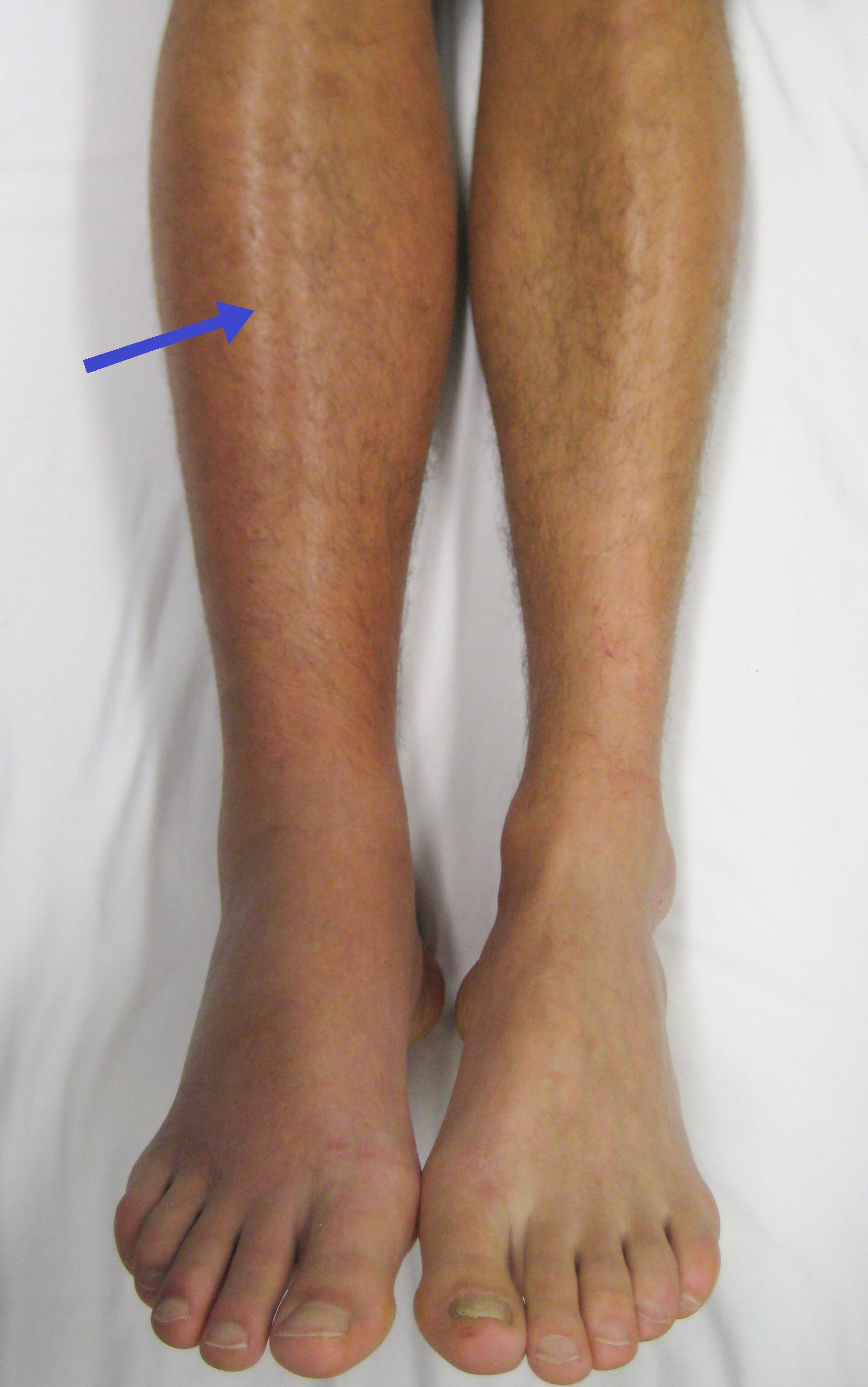
Deep Vein Thrombosis
Deep vein thrombosis (DVT) is a type of venous thrombosis involving the formation of a blood clot in a deep vein, most commonly in the legs or pelvis. A minority of DVTs occur in the arms. Symptoms can include pain, swelling, redness, and enlarged veins in the affected area, but some DVTs have no symptoms. The most common life-threatening concern with DVT is the potential for a clot to embolize (detach from the veins), travel as an embolus through the right side of the heart, and become lodged in a pulmonary artery that supplies blood to the lungs. This is called a pulmonary embolism (PE). DVT and PE comprise the cardiovascular disease of venous thromboembolism (VTE). About two-thirds of VTE manifests as DVT only, with one-third manifesting as PE with or without DVT. The most frequent long-term DVT complication is post-thrombotic syndrome, which can cause pain, swelling, a sensation of heaviness, itching, and in severe cases, ulcers. Recurrent VTE occurs in about 30% ... [...More Info...] [...Related Items...] OR: [Wikipedia] [Google] [Baidu] |
Femoral Head
The femoral head (femur head or head of the femur) is the highest part of the thigh bone ( femur). It is supported by the femoral neck. Structure The head is globular and forms rather more than a hemisphere, is directed upward, medialward, and a little forward, the greater part of its convexity being above and in front. The femoral head's surface is smooth. It is coated with cartilage in the fresh state, except over an ovoid depression, the fovea capitis, which is situated a little below and behind the center of the femoral head, and gives attachment to the ligament of head of femur. The thickest region of the articular cartilage is at the centre of the femoral head, measuring up to 2.8 mm. The diameter of the femoral head is usually larger in men than in women. Fovea capitis The fovea capitis is a small, concave depression within the head of the femur that serves as an attachment point for the ligamentum teres (Saladin). It is slightly ovoid in shape and is oriented "super ... [...More Info...] [...Related Items...] OR: [Wikipedia] [Google] [Baidu] |


