|
Acetabular Branch Of Medial Circumflex Femoral Artery
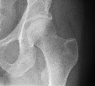
The acetabular branch is an artery in the hip that arises from the medial circumflex femoral artery opposite the acetabular notch and enters the hip-joint beneath the transverse ligament in company with an articular branch from the obturator artery. It supplies the fat in the bottom of the acetabulum, and is continued along the ligament to the head of the femur The femur (; ), or thigh bone, is the proximal bone of the hindlimb in tetrapod vertebrates. The head of the femur articulates with the acetabulum in the pelvic bone forming the hip joint, while the distal part of the femur articulates wit .... References Arteries of the lower limb {{circulatory-stub ... [...More Info...] [...Related Items...] OR: [Wikipedia] [Google] [Baidu] |
Profunda Femoris Artery
The deep artery of the thigh, (profunda femoris artery or deep femoral artery) is a large branch of the femoral artery. It travels more deeply (posteriorly) than the rest of the femoral artery. Structure The deep artery of the thigh branches off the posterolateral side of the femoral artery soon after its origin. It travels down the thigh closer to the femur than the femoral artery. It runs between the pectineus muscle and the adductor longus muscle. It runs on the posterior side of adductor longus muscle. It pierces the adductor magnus muscle, and may be known as the fourth perforating artery as it continues. The deep femoral artery does not leave the thigh. Branches The deep artery of the thigh gives off the following branches: * Lateral circumflex femoral artery. * Medial circumflex femoral artery. * 3 Perforating arteries - perforate the adductor magnus muscle to the posterior and medial compartments of the thigh to connect with the branches of the popliteal artery behi ... [...More Info...] [...Related Items...] OR: [Wikipedia] [Google] [Baidu] |
Femoral Artery
The femoral artery is a large artery in the thigh and the main arterial supply to the thigh and leg. The femoral artery gives off the deep femoral artery or profunda femoris artery and descends along the anteromedial part of the thigh in the femoral triangle. It enters and passes through the adductor canal, and becomes the popliteal artery as it passes through the adductor hiatus in the adductor magnus near the junction of the middle and distal thirds of the thigh. Structure The femoral artery enters the thigh from behind the inguinal ligament as the continuation of the external iliac artery. Here, it lies midway between the anterior superior iliac spine and the symphysis pubis (Mid-inguinal point). Segments In clinical parlance, the femoral artery has the following segments: *The common femoral artery (CFA) is the segment of the femoral artery between the inferior margin of the inguinal ligament and the branching point of the deep femoral artery/profunda femoris arte ... [...More Info...] [...Related Items...] OR: [Wikipedia] [Google] [Baidu] |
Thigh
In human anatomy, the thigh is the area between the hip ( pelvis) and the knee. Anatomically, it is part of the lower limb. The single bone in the thigh is called the femur. This bone is very thick and strong (due to the high proportion of bone tissue), and forms a ball and socket joint at the hip, and a modified hinge joint at the knee. Structure Bones The femur is the only bone in the thigh and serves as an attachment site for all muscles in the thigh. The head of the femur articulates with the acetabulum in the pelvic bone forming the hip joint, while the distal part of the femur articulates with the tibia and patella forming the knee. By most measures, the femur is the strongest bone in the body. The femur is also the longest bone in the body. The femur is categorised as a long bone and comprises a diaphysis, the shaft (or body) and two epiphysis or extremities that articulate with adjacent bones in the hip and knee. Muscular compartments In cross-section, ... [...More Info...] [...Related Items...] OR: [Wikipedia] [Google] [Baidu] |
Medial Circumflex Femoral Artery
The medial circumflex femoral artery (internal circumflex artery, medial femoral circumflex artery) is an artery in the upper thigh that arises from the profunda femoris artery''.'' Damage to the artery following a femoral neck fracture may lead to avascular necrosis (ischemic) of the femoral neck/head. Structure Origin The medial femoral circumflex artery arises from the posterior medial aspect of the profunda femoris artery''.'' The medial femoral circumflex artery may occasionally arise directly from the femoral artery. Course and relations It winds around the medial side of the femur, passing first between the pectineus and iliopsoas muscles, and then between the obturator externus and the adductor brevis muscles. Branches At the upper border of the adductor brevis it gives off two branches: * The '' ascending branch'' * The ''descending branch'' descends beneath the adductor brevis, to supply it and the adductor magnus; the continuation of the vessel passes backward ... [...More Info...] [...Related Items...] OR: [Wikipedia] [Google] [Baidu] |
Artery
An artery (plural arteries) () is a blood vessel in humans and most animals that takes blood away from the heart to one or more parts of the body (tissues, lungs, brain etc.). Most arteries carry oxygenated blood; the two exceptions are the pulmonary and the umbilical arteries, which carry deoxygenated blood to the organs that oxygenate it (lungs and placenta, respectively). The effective arterial blood volume is that extracellular fluid which fills the arterial system. The arteries are part of the circulatory system, that is responsible for the delivery of oxygen and nutrients to all cells, as well as the removal of carbon dioxide and waste products, the maintenance of optimum blood pH, and the circulation of proteins and cells of the immune system. Arteries contrast with veins, which carry blood back towards the heart. Structure The anatomy of arteries can be separated into gross anatomy, at the macroscopic level, and microanatomy, which must be studied with a ... [...More Info...] [...Related Items...] OR: [Wikipedia] [Google] [Baidu] |
Medial Circumflex Femoral Artery
The medial circumflex femoral artery (internal circumflex artery, medial femoral circumflex artery) is an artery in the upper thigh that arises from the profunda femoris artery''.'' Damage to the artery following a femoral neck fracture may lead to avascular necrosis (ischemic) of the femoral neck/head. Structure Origin The medial femoral circumflex artery arises from the posterior medial aspect of the profunda femoris artery''.'' The medial femoral circumflex artery may occasionally arise directly from the femoral artery. Course and relations It winds around the medial side of the femur, passing first between the pectineus and iliopsoas muscles, and then between the obturator externus and the adductor brevis muscles. Branches At the upper border of the adductor brevis it gives off two branches: * The '' ascending branch'' * The ''descending branch'' descends beneath the adductor brevis, to supply it and the adductor magnus; the continuation of the vessel passes backward ... [...More Info...] [...Related Items...] OR: [Wikipedia] [Google] [Baidu] |
Acetabular Notch by the transverse acetabular ligament; through the foramen nutrient vessels and nerves enter the joint; the margins of the notch serve for the attachment of the The acetabular notch is a deep notch in the acetabulum of the hip bone. The acetabular notch is continuous with a circular non-articular depression, the acetabular fossa, at the bottom of the cavity: this depression is perforated by numerous apertures, and lodges a mass of fat. The notch is converted into a foramen In anatomy and osteology, a foramen (; in [...More Info...] [...Related Items...] OR: [Wikipedia] [Google] [Baidu] |
Hip-joint
In vertebrate anatomy, hip (or "coxa"Latin ''coxa'' was used by Celsus in the sense "hip", but by Pliny the Elder in the sense "hip bone" (Diab, p 77) in medical terminology) refers to either an anatomical region or a joint. The hip region is located lateral and anterior to the gluteal region, inferior to the iliac crest, and overlying the greater trochanter of the femur, or "thigh bone". In adults, three of the bones of the pelvis have fused into the hip bone or acetabulum which forms part of the hip region. The hip joint, scientifically referred to as the acetabulofemoral joint (''art. coxae''), is the joint between the head of the femur and acetabulum of the pelvis and its primary function is to support the weight of the body in both static (e.g., standing) and dynamic (e.g., walking or running) postures. The hip joints have very important roles in retaining balance, and for maintaining the pelvic inclination angle. Pain of the hip may be the result of numerous cau ... [...More Info...] [...Related Items...] OR: [Wikipedia] [Google] [Baidu] |
Obturator Artery
The obturator artery is a branch of the internal iliac artery that passes antero-inferiorly (forwards and downwards) on the lateral wall of the pelvis, to the upper part of the obturator foramen, and, escaping from the pelvic cavity through the obturator canal, it divides into both an anterior and a posterior branch. Structure In the pelvic cavity this vessel is in relation, laterally, with the obturator fascia; medially, with the ureter, ductus deferens, and peritoneum; while a little below it is the obturator nerve. The obturator artery usually arises from the internal iliac artery. Inside the pelvis the obturator artery gives off iliac branches to the iliac fossa, which supply the bone and the Iliacus, and anastomose with the ilio-lumbar artery; a vesical branch, which runs backward to supply the bladder; and a pubic branch, which is given off from the vessel just before it leaves the pelvic cavity. The pubic branch ascends upon the back of the pubis, communicating with the c ... [...More Info...] [...Related Items...] OR: [Wikipedia] [Google] [Baidu] |
Acetabulum
The acetabulum (), also called the cotyloid cavity, is a concave surface of the pelvis. The head of the femur meets with the pelvis at the acetabulum, forming the hip joint. Structure There are three bones of the ''os coxae'' (hip bone) that come together to form the ''acetabulum''. Contributing a little more than two-fifths of the structure is the ischium, which provides lower and side boundaries to the acetabulum. The ilium forms the upper boundary, providing a little less than two-fifths of the structure of the acetabulum. The rest is formed by the pubis, near the midline. It is bounded by a prominent uneven rim, which is thick and strong above, and serves for the attachment of the acetabular labrum, which reduces its opening, and deepens the surface for formation of the hip joint. At the lower part of the ''acetabulum'' is the acetabular notch, which is continuous with a circular depression, the acetabular fossa, at the bottom of the cavity of the ''acetabulum''. The ... [...More Info...] [...Related Items...] OR: [Wikipedia] [Google] [Baidu] |
Ligament Of Head Of Femur
In human anatomy, the ligament of the head of the femur (round ligament of the femur, ligamentum teres femoris, the foveal ligament, or Fillmore’s ligament) is a ligament located in the hip. It is triangular in shape and somewhat flattened. The ligament is implanted by its apex into the antero-superior part of the fovea capitis femoris and its base is attached by two bands, one into either side of the acetabular notch, and between these bony attachments it blends with the transverse ligament.'' Gray's Anatomy'' (1918), see infobox It is ensheathed by the synovial membrane The synovial membrane (also known as the synovial stratum, synovium or stratum synoviale) is a specialized connective tissue that lines the inner surface of capsules of synovial joints and tendon sheath A tendon sheath is a layer of synovial m ..., and varies greatly in strength in different subjects; occasionally only the synovial fold exists, and in rare cases even this is absent. The ligament of t ... [...More Info...] [...Related Items...] OR: [Wikipedia] [Google] [Baidu] |
Femur Head
The femoral head (femur head or head of the femur) is the highest part of the thigh bone (femur). It is supported by the femoral neck. Structure The head is globular and forms rather more than a hemisphere, is directed upward, medialward, and a little forward, the greater part of its convexity being above and in front. The femoral head's surface is smooth. It is coated with cartilage in the fresh state, except over an ovoid depression, the fovea capitis, which is situated a little below and behind the center of the femoral head, and gives attachment to the ligament of head of femur. The thickest region of the articular cartilage is at the centre of the femoral head, measuring up to 2.8 mm. The diameter of the femoral head is usually larger in men than in women. Fovea capitis The fovea capitis is a small, concave depression within the head of the femur that serves as an attachment point for the ligamentum teres (Saladin). It is slightly ovoid in shape and is oriented "superior ... [...More Info...] [...Related Items...] OR: [Wikipedia] [Google] [Baidu] |


