|
Acardiac Twin
Twin reversed arterial perfusion sequence, also called TRAP sequence, TRAPS, or acardiac twinning, is a rare complication of monochorionic twin pregnancies. It is a severe variant of twin-to-twin transfusion syndrome (TTTS). In addition to the twins' blood systems being connected instead of independent, one twin, called the acardiac twin, TRAP fetus or acardius, is severely malformed. The heart is absent or deformed, hence the name "acardiac", as are the upper structures of the body. The other limbs may be partially present or missing, and internal structures of the torso are often poorly formed. The other twin is usually normal in appearance. The normal twin, called the pump twin, drives blood through both fetuses. It is called "reversed arterial perfusion" because in the acardiac twin the blood flows in a reversed direction. TRAP sequence occurs in 1% of monochorionic twin pregnancies and 1 in 35,000 pregnancies overall. Acardiac twin The acardiac twin is a parasitic twin th ... [...More Info...] [...Related Items...] OR: [Wikipedia] [Google] [Baidu] |
Selective Reduction
Selective reduction is the practice of reducing the number of fetuses in a multiple pregnancy, such as quadruplets, to a twin or singleton pregnancy. The procedure is also called multifetal pregnancy reduction. The procedure is most commonly done to reduce the number of fetuses in a multiple pregnancy to a safe number, when the multiple pregnancy is the result of use of assisted reproductive technology; outcomes for both the mother and the babies are generally worse the higher the number of fetuses. The procedure is also used in multiple pregnancies when one of the fetuses has a serious and incurable disease, or in the case where one of the fetuses is outside the uterus, in which case it is called selective termination. The procedure generally takes two days; the first day for testing to select which fetuses to reduce, and the 2nd day for the procedure itself, in which potassium chloride is injected into the heart of each selected fetus under the guidance of ultrasound imaging. ... [...More Info...] [...Related Items...] OR: [Wikipedia] [Google] [Baidu] |
Conjoined Twins
Conjoined twins, popularly referred to as Siamese twins, are twins joined '' in utero''. It is a very rare phenomenon, estimated to occur in anywhere between one in 50,000 births to one in 200,000 births, with a somewhat higher incidence in southwest Asia and Africa. Approximately half are stillborn, and an additional one-third die within 24 hours. Most live births are female, with a ratio of 3:1. Two possible explanations of the cause of conjoined twins have been proposed. The one that is generally accepted is ''fission'', in which the fertilized egg splits partially. The other explanation, no longer believed to be accurate, is ''fusion'', in which the fertilized egg completely separates, but stem cells (that search for similar cells) find similar stem cells on the other twin and fuse the twins together. Conjoined twins and some monozygotic, but not conjoined, twins share a single common chorion, placenta, and amniotic sac '' in utero''. Chang and Eng Bunker (1811–1874) we ... [...More Info...] [...Related Items...] OR: [Wikipedia] [Google] [Baidu] |
Neonatal Intensive Care Unit
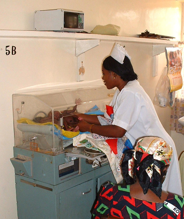
A neonatal intensive care unit (NICU), also known as an intensive care nursery (ICN), is an intensive care unit (ICU) specializing in the care of ill or premature newborn infants. The NICU is divided into several areas, including a critical care area for babies who require close monitoring and intervention, an intermediate care area for infants who are stable but still require specialized care, and a step down unit where babies who are ready to leave the hospital can receive additional care before being discharged. Neonatal refers to the first 28 days of life. Neonatal care, as known as specialized nurseries or intensive care, has been around since the 1960s. The first American newborn intensive care unit, designed by Louis Gluck, was opened in October 1960 at Yale New Haven Hospital. An NICU is typically directed by one or more neonatologists and staffed by resident physicians, nurses, nurse practitioners, pharmacists, physician assistants, respiratory therapists, and ... [...More Info...] [...Related Items...] OR: [Wikipedia] [Google] [Baidu] |
Obstetrician
Obstetrics is the field of study concentrated on pregnancy, childbirth and the postpartum period. As a medical specialty, obstetrics is combined with gynecology under the discipline known as obstetrics and gynecology (OB/GYN), which is a surgical field. Main areas Prenatal care Prenatal care is important in screening for various complications of pregnancy. This includes routine office visits with physical exams and routine lab tests along with telehealth care for women with low-risk pregnancies: Image:Ultrasound_image_of_a_fetus.jpg, 3D ultrasound of fetus (about 14 weeks gestational age) Image:Sucking his thumb and waving.jpg, Fetus at 17 weeks Image:3dultrasound 20 weeks.jpg, Fetus at 20 weeks First trimester Routine tests in the first trimester of pregnancy generally include: * Complete blood count * Blood type ** Rh-negative antenatal patients should receive RhoGAM at 28 weeks to prevent Rh disease. * Indirect Coombs test (AGT) to assess risk of hemoly ... [...More Info...] [...Related Items...] OR: [Wikipedia] [Google] [Baidu] |
Echocardiogram
Echocardiography, also known as cardiac ultrasound, is the use of ultrasound to examine the heart. It is a type of medical imaging, using standard ultrasound or Doppler ultrasound. The visual image formed using this technique is called an echocardiogram, a cardiac echo, or simply an echo. Echocardiography is routinely used in the diagnosis, management, and follow-up of patients with any suspected or known heart diseases. It is one of the most widely used diagnostic imaging modalities in cardiology. It can provide a wealth of helpful information, including the size and shape of the heart (internal chamber size quantification), pumping capacity, location and extent of any tissue damage, and assessment of valves. An echocardiogram can also give physicians other estimates of heart function, such as a calculation of the cardiac output, ejection fraction, and diastolic function (how well the heart relaxes). Echocardiography is an important tool in assessing wall motion abnormality ... [...More Info...] [...Related Items...] OR: [Wikipedia] [Google] [Baidu] |
Radiofrequency Ablation
Radiofrequency ablation (RFA), also called fulguration, is a medical procedure in which part of the electrical conduction system of the heart, tumor, sensory nerves or a dysfunctional tissue is ablated using the heat generated from medium frequency alternating current (in the range of 350–500 kHz). RFA is generally conducted in the outpatient setting, using either a local anesthetic or twilight anesthesia. When it is delivered via catheter, it is called radiofrequency catheter ablation. Two advantages of radio frequency current (over previously used low frequency AC or pulses of DC) are that it does not directly stimulate nerves or heart muscle, and therefore can often be used without the need for general anesthesia, and that it is specific for treating the desired tissue without significant collateral damage. Due to this, RFA is an alternative for eligible patients who have comorbidities or do not want to undergo surgery. Documented benefits have led to RFA becomin ... [...More Info...] [...Related Items...] OR: [Wikipedia] [Google] [Baidu] |
Amniocentesis
Amniocentesis is a medical procedure used primarily in the prenatal diagnosis of genetic conditions. It has other uses such as in the assessment of infection and fetal lung maturity. Prenatal diagnostic testing, which includes amniocentesis, is necessary to conclusively diagnose the majority of genetic disorders, with amniocentesis being the gold-standard procedure after 15 weeks' gestation. In this procedure, a thin needle is inserted into the abdomen of the pregnant woman. The needle punctures the amnion, which is the membrane that surrounds the developing fetus. The fluid within the amnion is called amniotic fluid, and because this fluid surrounds the developing fetus, it contains fetal cells. The amniotic fluid is sampled and analyzed via methods such as karyotyping and DNA analysis technology for genetic abnormalities. An amniocentesis is typically performed in the second trimester between the 15th and 20th week of gestation. Women who choose to have this test are prima ... [...More Info...] [...Related Items...] OR: [Wikipedia] [Google] [Baidu] |
Medical Ultrasonography
Medical ultrasound includes Medical diagnosis, diagnostic techniques (mainly medical imaging, imaging) using ultrasound, as well as therapeutic ultrasound, therapeutic applications of ultrasound. In diagnosis, it is used to create an image of internal body structures such as tendons, muscles, joints, blood vessels, and internal organs, to measure some characteristics (e.g., distances and velocities) or to generate an informative audible sound. The usage of ultrasound to produce visual images for medicine is called medical ultrasonography or simply sonography, or echography. The practice of examining pregnant women using ultrasound is called obstetric ultrasonography, and was an early development of clinical ultrasonography. The machine used is called an ultrasound machine, a sonograph or an echograph. The visual image formed using this technique is called an ultrasonogram, a sonogram or an echogram. Ultrasound is composed of sound waves with frequency, frequencies greater than ... [...More Info...] [...Related Items...] OR: [Wikipedia] [Google] [Baidu] |
Monochorionic
Monochorionic twins are monozygotic (identical) twins that share the same placenta. If the placenta is shared by more than two twins (see multiple birth), these are monochorionic multiples. Monochorionic twins occur in 0.3% of all pregnancies. Seventy-five percent of monozygotic twin pregnancies are monochorionic; the remaining 25% are ''dichorionic diamniotic''. If the placenta divides, this takes place before the third day after fertilization. Amniocity and zygosity Monochorionic twins generally have two amniotic sacs (called Monochorionic-Diamniotic "MoDi"), but sometimes, in the case of monoamniotic twins (Monochorionic-Monoamniotic "MoMo"), they also share the same amniotic sac. Monoamniotic twins occur when the split takes place after the ninth day after fertilization. Monoamniotic twins are ''always'' monozygotic (identical twins).Pregnancy-Info -- > Monoamniotic TwinsRetrieved on July 9, 2009 Monochorionic-Diamniotic twins are always monozygotic. Diagnosis By per ... [...More Info...] [...Related Items...] OR: [Wikipedia] [Google] [Baidu] |
Polyhydramnios
Polyhydramnios is a medical condition describing an excess of amniotic fluid in the amniotic sac. It is seen in about 1% of pregnancies. It is typically diagnosed when the amniotic fluid index (AFI) is greater than 24 cm. There are two clinical varieties of polyhydramnios: chronic polyhydramnios where excess amniotic fluid accumulates gradually, and acute polyhydramnios where excess amniotic fluid collects rapidly. The opposite to polyhydramnios is oligohydramnios, not enough amniotic fluid. Presentation Associated conditions Fetuses with polyhydramnios are at risk for a number of other problems including cord prolapse, placental abruption, premature birth and perinatal death. At delivery the baby should be checked for congenital abnormalities. Causes In most cases, the exact cause cannot be identified. A single case may have one or more causes, including intrauterine infection (TORCH), rh-isoimmunisation, or chorioangioma of the placenta. In a multiple gestation pregna ... [...More Info...] [...Related Items...] OR: [Wikipedia] [Google] [Baidu] |
Teratoma
A teratoma is a neoplasia, tumor made up of several types of biological tissue, tissue, such as hair, muscle, Human tooth, teeth, or bone. Teratomata typically form in the tailbone (where it is known as a sacrococcygeal teratoma), ovary, or testicle. Symptoms Symptoms may be minimal if the tumor is small. A testicular teratoma may present as a painless lump. Complications may include ovarian torsion, testicular torsion, or hydrops fetalis. They are a type of germ cell tumor (a tumor that begins in the cells that give rise to sperm or egg cell, eggs). They are divided into two types: mature and immature. Mature teratomas include dermoid cysts and are generally benign tumor, benign. Immature teratomas may be cancerous. Most ovarian teratomas are mature. In adults, testicular teratomas are generally cancerous. Definitive diagnosis is based on a tissue biopsy. Treatment of coccyx, testicular, and ovarian teratomas is generally by surgery. Testicular and immature ovarian terato ... [...More Info...] [...Related Items...] OR: [Wikipedia] [Google] [Baidu] |


