|
AV Fistula
An arteriovenous fistula is an abnormal connection or passageway between an artery and a vein. It may be congenital, surgically created for hemodialysis treatments, or acquired due to pathologic process, such as trauma or erosion of an arterial aneurysm. Clinical features Pathological Hereditary hemorrhagic telangiectasia is a condition where there is direct connection between arterioles and venules without intervening capillary beds, at the mucocutaneous region and internal bodily organs. Those who are affected by this conditions usually do not experience any symptoms. Difficulty in breathing is the most common symptom for those who experience symptoms. Just like berry aneurysm, a cerebral arteriovenous malformation can rupture causing subarachnoid hemorrhage. Causes The cause of this condition include * Congenital (developmental defect) * Rupture of arterial aneurysm into an adjacent vein * Penetrating injuries * Inflammatory necrosis of adjacent vessels * Complication of ... [...More Info...] [...Related Items...] OR: [Wikipedia] [Google] [Baidu] |
Artery
An artery () is a blood vessel in humans and most other animals that takes oxygenated blood away from the heart in the systemic circulation to one or more parts of the body. Exceptions that carry deoxygenated blood are the pulmonary arteries in the pulmonary circulation that carry blood to the lungs for oxygenation, and the umbilical arteries in the fetal circulation that carry deoxygenated blood to the placenta. It consists of a multi-layered artery wall wrapped into a tube-shaped channel. Arteries contrast with veins, which carry deoxygenated blood back towards the heart; or in the pulmonary and fetal circulations carry oxygenated blood to the lungs and fetus respectively. Structure The anatomy of arteries can be separated into gross anatomy, at the macroscopic scale, macroscopic level, and histology, microanatomy, which must be studied with a microscope. The arterial system of the human body is divided into systemic circulation, systemic arteries, carrying blood from the ... [...More Info...] [...Related Items...] OR: [Wikipedia] [Google] [Baidu] |
Cardiac Output
In cardiac physiology, cardiac output (CO), also known as heart output and often denoted by the symbols Q, \dot Q, or \dot Q_ , edited by Catherine E. Williamson, Phillip Bennett is the volumetric flow rate of the heart's pumping output: that is, the volume of blood being pumped by a single Ventricle (heart), ventricle of the heart, per unit time (usually measured per minute). Cardiac output (CO) is the product of the heart rate (HR), i.e. the number of heartbeats per minute (bpm), and the stroke volume (SV), which is the volume of blood pumped from the left ventricle per beat; thus giving the formula: :CO = HR \times SV Values for cardiac output are usually denoted as L/min. For a healthy individual weighing 70 kg, the cardiac output at rest averages about 5 L/min; assuming a heart rate of 70 beats/min, the stroke volume would be approximately 70 mL. Because cardiac output is related to the quantity of blood delivered to various parts of the body, it is an important com ... [...More Info...] [...Related Items...] OR: [Wikipedia] [Google] [Baidu] |
Vascular Bypass
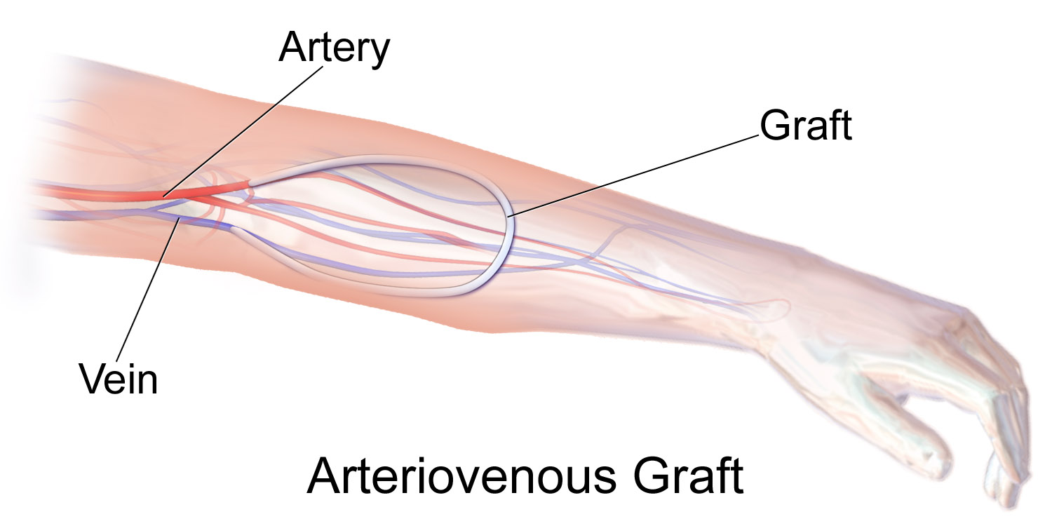
A vascular bypass is a surgical procedure performed to redirect blood flow from one area to another by reconnecting blood vessels. Often, this is done to bypass around a diseased artery, from an area of normal blood flow to another relatively normal area. It is commonly performed due to inadequate blood flow (ischemia) caused by atherosclerosis, as a part of organ transplantation, or for vascular access in hemodialysis. In general, someone's own vein (autograft) is the preferred graft material (or conduit) for a vascular bypass, but other types of grafts such as polytetrafluoroethylene (Teflon), polyethylene terephthalate (Dacron), or a different person's vein (allograft) are also commonly used. Arteries can also serve as vascular grafts. A surgeon sews the graft to the source and target vessels by hand using surgical suture, creating a surgical anastomosis. Common bypass sites include the heart (coronary artery bypass surgery) to treat coronary artery disease, and the legs ... [...More Info...] [...Related Items...] OR: [Wikipedia] [Google] [Baidu] |
Pseudoaneurysm
A pseudoaneurysm, also known as a false aneurysm, is a locally contained hematoma outside an artery or the heart due to damage to the vessel wall. The injury passes through all three layers of the arterial wall, causing a leak, which is contained by a new, weak "wall" formed by the products of the clotting cascade. A pseudoaneurysm (PSA) does not contain any layer of the vessel wall. This differentiates it from a true aneurysm, which is contained by all three layers of the vessel wall, and a dissecting aneurysm, which has a breach in the innermost layer of an artery and subsequent dissection/separation of the tunica intima from the tunica media. A pseudoaneurysm, being associated with a vessel, can be pulsatile; it may be confused with a true aneurysm or dissecting aneurysm. The most common presentation of pseudoaneurysm is femoral artery pseudoaneurysm following access for an endovascular procedure, and this event may complicate up to 8% of vascular interventional proc ... [...More Info...] [...Related Items...] OR: [Wikipedia] [Google] [Baidu] |
Human Umbilical Vein Graft
A vascular bypass is a surgical procedure performed to redirect blood flow from one area to another by reconnecting blood vessels. Often, this is done to bypass around a diseased artery, from an area of normal blood flow to another relatively normal area. It is commonly performed due to inadequate blood flow (ischemia) caused by atherosclerosis, as a part of organ transplantation, or for vascular access in hemodialysis. In general, someone's own vein (autograft) is the preferred graft material (or conduit) for a vascular bypass, but other types of grafts such as polytetrafluoroethylene (Teflon), polyethylene terephthalate (Dacron), or a different person's vein (allograft) are also commonly used. Arteries can also serve as vascular grafts. A surgeon sews the graft to the source and target vessels by hand using surgical suture, creating a surgical anastomosis. Common bypass sites include the heart (coronary artery bypass surgery) to treat coronary artery disease, and the legs, where ... [...More Info...] [...Related Items...] OR: [Wikipedia] [Google] [Baidu] |
Fistula
In anatomy, a fistula (: fistulas or fistulae ; from Latin ''fistula'', "tube, pipe") is an abnormal connection (i.e. tube) joining two hollow spaces (technically, two epithelialized surfaces), such as blood vessels, intestines, or other hollow organs to each other, often resulting in an abnormal flow of fluid from one space to the other. An anal fistula connects the anal canal to the perianal skin. An anovaginal or rectovaginal fistula is a hole joining the anus or rectum to the vagina. A colovaginal fistula joins the space in the colon to that in the vagina. A urinary tract fistula is an abnormal opening in the urinary tract or an abnormal connection between the urinary tract and another organ. An abnormal communication (i.e. hole or tube) between the bladder and the uterus is called a vesicouterine fistula, while if it is between the bladder and the vagina it is known as a vesicovaginal fistula, and if between the urethra and the vagina: a urethrovaginal fistu ... [...More Info...] [...Related Items...] OR: [Wikipedia] [Google] [Baidu] |
Carotid-cavernous Fistula
A carotid-cavernous fistula results from an abnormal communication between the arterial and venous systems within the cavernous sinus in the skull. It is a type of arteriovenous fistula. As arterial blood under high pressure enters the cavernous sinus, the normal venous return to the cavernous sinus is impeded and this causes engorgement of the draining veins, manifesting most dramatically as a sudden engorgement and redness of the eye of the same side. Presentation CCF symptoms include bruit (a humming sound within the skull due to high blood flow through the arteriovenous fistula), progressive visual loss, and pulsatile proptosis Exophthalmos (also called exophthalmus, exophthalmia, proptosis, or exorbitism) is a bulging of the eye anteriorly out of the orbit. Exophthalmos can be either bilateral (as is often seen in Graves' disease) or unilateral (as is often seen in ... or progressive bulging of the eye due to dilatation of the veins draining the eye. Pain is the sympt ... [...More Info...] [...Related Items...] OR: [Wikipedia] [Google] [Baidu] |
Branham Sign
Branham may refer to: * Branham (surname) * 4140 Branham, a main-belt asteroid * Branham (VTA), a light rail station operated by Santa Clara Valley Transportation Authority * Branham High School, a secondary school located in San Jose, California See also * Barnham (other) * Branham House (other) * Branham sign {{disambiguation ... [...More Info...] [...Related Items...] OR: [Wikipedia] [Google] [Baidu] |
Arteriovenous Malformation
An arteriovenous malformation (AVM) is an abnormal connection between arteries and veins, bypassing the capillary system. Usually congenital, this vascular anomaly is widely known because of its occurrence in the central nervous system (usually as a cerebral AVM), but can appear anywhere in the body. The symptoms of AVMs can range from none at all to intense pain or bleeding, and they can lead to other serious medical problems. Signs and symptoms Symptoms of AVMs vary according to their location. Most neurological AVMs produce few to no symptoms. Often the malformation is discovered as part of an autopsy or during treatment of an unrelated disorder (an " incidental finding"); in rare cases, its expansion or a micro-bleed from an AVM in the brain can cause epilepsy, neurological deficit, or pain. The most general symptoms of a cerebral AVM include headaches and epileptic seizures, with more specific symptoms that normally depend on its location and the individual, in ... [...More Info...] [...Related Items...] OR: [Wikipedia] [Google] [Baidu] |
Preload (cardiology)
In cardiac physiology, preload is the amount of sarcomere stretch experienced by cardiac muscle cells, called cardiomyocytes, at the end of ventricular filling during diastole. Preload is directly related to ventricular filling. As the relaxed ventricle fills during diastole, the walls are stretched and the length of sarcomeres increases. Sarcomere length can be approximated by the volume of the ventricle because each shape has a conserved surface-area-to-volume ratio. This is useful clinically because measuring the sarcomere length is destructive to heart tissue. It requires cutting out a piece of cardiac muscle to look at the sarcomeres under a microscope. It is currently not possible to directly measure preload in the beating heart of a living animal. Preload is estimated from end-diastolic ventricular pressure and is measured in millimeters of mercury (mmHg). Estimating preload Though not exactly equivalent to the strict definition of ''preload,'' end-diastolic volume ... [...More Info...] [...Related Items...] OR: [Wikipedia] [Google] [Baidu] |
Cannulation
A cannula (; Latin meaning 'little reed'; : cannulae or cannulas) is a tube that can be inserted into the body, often for the delivery or removal of fluid or for the gathering of samples. In simple terms, a cannula can surround the inner or outer surfaces of a trocar needle thus extending the effective needle length by at least half the length of the original needle. Its size mainly ranges from 14 to 26 gauge. Different-sized cannula have different colours as coded. Decannulation is the permanent removal of a cannula ( extubation), especially of a tracheostomy cannula, once a physician determines it is no longer needed for breathing. Medicine Cannulas normally come with a trocar inside. The trocar is a needle, which punctures the body in order to get into the intended space. Intravenous cannulas are the most common in hospital use. A variety of cannulas are used to establish cardiopulmonary bypass in cardiac surgery. A nasal cannula is a piece of plastic tubing that runs un ... [...More Info...] [...Related Items...] OR: [Wikipedia] [Google] [Baidu] |
Cephalic Vein
In human anatomy, the cephalic vein (also called the antecubital vein) is a superficial vein in the arm. It is the longest vein of the upper limb. It starts at the anatomical snuffbox from the radial end of the dorsal venous network of hand, and ascends along the radial (lateral) side of the arm before emptying into the axillary vein. At the elbow, it communicates with the basilic vein via the median cubital vein. Anatomy The cephalic vein is situated within the superficial fascia along the anterolateral surface of the biceps. Origin The cephalic vein forms at the roof of the anatomical snuffbox at the radial end of the dorsal venous network of hand. Course and relations From its origin, it ascends up the lateral aspect of the radius. Near the shoulder, the cephalic vein passes between the deltoid and pectoralis major muscles ( deltopectoral groove) through the clavipectoral triangle, where it empties into the axillary vein. Anastomoses It communicates wit ... [...More Info...] [...Related Items...] OR: [Wikipedia] [Google] [Baidu] |


