|
5α-Reductase 2 Deficiency
5α-Reductase 2 deficiency (5αR2D) is an autosomal recessive condition caused by mutations impairing the function of ''SRD5A2'', a gene located on chromosome 2 and encoding the enzyme 5α-reductase type 2 (5αR2). 5αR2 is expressed in specific tissues and catalyzes the transformation of testosterone (T) to 5α-dihydrotestosterone (DHT). DHT plays a key role in the process of sexual differentiation. This rare deficiency causes atypical sex development in genetic males (people with a 46XY karyotype), with a broad spectrum of presentations most apparent in the genitalia. Many people with 5-alpha reductase deficiency are assigned female at birth based on their external genitalia. In other cases, affected infants are assigned male at birth based on their external genitalia, often an unusually small penis (micropenis) and the urethra opening on the underside of the penis (hypospadias). Still other affected infants may be assigned either female or male at birth as their external ... [...More Info...] [...Related Items...] OR: [Wikipedia] [Google] [Baidu] |
Autosomal Recessive
In genetics, dominance is the phenomenon of one variant (allele) of a gene on a chromosome masking or overriding the Phenotype, effect of a different variant of the same gene on Homologous chromosome, the other copy of the chromosome. The first variant is termed dominant and the second is called recessive. This state of having Heterozygosity, two different variants of the same gene on each chromosome is originally caused by a mutation in one of the genes, either new (''de novo'') or Heredity, inherited. The terms autosomal dominant or autosomal recessive are used to describe gene variants on non-sex chromosomes (autosomes) and their associated traits, while those on sex chromosomes (allosomes) are termed X-linked dominant, X-linked recessive or Y-linked; these have an inheritance and presentation pattern that depends on the sex of both the parent and the child (see Sex linkage). Since there is only one Y chromosome, Y-linked traits cannot be dominant or recessive. Additionally, ... [...More Info...] [...Related Items...] OR: [Wikipedia] [Google] [Baidu] |
Micropenis
A micropenis or microphallus is an unusually small Human penis, penis. A common criterion is a dorsal (measured on top) Human penis size, penile length of at least 2.5 standard deviations smaller than the mean human penis size for age. A micropenis is stretched penile length equal to or less than 1.9 Centimetre, cm (0.75 Inch, in) in term Infant, infants, and 9.3 cm (3.67 in) in adults. The condition is usually recognized shortly after Childbirth, birth. The term is most often used medically when the rest of the penis, scrotum, and perineum are without Ambiguous genitalia, ambiguity, such as hypospadias. Traditionally, a microphallus describes a micropenis with hypospadias. Micropenis incidence is about 1.5 in 10,000 male newborns in North America.ScienceDaily.com (2004).Surgeons Pinch More Than An Inch From The Arm To Rebuild A Micropenis" 6 Dec. 2004, retrieved 2 April 2012. Causes Of the abnormal conditions associated with micropenis, most are conditions of reduced prenata ... [...More Info...] [...Related Items...] OR: [Wikipedia] [Google] [Baidu] |
Male Pattern Baldness
Pattern hair loss (also known as androgenetic alopecia (AGA)) is a hair loss condition that primarily affects the top and front of the scalp. In male-pattern hair loss (MPHL), the hair loss typically presents itself as either a receding front hairline, loss of hair on the crown and vertex of the scalp, or a combination of both. Female-pattern hair loss (FPHL) typically presents as a diffuse thinning of the hair across the entire scalp. The condition is caused by a combination of male sex hormones (balding never occurs in castrated men) and genetic factors. Some research has found evidence for the role of oxidative stress in hair loss, the microbiome of the scalp, genetics, and circulating androgens; particularly dihydrotestosterone (DHT). Men with early onset androgenic alopecia (before the age of 35) have been deemed the male phenotypic equivalent for polycystic ovary syndrome (PCOS). The cause in female pattern hair loss remains unclear; androgenetic alopecia for wom ... [...More Info...] [...Related Items...] OR: [Wikipedia] [Google] [Baidu] |
17β-hydroxysteroid Dehydrogenase 3 Deficiency
A hydroxysteroid is a molecule derived from a steroid with a hydrogen replaced with a hydroxy group. When the hydroxy group is specifically at the C3 position, hydroxysteroids are referred to as sterols, with an example being cholesterol. See also * Hydroxysteroid dehydrogenase * Ketosteroid class=skin-invert-image, 150px, Androstenedione class=skin-invert-image, 150px, Androsterone class=skin-invert-image, 150px, Estrone A ketosteroid, or an oxosteroid, is a steroid in which a hydrogen atom has been replaced with a ketone (C=O) g ... External links * Alcohols Steroids {{steroid-stub ... [...More Info...] [...Related Items...] OR: [Wikipedia] [Google] [Baidu] |
Partial Androgen Insensitivity Syndrome
Partial androgen insensitivity syndrome (PAIS) is a condition that results in the partial inability of the Eukaryote#Animal cell, cell to respond to androgens. It is an X linked recessive condition. The partial unresponsiveness of the cell to the presence of androgenic hormones impairs the Development of the reproductive system#External genitalia, masculinization of male genitalia in the developing fetus, as well as the development of male Secondary sex characteristics, secondary sexual characteristics at puberty, but does not significantly impair female genital or sexual development. As such, the insensitivity to androgens is clinically significant only when it occurs in individuals with a Y chromosome (or more specifically, an SRY, SRY gene). Clinical features include ambiguous genitalia at birth and primary amenhorrhoea with clitoromegaly with inguinal masses. Müllerian duct, Müllerian structures are not present in the individual. PAIS is one of three types of androgen insen ... [...More Info...] [...Related Items...] OR: [Wikipedia] [Google] [Baidu] |
Gynecomastia
Gynecomastia (also spelled gynaecomastia) is the non-cancerous enlargement of one or both breasts in men due to the growth of breast tissue as a result of a hormone imbalance between estrogens and androgens. Updated by Brent Wisse (10 November 2018) Physically speaking, gynecomastia is completely benign, but it is associated with significant psychological distress, social stigma, and dysphoria. Gynecomastia can be normal in newborn male babies due to exposure to estrogen from the mother, in adolescent boys going through puberty, in older men over the age of 50, and in obese men. Most occurrences of gynecomastia do not require diagnostic tests. Gynecomastia may be caused by abnormal hormone changes, any condition that leads to an increase in the ratio of estrogens/androgens such as liver disease, kidney failure, thyroid disease and some non-breast tumors. Alcohol and some drugs can also cause breast enlargement. Other causes may include Klinefelter syndrome, metabolic dysf ... [...More Info...] [...Related Items...] OR: [Wikipedia] [Google] [Baidu] |
Virilization
Virilization or masculinization is the biological development of adult male characteristics in young males or females. Most of the changes of virilization are produced by androgens. Virilization is a medical term commonly used in three medical and biology of sex contexts: prenatal biological sexual differentiation, the postnatal changes of typical chromosomal male (46, XY) puberty, and excessive androgen effects in typical chromosomal females (46, XX). It is also the intended result of androgen replacement therapy in males with delayed puberty and low testosterone. Prenatal virilization In the prenatal period, virilization refers to closure of the perineum, thinning and wrinkling (rugation) of the scrotum, growth of the penis, and closure of the urethral groove to the tip of the penis. In this context, ''masculinization'' is synonymous with ''virilization''. Prenatal virilization of XX fetuses and undervirilization of XY fetuses are common causes of ambiguous genitalia suc ... [...More Info...] [...Related Items...] OR: [Wikipedia] [Google] [Baidu] |
Prostate Hypoplasia
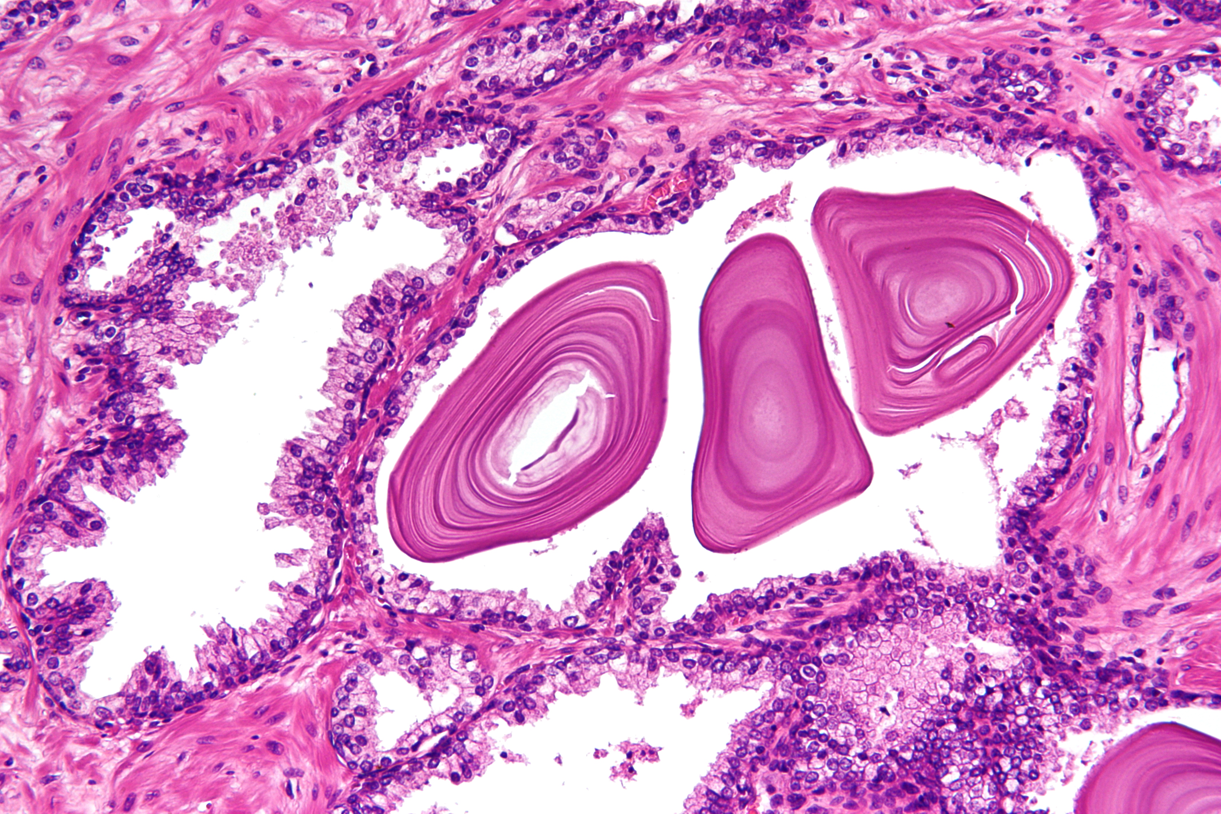
The prostate is an accessory gland of the male reproductive system and a muscle-driven mechanical switch between urination and ejaculation. It is found in all male mammals. It differs between species anatomically, chemically, and physiologically. Anatomically, the prostate is found below the bladder, with the urethra passing through it. It is described in gross anatomy as consisting of lobes and in microanatomy by zone. It is surrounded by an elastic, fibromuscular capsule and contains glandular tissue, as well as connective tissue. The prostate produces and contains fluid that forms part of semen, the substance emitted during ejaculation as part of the male sexual response. This prostatic fluid is slightly alkaline, milky or white in appearance. The alkalinity of semen helps neutralize the acidity of the vaginal tract, prolonging the lifespan of sperm. The prostatic fluid is expelled in the first part of ejaculate, together with most of the sperm, because of the action of sm ... [...More Info...] [...Related Items...] OR: [Wikipedia] [Google] [Baidu] |
Testes
A testicle or testis ( testes) is the gonad in all male bilaterians, including humans, and is homologous to the ovary in females. Its primary functions are the production of sperm and the secretion of androgens, primarily testosterone. The release of testosterone is regulated by luteinizing hormone (LH) from the anterior pituitary gland. Sperm production is controlled by follicle-stimulating hormone (FSH) from the anterior pituitary gland and by testosterone produced within the gonads. Structure Appearance Males have two testicles of similar size contained within the scrotum, which is an extension of the abdominal wall. Scrotal asymmetry, in which one testicle extends farther down into the scrotum than the other, is common. This is because of the differences in the vasculature's anatomy. For 85% of men, the right testis hangs lower than the left one. Measurement and volume The volume of the testicle can be estimated by palpating it and comparing it to ellipsoids (an ... [...More Info...] [...Related Items...] OR: [Wikipedia] [Google] [Baidu] |
Ejaculatory Ducts
The ejaculatory ducts (''ductus ejaculatorii'') are paired structures in the male reproductive system. Each ejaculatory duct is formed by the union of the vas deferens with the duct of the seminal vesicle. They pass through the prostate, and open into the urethra above the seminal colliculus. During ejaculation, semen passes through the prostate gland, enters the urethra and exits the body via the urinary meatus. Function Ejaculation Ejaculation occurs in two stages, the emission stage and the expulsion stage.Rathus, S. A., Nevid, J. S., Fichner-Rathus, L., Herold, E. S. (2010). ''Human Sexuality in a World of Diversity''. Pearsons Education Canada, Pearson Canada Inc. Toronto, ON. The emission stage involves the workings of several structures of the ejaculatory duct; contractions of the prostate gland, the seminal vesicles, the bulbourethral gland and the vas deferens push fluids into the prostatic urethra. The semen is stored here until ejaculation occurs. Muscles at the base ... [...More Info...] [...Related Items...] OR: [Wikipedia] [Google] [Baidu] |
Epididymides
The epididymis (; : epididymides or ) is an elongated tubular genital organ attached to the posterior side of each one of the two male reproductive glands, the testicles. It is a single, narrow, tightly coiled tube in adult humans, in length; uncoiled the tube would be approximately 6 m (20 feet) long. It connects the testicle to the vas deferens in the male reproductive system. The epididymis serves as an interconnection between the multiple efferent ducts at the rear of a testicle (proximally), and the vas deferens (distally). Its primary function is the storage, maturation and transport of sperm cells. Structure The human epididymis is situated posterior and somewhat lateral to the testis. The epididymis is invested completely by the tunica vaginalis (which is continuous with the tunica vaginalis covering the testis). The epididymis can be divided into three main regions: * The head (). The head of the epididymis receives spermatozoa via the efferent ducts of the m ... [...More Info...] [...Related Items...] OR: [Wikipedia] [Google] [Baidu] |
Seminal Vesicles
The seminal vesicles (also called vesicular glands or seminal glands) are a pair of convoluted tubular accessory glands that lie behind the urinary bladder of male mammals. They secrete fluid that largely composes the semen. The vesicles are 5–10 cm in size, 3–5 cm in diameter, and are located between the bladder and the rectum. They have multiple outpouchings, which contain secretory glands, which join together with the vasa deferentia at the ejaculatory ducts. They receive blood from the vesiculodeferential artery, and drain into the vesiculodeferential veins. The glands are lined with column-shaped and cuboidal cells. The vesicles are present in many groups of mammals, but not marsupials, monotremes or carnivores. Inflammation of the seminal vesicles is called seminal vesiculitis and most often is due to bacterial infection as a result of a sexually transmitted infection or following a surgical procedure. Seminal vesiculitis can cause pain in the lower abdo ... [...More Info...] [...Related Items...] OR: [Wikipedia] [Google] [Baidu] |


