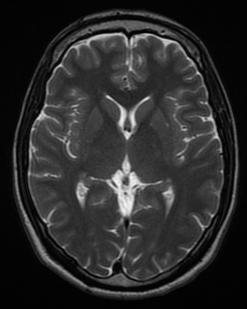
MRI of brain and brain stem on:
[Wikipedia]
[Google]
[Amazon]
Magnetic resonance imaging of the brain uses magnetic resonance imaging (MRI) to produce high quality two-dimensional or three-dimensional images of the
 In the early 1980s to the early 1990s, 'Jedi' helmets, inspired by the '
 The record for the highest spatial resolution of a whole intact brain (postmortem) is 100 microns, from Massachusetts General Hospital. The data was published in NATURE on 30th of October 2019.
 * Fluid attenuation inversion recovery ( FLAIR): useful for evaluation of
Image:Brain regions on T1 MRI.png, Brain regions on T1 MRI
Image:Falxmeningeom MRT T1 mit Kontrastmittel.jpg, T1 (note CSF is dark) with contrast (arrow pointing to meningioma of the falx)
Image:Brain-T2-axial.png, Normal axial T2-weighted MR image of the brain
Image:MRI brain surface normal.jpg, MRI image of the surface of the brain.
brain
A brain is an organ that serves as the center of the nervous system in all vertebrate and most invertebrate animals. It is located in the head, usually close to the sensory organs for senses such as vision. It is the most complex organ in a ve ...
and brainstem as well as the cerebellum without the use of ionizing radiation (X-rays
An X-ray, or, much less commonly, X-radiation, is a penetrating form of high-energy electromagnetic radiation. Most X-rays have a wavelength ranging from 10 picometers to 10 nanometers, corresponding to frequencies in the range 30&nbs ...
) or radioactive tracer
A radioactive tracer, radiotracer, or radioactive label is a chemical compound in which one or more atoms have been replaced by a radionuclide so by virtue of its radioactive decay it can be used to explore the mechanism of chemical reactions by ...
s.
History
The first MR images of a human brain were obtained in 1978 by two groups of researchers at EMI Laboratories led byIan Robert Young
Ian Robert Young (11 January 1932 – 27 September 2019) was a British medical physicist, known for his work in the field of magnetic resonance imaging (MRI).
Life
He was educated at Sedbergh School and later studied physics at Aberdeen Un ...
and Hugh Clow. In 1986, Charles L. Dumoulin and Howard R. Hart at General Electric
General Electric Company (GE) is an American multinational conglomerate founded in 1892, and incorporated in New York state and headquartered in Boston. The company operated in sectors including healthcare, aviation, power, renewable en ...
developed MR angiography, and Denis Le Bihan obtained the first images and later patented diffusion MRI
Diffusion-weighted magnetic resonance imaging (DWI or DW-MRI) is the use of specific MRI sequences as well as software that generates images from the resulting data that uses the diffusion of water molecules to generate contrast in MR images. It ...
. In 1988, Arno Villringer
Arno Villringer (born 1958, Schopfheim, Germany) is a Director at the Department of Neurology at the Max Planck Institute for Human Cognitive and Brain Sciences in Leipzig, Germany; Director of the Department of Cognitive Neurology at Universi ...
and colleagues demonstrated that susceptibility contrast agents
A contrast agent (or contrast medium) is a substance used to increase the contrast of structures or fluids within the body in medical imaging. Contrast agents absorb or alter external electromagnetism or ultrasound, which is different from radiop ...
may be employed in perfusion MRI. In 1990, Seiji Ogawa
Seiji Ogawa (小川 誠二 ''Ogawa Seiji'', born January 19, 1934) is a Japanese biophysicist and neuroscientist known for discovering the technique that underlies Functional Magnetic Resonance Imaging (fMRI). He is regarded as the father of moder ...
at AT&T Bell labs
Nokia Bell Labs, originally named Bell Telephone Laboratories (1925–1984),
then AT&T Bell Laboratories (1984–1996)
and Bell Labs Innovations (1996–2007),
is an American industrial research and scientific development company owned by mult ...
recognized that oxygen-depleted blood with dHb was attracted to a magnetic field, and discovered the technique that underlies Functional Magnetic Resonance Imaging (fMRI).
 In the early 1980s to the early 1990s, 'Jedi' helmets, inspired by the '
Return of the Jedi
''Return of the Jedi'' (also known as ''Star Wars: Episode VI – Return of the Jedi'' is a 1983 American epic space opera film directed by Richard Marquand. The screenplay is by Lawrence Kasdan and George Lucas from a story by Lucas, who ...
' Starwars file, were sometimes worn by children in order to obtain good image quality. The copper coils of the helmet were used as a radio aerial to detect the signals while the 'Jedi' association encouraged children to wear the helmets and not be frightened by the procedure. These helmets were no longer needed as MR scanners improved.
In the early 1990s, Peter Basser and Le Bihan, working at NIH
The National Institutes of Health, commonly referred to as NIH (with each letter pronounced individually), is the primary agency of the United States government responsible for biomedical and public health research. It was founded in the late ...
, and Aaron Filler, Franklyn Howe, and colleagues developed diffusion tensor imaging (DTI). Joseph Hajnal, Young and Graeme Bydder described the use of FLAIR pulse sequence to demonstrate high signal regions in normal white matter in 1992. In the same year, John Detre, Alan P. Koretsky and coworkers developed arterial spin labeling Arterial spin labeling (ASL), also known as arterial spin tagging, is a magnetic resonance imaging technique used to quantify cerebral blood perfusion by labelling blood water as it flows throughout the brain. ASL specifically refers to magnetic lab ...
. In 1997, Jürgen R. Reichenbach, E. Mark Haacke and coworkers at Washington University
Washington University in St. Louis (WashU or WUSTL) is a private research university with its main campus in St. Louis County, and Clayton, Missouri. Founded in 1853, the university is named after George Washington. Washington University is r ...
developed Susceptibility weighted imaging
Susceptibility weighted imaging (SWI), originally called BOLD venographic imaging, is an MRI sequence that is exquisitely sensitive to venous blood, hemorrhage and iron storage. SWI uses a fully flow compensated, long echo, gradient recalled echo ...
.
The first study of the human brain at 3.0 T was published in 1994, and in 1998 at 8 T. Studies of the human brain have been performed at 9.4 T (2006) and up to 10.5 T (2019).
Paul Lauterbur
Paul Christian Lauterbur (May 6, 1929 – March 27, 2007) was an American chemist who shared the Nobel Prize in Physiology or Medicine in 2003 with Peter Mansfield for his work which made the development of magnetic resonance imaging (MRI) poss ...
and Sir Peter Mansfield
Sir Peter Mansfield (9 October 1933 – 8 February 2017) was an English physicist who was awarded the 2003 Nobel Prize in Physiology or Medicine, shared with Paul Lauterbur, for discoveries concerning Magnetic Resonance Imaging (MRI). Mansfi ...
were awarded the 2003 Nobel Prize in Physiology or Medicine
The Nobel Prize in Physiology or Medicine is awarded yearly by the Nobel Assembly at the Karolinska Institute for outstanding discoveries in physiology or medicine. The Nobel Prize is not a single prize, but five separate prizes that, accord ...
for their discoveries concerning MRI. The record for the highest spatial resolution of a whole intact brain (postmortem) is 100 microns, from Massachusetts General Hospital. The data was published in NATURE on 30th of October 2019.
Applications
One advantage of MRI of the brain overcomputed tomography of the head
Computed tomography of the head uses a series of X-rays in a CT scan of the head taken from many different directions; the resulting data is transformed into a series of cross sections of the brain using a computer program. CT images of the head ...
is better tissue contrast, and it has fewer artifacts than CT when viewing the brainstem. MRI is also superior for pituitary
In vertebrate anatomy, the pituitary gland, or hypophysis, is an endocrine gland, about the size of a chickpea and weighing, on average, in humans. It is a protrusion off the bottom of the hypothalamus at the base of the brain. The hypoph ...
imaging. It may however be less effective at identifying early cerebritis
Cerebritis is the inflammation of the cerebrum, which performs a number of important functions, such as memory and speech. It is also defined as a purulent nonencapsulated parenchymal infection of brain which is characterized by nonspecific featu ...
.
In the case of a concussion, an MRI should be avoided unless there are progressive neurological symptoms, focal neurological findings or concern of skull fracture on exam. In the analysis of a concussion, measurements of Fractional Anisotropy, Mean Diffusivity, Cerebral Blood Flow, and Global Connectivity can be taken to observe the pathophysiological mechanisms being made while in recovery.
In analysis of the fetal brain, MRI provides more information about gyration than ultrasound
Ultrasound is sound waves with frequencies higher than the upper audible limit of human hearing. Ultrasound is not different from "normal" (audible) sound in its physical properties, except that humans cannot hear it. This limit varies ...
.
MRI is sensitive for the detection of brain abscess.
A number of different imaging modalities or sequences
In mathematics, a sequence is an enumerated collection of objects in which repetitions are allowed and order matters. Like a set, it contains members (also called ''elements'', or ''terms''). The number of elements (possibly infinite) is called t ...
can be used with imaging the nervous system:
* ''T''1-weighted (T1W) images: Cerebrospinal fluid
Cerebrospinal fluid (CSF) is a clear, colorless body fluid found within the tissue that surrounds the brain and spinal cord of all vertebrates.
CSF is produced by specialised ependymal cells in the choroid plexus of the ventricles of the ...
is dark. ''T''1-weighted images are useful for visualizing normal anatomy.
* ''T''2-weighted (T2W) images: CSF is light, but fat (and thus white matter
White matter refers to areas of the central nervous system (CNS) that are mainly made up of myelinated axons, also called tracts. Long thought to be passive tissue, white matter affects learning and brain functions, modulating the distributi ...
) is darker than with ''T''1. ''T''2-weighted images are useful for visualizing pathology.
* Diffusion-weighted images (DWI): DWI uses the diffusion of water molecules to generate contrast in MR images.
* Proton density (PD) images: CSF has a relatively high level of protons, making CSF appear bright. Gray matter
Grey matter is a major component of the central nervous system, consisting of neuronal cell bodies, neuropil (dendrites and unmyelinated axons), glial cells (astrocytes and oligodendrocytes), synapses, and capillaries. Grey matter is distingui ...
is brighter than white matter.
 * Fluid attenuation inversion recovery ( FLAIR): useful for evaluation of
 white matter
White matter refers to areas of the central nervous system (CNS) that are mainly made up of myelinated axons, also called tracts. Long thought to be passive tissue, white matter affects learning and brain functions, modulating the distributi ...
plaques near the ventricles. It is useful in identifying demyelination
A demyelinating disease is any disease of the nervous system in which the myelin sheath of neurons is damaged. This damage impairs the conduction of signals in the affected nerves. In turn, the reduction in conduction ability causes deficiency i ...
.
Artificial intelligence
On the topic of diagnosis, MRI data may be used to identify brain tumors.See also
*Human Connectome Project
The Human Connectome Project (HCP) is a five-year project sponsored by sixteen components of the National Institutes of Health, split between two consortia of research institutions. The project was launched in July 2009 as the first of three Grand ...
* History of neuroimaging
The first neuroimaging technique ever is the so-called 'human circulation balance' invented by Angelo Mosso in the 1880s and able to non-invasively measure the redistribution of blood during emotional and intellectual activity.
Then, in the early ...
Gallery
References
{{Medical imaging Magnetic resonance imaging Neuroimaging