Connectogram on:
[Wikipedia]
[Google]
[Amazon]
Connectograms are graphical representations of connectomics, the field of study dedicated to mapping and interpreting all of the
 Connectograms are circular, with the left half depicting the left hemisphere and the right half depicting the right hemisphere. The hemispheres are further broken down into

white matter
White matter refers to areas of the central nervous system (CNS) that are mainly made up of myelinated axons, also called tracts. Long thought to be passive tissue, white matter affects learning and brain functions, modulating the distribu ...
fiber connections in the human brain. These circular graphs based on diffusion MRI
Diffusion-weighted magnetic resonance imaging (DWI or DW-MRI) is the use of specific MRI sequences as well as software that generates images from the resulting data that uses the diffusion of water molecules to generate contrast in MR images. It ...
data utilize graph theory
In mathematics, graph theory is the study of '' graphs'', which are mathematical structures used to model pairwise relations between objects. A graph in this context is made up of '' vertices'' (also called ''nodes'' or ''points'') which are conn ...
to demonstrate the white matter connections and cortical characteristics for single structures, single subjects, or populations.
Structure
Background and description
The ''connectogram'', as a graphical representation of brain connectomics, was proposed in 2012. Circular representations of connections have been used in a number of disciplines; examples include representation of aspects of epidemics, geographical networks, musical beats, diversity in bird populations, and genomic data. Connectograms were also cited as a source of inspiration for the heads-up display style of Tony Stark's helmet inIron Man 3
''Iron Man 3'' (titled onscreen as ''Iron Man Three'') is a 2013 American superhero film based on the Marvel Comics character Iron Man, produced by Marvel Studios and distributed by Walt Disney Studios Motion Pictures. It is the sequel to ''Ir ...
.
 Connectograms are circular, with the left half depicting the left hemisphere and the right half depicting the right hemisphere. The hemispheres are further broken down into
 frontal lobe
The frontal lobe is the largest of the four major lobes of the brain in mammals, and is located at the front of each cerebral hemisphere (in front of the parietal lobe and the temporal lobe). It is parted from the parietal lobe by a groove be ...
, insular cortex
The insular cortex (also insula and insular lobe) is a portion of the cerebral cortex folded deep within the lateral sulcus (the fissure separating the temporal lobe from the parietal and frontal lobes) within each hemisphere of the mammalian b ...
, limbic lobe, temporal lobe
The temporal lobe is one of the four major lobes of the cerebral cortex in the brain of mammals. The temporal lobe is located beneath the lateral fissure on both cerebral hemispheres of the mammalian brain.
The temporal lobe is involved i ...
, parietal lobe
The parietal lobe is one of the four major lobes of the cerebral cortex in the brain of mammals. The parietal lobe is positioned above the temporal lobe and behind the frontal lobe and central sulcus.
The parietal lobe integrates sensory informa ...
, occipital lobe
The occipital lobe is one of the four major lobes of the cerebral cortex in the brain of mammals. The name derives from its position at the back of the head, from the Latin ''ob'', "behind", and ''caput'', "head".
The occipital lobe is the vi ...
, subcortical structures, and cerebellum
The cerebellum (Latin for "little brain") is a major feature of the hindbrain of all vertebrates. Although usually smaller than the cerebrum, in some animals such as the mormyrid fishes it may be as large as or even larger. In humans, the cerebe ...
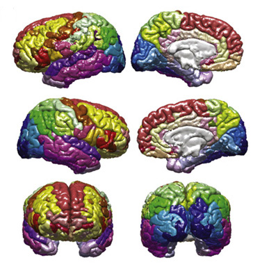
. At the bottom the brain stem is also represented between the two hemispheres. Within these lobes, each cortical area is labeled with an abbreviation and assigned its own color, which can be used to designate these same cortical regions in other figures, such as the parcellated brain surfaces in the adjacent image, so that the reader can find the corresponding cortical areas on a geometrically accurate surface and see exactly how disparate the connected regions may be. Inside the cortical surface ring, the concentric circles each represent different attributes of the corresponding cortical regions. In order from outermost to innermost, these metric rings represent the grey matter
Grey matter is a major component of the central nervous system, consisting of neuronal cell bodies, neuropil ( dendrites and unmyelinated axons), glial cells ( astrocytes and oligodendrocytes), synapses, and capillaries. Grey matter is ...
volume, surface area
The surface area of a solid object is a measure of the total area that the surface of the object occupies. The mathematical definition of surface area in the presence of curved surfaces is considerably more involved than the definition of ...
, cortical thickness, curvature, and degree of connectivity (the relative proportion of fibers initiating or terminating in the region compared to the whole brain). Inside these circles, lines connect regions that are found to be structurally connected. The relative density (number of fibers) of these connections is reflected in the opacity of the lines, so that one can easily compare various connections and their structural importance. The fractional anisotropy Fractional anisotropy (FA) is a scalar value between zero and one that describes the degree of anisotropy of a diffusion process. A value of zero means that diffusion is isotropic, i.e. it is unrestricted (or equally restricted) in all directions. A ...
of each connection is reflected in its color.
Uses
Brain mapping
With the recent concerted push to map all of the human brain and its connections, it has become increasingly important to find ways to graphically represent the large amounts of data involved in connectomics. Most other representations of the connectome use 3 dimensions, and therefore require an interactive graphical user interface. The connectogram can display 83 cortical regions within each hemisphere, and visually display which areas are structurally connected, all on a flat surface. It is therefore conveniently filed in patient records, or to display in print. The graphs were originally developed using the visualization tool called Circos,.Clinical use
On an individual level, connectograms can be used to inform the treatment of patients with neuroanatomical abnormalities. Connectograms have been used to monitor the progression of neurological recovery of patients who suffered atraumatic brain injury
A traumatic brain injury (TBI), also known as an intracranial injury, is an injury to the brain caused by an external force. TBI can be classified based on severity (ranging from mild traumatic brain injury TBI/concussionto severe traumatic br ...
(TBI). They have also been applied to famous patient Phineas Gage
Phineas P. Gage (18231860) was an American railroad construction foreman known for his improbable survival of an accident in which a large iron rod was driven completely through his head, destroying much of his brain's left frontal lobe, and ...
, to estimate damage to his neural network
A neural network is a network or circuit of biological neurons, or, in a modern sense, an artificial neural network, composed of artificial neurons or nodes. Thus, a neural network is either a biological neural network, made up of biological ...
(as well as the damage at the cortical level—the primary focus of earlier studies on Gage).
Empirical study
Connectograms can represent the averages of cortical metrics (grey matter volume, surface area, cortical thickness, curvature, and degree of connectivity), as well astractography
In neuroscience, tractography is a 3D modeling technique used to visually represent nerve tracts using data collected by diffusion MRI. It uses special techniques of magnetic resonance imaging (MRI) and computer-based diffusion MRI. The results ...
data, such as the average densities and fractional anisotropy of the connections, across populations of any size. This allows for visual and statistical comparison between groups such as males and females, differing age cohorts, or healthy controls and patients. Some versions have been used to analyze how partitioned networks are in patient populations or the relative balance between inter- and intra-hemispheric connections.
Modified versions
There are many possibilities for which measures are included in the rings of a connectogram. Irimia and Van Horn (2012) have published connectograms which examine the correlative relationships between regions and uses the figures to compare the approaches of graph theory and connectomics. Some have been published without the inner circles of cortical metrics. Others include additional measures relating toneural network
A neural network is a network or circuit of biological neurons, or, in a modern sense, an artificial neural network, composed of artificial neurons or nodes. Thus, a neural network is either a biological neural network, made up of biological ...
s, which can be added as additional rings to the inside to show metrics of graph theory
In mathematics, graph theory is the study of '' graphs'', which are mathematical structures used to model pairwise relations between objects. A graph in this context is made up of '' vertices'' (also called ''nodes'' or ''points'') which are conn ...
, as in the extended connectogram here:

Regions and their abbreviations
See also
*Connectome
A connectome () is a comprehensive map of neural connections in the brain, and may be thought of as its "wiring diagram". An organism's nervous system is made up of neurons which communicate through synapses. A connectome is constructed by tr ...
* Connectomics
* Human Connectome Project
The Human Connectome Project (HCP) is a five-year project sponsored by sixteen components of the National Institutes of Health, split between two consortia of research institutions. The project was launched in July 2009 as the first of three Grand ...
* Brain mapping
Brain mapping is a set of neuroscience techniques predicated on the mapping of (biological) quantities or properties onto spatial representations of the (human or non-human) brain resulting in maps.
According to the definition established in ...
* Tractography
In neuroscience, tractography is a 3D modeling technique used to visually represent nerve tracts using data collected by diffusion MRI. It uses special techniques of magnetic resonance imaging (MRI) and computer-based diffusion MRI. The results ...
* Chord diagram (information visualization)
References
Further reading
{{Reflist, group="further" Neuroimaging Articles containing video clips