Beta sheet on:
[Wikipedia]
[Google]
[Amazon]
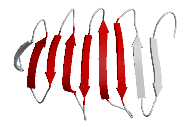
 The beta sheet, (β-sheet) (also β-pleated sheet) is a common motif of the regular protein secondary structure. Beta sheets consist of beta strands (β-strands) connected laterally by at least two or three
 The first β-sheet structure was proposed by
 However, β-strands are rarely perfectly extended; rather, they exhibit a twist. The energetically preferred


 A β-helix is formed from repeating structural units consisting of two or three short β-strands linked by short loops. These units "stack" atop one another in a helical fashion so that successive repetitions of the same strand hydrogen-bond with each other in a parallel orientation. See the β-helix article for further information.
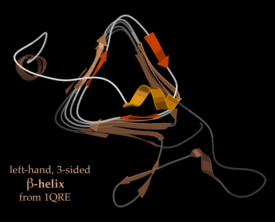
In lefthanded β-helices, the strands themselves are quite straight and untwisted; the resulting helical surfaces are nearly flat, forming a regular
  Righthanded β-helices, typified by the pectate lyase enzyme shown at left or P22 phage tailspike protein, have a less regular cross-section, longer and indented on one of the sides; of the three linker loops, one is consistently just two residues long and the others are variable, often elaborated to form a binding or active site.
A two-sided β-helix (right-handed) is found in some bacterial
Anatomy & Taxonomy of Protein Structures -survey
NetSurfP - Secondary Structure and Surface Accessibility predictor
{{DEFAULTSORT:Beta Sheet Protein structural motifs
 The beta sheet, (β-sheet) (also β-pleated sheet) is a common motif of the regular protein secondary structure. Beta sheets consist of beta strands (β-strands) connected laterally by at least two or three
 backbone
The backbone is the vertebral column of a vertebrate.
Arts, entertainment, and media Film
* ''Backbone'' (1923 film), a 1923 lost silent film starring Alfred Lunt
* ''Backbone'' (1975 film), a 1975 Yugoslavian drama directed by Vlatko Gilić
...
hydrogen bonds, forming a generally twisted, pleated sheet. A β-strand is a stretch of polypeptide chain typically 3 to 10 amino acid
Amino acids are organic compounds that contain both amino and carboxylic acid functional groups. Although hundreds of amino acids exist in nature, by far the most important are the alpha-amino acids, which comprise proteins. Only 22 alpha a ...
s long with backbone in an extended conformation. The supramolecular association of β-sheets has been implicated in the formation of the fibrils and protein aggregates observed in amyloidosis, notably Alzheimer's disease.
History
 The first β-sheet structure was proposed by
 William Astbury
William Thomas Astbury FRS (25 February 1898 – 4 June 1961) was an English physicist and molecular biologist who made pioneering X-ray diffraction studies of biological molecules. His work on keratin provided the foundation for Linus Pauling ...
in the 1930s. He proposed the idea of hydrogen bonding between the peptide bonds of parallel or antiparallel extended β-strands. However, Astbury did not have the necessary data on the bond geometry of the amino acids in order to build accurate models, especially since he did not then know that the peptide bond was planar. A refined version was proposed by Linus Pauling and Robert Corey
Robert Brainard Corey (August 19, 1897 – April 23, 1971) was an American biochemist, mostly known for his role in discovery of the α-helix and the β-sheet with Linus Pauling. Also working with Pauling was Herman Branson. Their discoveries w ...
in 1951. Their model incorporated the planarity of the peptide bond which they previously explained as resulting from keto-enol tautomerization
Tautomers () are structural isomers (constitutional isomers) of chemical compounds that readily interconvert.
The chemical reaction interconverting the two is called tautomerization. This conversion commonly results from the relocation of a hyd ...
.
Structure and orientation
Geometry
The majority of β-strands are arranged adjacent to other strands and form an extensive hydrogen bond network with their neighbors in which the N−H groups in the backbone of one strand establish hydrogen bonds with the C=O groups in the backbone of the adjacent strands. In the fully extended β-strand, successive side chains point straight up and straight down in an alternating pattern. Adjacent β-strands in a β-sheet are aligned so that their Cα atoms are adjacent and their side chains point in the same direction. The "pleated" appearance of β-strands arises from tetrahedral chemical bonding at the Cα atom; for example, if a side chain points straight up, then the bonds to the C′ must point slightly downwards, since its bond angle is approximately 109.5°. The pleating causes the distance between C and C to be approximately , rather than the expected from two fully extended ''trans
Trans- is a Latin prefix meaning "across", "beyond", or "on the other side of".
Used alone, trans may refer to:
Arts, entertainment, and media
* Trans (festival), a former festival in Belfast, Northern Ireland, United Kingdom
* ''Trans'' (fil ...
'' peptide
Peptides (, ) are short chains of amino acids linked by peptide bonds. Long chains of amino acids are called proteins. Chains of fewer than twenty amino acids are called oligopeptides, and include dipeptides, tripeptides, and tetrapeptides.
...
s. The "sideways" distance between adjacent Cα atoms in hydrogen-bonded β-strands is roughly .
 However, β-strands are rarely perfectly extended; rather, they exhibit a twist. The energetically preferred
 dihedral angle
A dihedral angle is the angle between two intersecting planes or half-planes. In chemistry, it is the clockwise angle between half-planes through two sets of three atoms, having two atoms in common. In solid geometry, it is defined as the un ...
s near (''φ'', ''ψ'') = (–135°, 135°) (broadly, the upper left region of the Ramachandran plot
In biochemistry, a Ramachandran plot (also known as a Rama plot, a Ramachandran diagram or a �,ψplot), originally developed in 1963 by G. N. Ramachandran, C. Ramakrishnan, and V. Sasisekharan, is a way to visualize energetically allowed regions ...
) diverge significantly from the fully extended conformation (''φ'', ''ψ'') = (–180°, 180°). The twist is often associated with alternating fluctuations in the dihedral angle
A dihedral angle is the angle between two intersecting planes or half-planes. In chemistry, it is the clockwise angle between half-planes through two sets of three atoms, having two atoms in common. In solid geometry, it is defined as the un ...
s to prevent the individual β-strands in a larger sheet from splaying apart. A good example of a strongly twisted β-hairpin can be seen in the protein BPTI.
The side chains point outwards from the folds of the pleats, roughly perpendicularly to the plane of the sheet; successive amino acid residues point outwards on alternating faces of the sheet.
Hydrogen bonding patterns
Because peptide chains have a directionality conferred by their N-terminus and C-terminus, β-strands too can be said to be directional. They are usually represented in protein topology diagrams by an arrow pointing toward the C-terminus. Adjacent β-strands can form hydrogen bonds in antiparallel, parallel, or mixed arrangements. In an antiparallel arrangement, the successive β-strands alternate directions so that the N-terminus of one strand is adjacent to the C-terminus of the next. This is the arrangement that produces the strongest inter-strand stability because it allows the inter-strand hydrogen bonds between carbonyls and amines to be planar, which is their preferred orientation. The peptide backbone dihedral angles (''φ'', ''ψ'') are about (–140°, 135°) in antiparallel sheets. In this case, if two atoms C and C are adjacent in two hydrogen-bonded β-strands, then they form two mutual backbone hydrogen bonds to each other's flanking peptide groups; this is known as a close pair of hydrogen bonds. In a parallel arrangement, all of the N-termini of successive strands are oriented in the same direction; this orientation may be slightly less stable because it introduces nonplanarity in the inter-strand hydrogen bonding pattern. The dihedral angles (''φ'', ''ψ'') are about (–120°, 115°) in parallel sheets. It is rare to find less than five interacting parallel strands in a motif, suggesting that a smaller number of strands may be unstable, however it is also fundamentally more difficult for parallel β-sheets to form because strands with N and C termini aligned necessarily must be very distant in sequence . There is also evidence that parallel β-sheet may be more stable since small amyloidogenic sequences appear to generally aggregate into β-sheet fibrils composed of primarily parallel β-sheet strands, where one would expect anti-parallel fibrils if anti-parallel were more stable. In parallel β-sheet structure, if two atoms C and C are adjacent in two hydrogen-bonded β-strands, then they do ''not'' hydrogen bond to each other; rather, one residue forms hydrogen bonds to the residues that flank the other (but not vice versa). For example, residue ''i'' may form hydrogen bonds to residues ''j'' − 1 and ''j'' + 1; this is known as a wide pair of hydrogen bonds. By contrast, residue ''j'' may hydrogen-bond to different residues altogether, or to none at all. The hydrogen bond arrangement in parallel beta sheet resembles that in anamide ring
Amide Rings are small motifs in proteins and polypeptides. They consist of 9-atom or 11-atom rings formed by two CO...HN hydrogen bonds between a side chain amide group and the main chain atoms of a short polypeptide. They are observed with glut ...
motif with 11 atoms.
Finally, an individual strand may exhibit a mixed bonding pattern, with a parallel strand on one side and an antiparallel strand on the other. Such arrangements are less common than a random distribution of orientations would suggest, suggesting that this pattern is less stable than the anti-parallel arrangement, however bioinformatic analysis always struggles with extracting structural thermodynamics since there are always numerous other structural features present in whole proteins. Also proteins are inherently constrained by folding kinetics as well as folding thermodynamics, so one must always be careful in concluding stability from bioinformatic analysis.
The hydrogen bonding of β-strands need not be perfect, but can exhibit localized disruptions known as β-bulges.
The hydrogen bonds lie roughly in the plane of the sheet, with the peptide
Peptides (, ) are short chains of amino acids linked by peptide bonds. Long chains of amino acids are called proteins. Chains of fewer than twenty amino acids are called oligopeptides, and include dipeptides, tripeptides, and tetrapeptides.
...
carbonyl
In organic chemistry, a carbonyl group is a functional group composed of a carbon atom double-bonded to an oxygen atom: C=O. It is common to several classes of organic compounds, as part of many larger functional groups. A compound containi ...
groups pointing in alternating directions with successive residues; for comparison, successive carbonyls point in the ''same'' direction in the alpha helix.
Amino acid propensities
Large aromatic residues (tyrosine
-Tyrosine or tyrosine (symbol Tyr or Y) or 4-hydroxyphenylalanine is one of the 20 standard amino acids that are used by cells to synthesize proteins. It is a non-essential amino acid with a polar side group. The word "tyrosine" is from the G ...
, phenylalanine, tryptophan
Tryptophan (symbol Trp or W)
is an α-amino acid that is used in the biosynthesis of proteins. Tryptophan contains an α-amino group, an α-carboxylic acid group, and a side chain indole, making it a polar molecule with a non-polar aromatic ...
) and β-branched amino acids ( threonine, valine
Valine (symbol Val or V) is an α-amino acid that is used in the biosynthesis of proteins. It contains an α- amino group (which is in the protonated −NH3+ form under biological conditions), an α- carboxylic acid group (which is in the deprotona ...
, isoleucine) are favored to be found in β-strands in the ''middle'' of β-sheets. Different types of residues (such as proline) are likely to be found in the ''edge'' strands in β-sheets, presumably to avoid the "edge-to-edge" association between proteins that might lead to aggregation and amyloid
Amyloids are aggregates of proteins characterised by a fibrillar morphology of 7–13 nm in diameter, a beta sheet (β-sheet) secondary structure (known as cross-β) and ability to be stained by particular dyes, such as Congo red. In the huma ...
formation.
Common structural motifs

β-hairpin motif
A very simple structural motif involving β-sheets is the β-hairpin, in which two antiparallel strands are linked by a short loop of two to five residues, of which one is frequently aglycine
Glycine (symbol Gly or G; ) is an amino acid that has a single hydrogen atom as its side chain. It is the simplest stable amino acid ( carbamic acid is unstable), with the chemical formula NH2‐ CH2‐ COOH. Glycine is one of the proteinog ...
or a proline, both of which can assume the dihedral-angle conformations required for a tight turn or a β-bulge loop. Individual strands can also be linked in more elaborate ways with longer loops that may contain α-helices
The alpha helix (α-helix) is a common motif in the secondary structure of proteins and is a right hand-helix conformation in which every backbone N−H group hydrogen bonds to the backbone C=O group of the amino acid located four residues ear ...
.
Greek key motif
The Greek key motif consists of four adjacent antiparallel strands and their linking loops. It consists of three antiparallel strands connected by hairpins, while the fourth is adjacent to the first and linked to the third by a longer loop. This type of structure forms easily during theprotein folding
Protein folding is the physical process by which a protein chain is translated to its native three-dimensional structure, typically a "folded" conformation by which the protein becomes biologically functional. Via an expeditious and reproduc ...
process. It was named after a pattern common to Greek ornamental artwork (see meander
A meander is one of a series of regular sinuous curves in the channel of a river or other watercourse. It is produced as a watercourse erodes the sediments of an outer, concave bank ( cut bank) and deposits sediments on an inner, convex ba ...
).
β-α-β motif
Due to the chirality of their component amino acids, all strands exhibit right-handed twist evident in most higher-order β-sheet structures. In particular, the linking loop between two parallel strands almost always has a right-handed crossover chirality, which is strongly favored by the inherent twist of the sheet. This linking loop frequently contains a helical region, in which case it is called a β-α-β motif. A closely related motif called a β-α-β-α motif forms the basic component of the most commonly observed proteintertiary structure
Protein tertiary structure is the three dimensional shape of a protein. The tertiary structure will have a single polypeptide chain "backbone" with one or more protein secondary structures, the protein domains. Amino acid side chains may i ...
, the TIM barrel
The TIM barrel (triose-phosphate isomerase), also known as an alpha/beta barrel, is a conserved protein fold consisting of eight alpha helices (α-helices) and eight parallel beta strands (β-strands) that alternate along the peptide backbone ...
.

β-meander motif
A simple supersecondary protein topology composed of two or more consecutive antiparallel β-strands linked together byhairpin
A hairpin or hair pin is a long device used to hold a person's hair in place. It may be used simply to secure long hair out of the way for convenience or as part of an elaborate hairstyle or coiffure. The earliest evidence for dressing the hai ...
loops. This motif is common in β-sheets and can be found in several structural architectures including β-barrels and β-propellers.
The vast majority of β-meander regions in proteins are found packed against other motifs or sections of the polypeptide chain, forming portions of the hydrophobic core that canonically drives formation of the folded structure. However, several notable exceptions include the Outer Surface Protein A (OspA) variants and the Single Layer β-sheet Proteins (SLBPs) which contain single-layer β-sheets in the absence of a traditional hydrophobic core. These β-rich proteins feature an extended single-layer β-meander β-sheets that are primarily stabilized via inter-β-strand interactions and hydrophobic interactions present in the turn regions connecting individual strands.
Psi-loop motif
The psi-loop (Ψ-loop) motif consists of two antiparallel strands with one strand in between that is connected to both by hydrogen bonds. There are four possible strand topologies for single Ψ-loops. This motif is rare as the process resulting in its formation seems unlikely to occur during protein folding. The Ψ-loop was first identified in theaspartic protease
Aspartic proteases are a catalytic type of protease enzymes that use an activated water molecule bound to one or more aspartate residues for catalysis of their peptide substrates. In general, they have two highly conserved aspartates in the activ ...
family.
Structural architectures of proteins with β-sheets
β-sheets are present in all-β, α+β and α/β domains, and in manypeptide
Peptides (, ) are short chains of amino acids linked by peptide bonds. Long chains of amino acids are called proteins. Chains of fewer than twenty amino acids are called oligopeptides, and include dipeptides, tripeptides, and tetrapeptides.
...
s or small proteins with poorly defined overall architecture. All-β domains may form β-barrels, β-sandwiches, β-prisms, β-propellers, and β-helices.
Structural topology
The topology of a β-sheet describes the order of hydrogen-bonded β-strands along the backbone. For example, theflavodoxin fold 300px, Ribbon diagram of CheY (a regulator of the chemotactic response in bacteria, PDB accession code 3CHY), which adopts the flavodoxin fold. Ribbon is colored from blue (N-terminus) to red (C-terminus).
The flavodoxin fold is a common α/β pr ...
has a five-stranded, parallel β-sheet with topology 21345; thus, the edge strands are β-strand 2 and β-strand 5 along the backbone. Spelled out explicitly, β-strand 2 is H-bonded to β-strand 1, which is H-bonded to β-strand 3, which is H-bonded to β-strand 4, which is H-bonded to β-strand 5, the other edge strand. In the same system, the Greek key motif described above has a 4123 topology. The secondary structure of a β-sheet can be described roughly by giving the number of strands, their topology, and whether their hydrogen bonds are parallel or antiparallel.
β-sheets can be ''open'', meaning that they have two edge strands (as in the flavodoxin fold 300px, Ribbon diagram of CheY (a regulator of the chemotactic response in bacteria, PDB accession code 3CHY), which adopts the flavodoxin fold. Ribbon is colored from blue (N-terminus) to red (C-terminus).
The flavodoxin fold is a common α/β pr ...
or the immunoglobulin fold) or they can be ''closed β-barrels'' (such as the TIM barrel
The TIM barrel (triose-phosphate isomerase), also known as an alpha/beta barrel, is a conserved protein fold consisting of eight alpha helices (α-helices) and eight parallel beta strands (β-strands) that alternate along the peptide backbone ...
). β-Barrels are often described by their ''stagger'' or ''shear''. Some open β-sheets are very curved and fold over on themselves (as in the SH3 domain
The SRC Homology 3 Domain (or SH3 domain) is a small protein domain of about 60 amino acid residues. Initially, SH3 was described as a conserved sequence in the viral adaptor protein v-Crk. This domain is also present in the molecules of phos ...
) or form horseshoe shapes (as in the ribonuclease inhibitor
Ribonuclease inhibitor (RI) is a large (~450 residues, ~49 kDa), acidic (pI ~4.7), leucine-rich repeat protein that forms extremely tight complexes with certain ribonucleases. It is a major cellular protein, comprising ~0.1% of all cellular pr ...
). Open β-sheets can assemble face-to-face (such as the β-propeller domain or immunoglobulin fold) or edge-to-edge, forming one big β-sheet.
Dynamic features
β-pleated sheet structures are made from extended β-strand polypeptide chains, with strands linked to their neighbours by hydrogen bonds. Due to this extended backbone conformation, β-sheets resiststretching
Stretching is a form of physical exercise in which a specific muscle or tendon (or muscle group) is deliberately flexed or stretched in order to improve the muscle's felt elasticity and achieve comfortable muscle tone. The result is a feeling ...
. β-sheets in proteins may carry out low-frequency accordion-like motion as observed by the Raman spectroscopy and analyzed with the quasi-continuum model.
Parallel β-helices
 A β-helix is formed from repeating structural units consisting of two or three short β-strands linked by short loops. These units "stack" atop one another in a helical fashion so that successive repetitions of the same strand hydrogen-bond with each other in a parallel orientation. See the β-helix article for further information.
In lefthanded β-helices, the strands themselves are quite straight and untwisted; the resulting helical surfaces are nearly flat, forming a regular
 triangular prism
In geometry, a triangular prism is a three-sided prism; it is a polyhedron made of a triangular base, a translated copy, and 3 faces joining corresponding sides. A right triangular prism has rectangular sides, otherwise it is ''oblique''. A ...
shape, as shown for the 1QRE archaeal carbonic anhydrase at right. Other examples are the lipid A synthesis enzyme LpxA and insect antifreeze proteins with a regular array of Thr sidechains on one face that mimic the structure of ice.
 Righthanded β-helices, typified by the pectate lyase enzyme shown at left or P22 phage tailspike protein, have a less regular cross-section, longer and indented on one of the sides; of the three linker loops, one is consistently just two residues long and the others are variable, often elaborated to form a binding or active site.
 A two-sided β-helix (right-handed) is found in some bacterial
metalloprotease
A metalloproteinase, or metalloprotease, is any protease enzyme whose catalytic mechanism involves a metal. An example is ADAM12 which plays a significant role in the fusion of muscle cells during embryo development, in a process known as myo ...
s; its two loops are each six residues long and bind stabilizing calcium ions to maintain the integrity of the structure, using the backbone and the Asp side chain oxygens of a GGXGXD sequence motif. This fold is called a β-roll in the SCOP classification.
In pathology
Some proteins that are disordered or helical as monomers, such as amyloid β (seeamyloid plaque
Amyloid plaques (also known as neuritic plaques, amyloid beta plaques or senile plaques) are extracellular deposits of the amyloid beta (Aβ) protein mainly in the grey matter of the brain. Degenerative neuronal elements and an abundance of mi ...
) can form β-sheet-rich oligomeric structures associated with pathological states. The amyloid β protein's oligomeric form is implicated as a cause of Alzheimer's. Its structure has yet to be determined in full, but recent data suggest that it may resemble an unusual two-strand β-helix.
The side chains from the amino acid residues found in a β-sheet structure may also be arranged such that many of the adjacent sidechains on one side of the sheet are hydrophobic, while many of those adjacent to each other on the alternate side of the sheet are polar or charged (hydrophilic), which can be useful if the sheet is to form a boundary between polar/watery and nonpolar/greasy environments.
See also
*Collagen helix
In molecular biology, the collagen triple helix or type-2 helix is the main secondary structure of various types of fibrous collagen, including type I collagen. In 1954, Ramachandran & Kartha (13, 14) advanced a structure for the collagen tripl ...
* Foldamers
* Folding (chemistry)
*Tertiary structure
Protein tertiary structure is the three dimensional shape of a protein. The tertiary structure will have a single polypeptide chain "backbone" with one or more protein secondary structures, the protein domains. Amino acid side chains may i ...
*α-helix
The alpha helix (α-helix) is a common motif in the secondary structure of proteins and is a right hand-helix conformation in which every backbone N−H group hydrogen bonds to the backbone C=O group of the amino acid located four residues ...
* Structural motif
References
Further reading
* *External links
Anatomy & Taxonomy of Protein Structures -survey
NetSurfP - Secondary Structure and Surface Accessibility predictor
{{DEFAULTSORT:Beta Sheet Protein structural motifs