|
Venography
Venography (also called phlebography or ascending phlebography) is a procedure in which an x-ray of the veins, a venogram, is taken after a special dye is injected into the bone marrow or veins. The dye has to be injected constantly via a catheter, making it an invasive procedure. Normally the catheter is inserted by the groin and moved to the appropriate site by navigating through the vascular system. Contrast venography is the gold standard for judging diagnostic imaging methods for deep venous thrombosis; although, because of its cost, invasiveness, and other limitations this test is rarely performed. Venography can also be used to distinguish blood clots from obstructions in the veins, to evaluate congenital vein problems, to see how the deep leg vein valves are working, or to identify a vein for arterial bypass grafting. Areas of the venous system that can be investigated include the lower extremities, the inferior vena cava, and the upper extremities. The United States Na ... [...More Info...] [...Related Items...] OR: [Wikipedia] [Google] [Baidu] |
Deep Venous Thrombosis
Deep vein thrombosis (DVT) is a type of venous thrombosis involving the formation of a blood clot in a deep vein, most commonly in the legs or pelvis. A minority of DVTs occur in the arms. Symptoms can include pain, swelling, redness, and enlarged veins in the affected area, but some DVTs have no symptoms. The most common life-threatening concern with DVT is the potential for a clot to embolize (detach from the veins), travel as an embolus through the right side of the heart, and become lodged in a pulmonary artery that supplies blood to the lungs. This is called a pulmonary embolism (PE). DVT and PE comprise the cardiovascular disease of venous thromboembolism (VTE). About two-thirds of VTE manifests as DVT only, with one-third manifesting as PE with or without DVT. The most frequent long-term DVT complication is post-thrombotic syndrome, which can cause pain, swelling, a sensation of heaviness, itching, and in severe cases, ulcers. Recurrent VTE occurs in about 30% of thos ... [...More Info...] [...Related Items...] OR: [Wikipedia] [Google] [Baidu] |
Deep Vein Thrombosis
Deep vein thrombosis (DVT) is a type of venous thrombosis involving the formation of a blood clot in a deep vein, most commonly in the legs or pelvis. A minority of DVTs occur in the arms. Symptoms can include pain, swelling, redness, and enlarged veins in the affected area, but some DVTs have no symptoms. The most common life-threatening concern with DVT is the potential for a clot to embolize (detach from the veins), travel as an embolus through the right side of the heart, and become lodged in a pulmonary artery that supplies blood to the lungs. This is called a pulmonary embolism (PE). DVT and PE comprise the cardiovascular disease of venous thromboembolism (VTE). About two-thirds of VTE manifests as DVT only, with one-third manifesting as PE with or without DVT. The most frequent long-term DVT complication is post-thrombotic syndrome, which can cause pain, swelling, a sensation of heaviness, itching, and in severe cases, ulcers. Recurrent VTE occurs in about 30% of th ... [...More Info...] [...Related Items...] OR: [Wikipedia] [Google] [Baidu] |
Chronic Venous Insufficiency
Chronic venous insufficiency (CVI) is a medical condition in which blood pools in the veins, straining the walls of the vein. The most common cause of CVI is superficial venous reflux which is a treatable condition. As functional venous valves are required to provide for efficient blood return from the lower extremities, this condition typically affects the legs. If the impaired vein function causes significant symptoms, such as swelling and ulcer formation, it is referred to as chronic venous disease. It is sometimes called ''chronic peripheral venous insufficiency'' and should not be confused with post-thrombotic syndrome in which the deep veins have been damaged by previous deep vein thrombosis. Most cases of CVI can be improved with treatments to the superficial venous system or stenting the deep system. Varicose veins for example can now be treated by local anesthetic endovenous surgery. Rates of CVI are higher in women than in men. Other risk factors include genetics, sm ... [...More Info...] [...Related Items...] OR: [Wikipedia] [Google] [Baidu] |
Bone Marrow
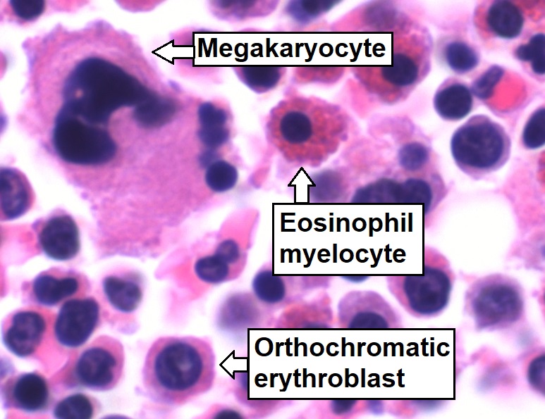
Bone marrow is a semi-solid biological tissue, tissue found within the Spongy bone, spongy (also known as cancellous) portions of bones. In birds and mammals, bone marrow is the primary site of new blood cell production (or haematopoiesis). It is composed of Blood cell, hematopoietic cells, marrow adipose tissue, and supportive stromal cells. In adult humans, bone marrow is primarily located in the Rib cage, ribs, vertebrae, sternum, and Pelvis, bones of the pelvis. Bone marrow comprises approximately 5% of total body mass in healthy adult humans, such that a man weighing 73 kg (161 lbs) will have around 3.7 kg (8 lbs) of bone marrow. Human marrow produces approximately 500 billion blood cells per day, which join the Circulatory system, systemic circulation via permeable vasculature sinusoids within the medullary cavity. All types of hematopoietic cells, including both Myeloid tissue, myeloid and Lymphocyte, lymphoid lineages, are created in bone marrow; howev ... [...More Info...] [...Related Items...] OR: [Wikipedia] [Google] [Baidu] |
Gold Standard (test)
In medicine and statistics, a gold standard test is usually the diagnostic test or benchmark that is the best available under reasonable conditions. In other words, a gold standard is the most accurate test possible without restrictions. Both meanings are different because for example, in medicine, dealing with conditions that would require an autopsy to have a perfect diagnosis, the gold standard test would be the best one that keeps the patient alive instead of the autopsy. In medicine "Gold standard" can refer to the criteria by which scientific evidence is evaluated. For example, in resuscitation research, the "gold standard" test of a medication or procedure is whether or not it leads to an increase in the number of neurologically intact survivors that walk out of the hospital.''ACLS: Principles and Practice''. p. 62. Dallas: American Heart Association, 2003. . Other types of medical research might regard a significant decrease in 30-day mortality as the gold standard. The ... [...More Info...] [...Related Items...] OR: [Wikipedia] [Google] [Baidu] |
Vein Valve
Veins are blood vessels in humans and most other animals that carry blood towards the heart. Most veins carry deoxygenated blood from the tissues back to the heart; exceptions are the pulmonary and umbilical veins, both of which carry oxygenated blood to the heart. In contrast to veins, arteries carry blood away from the heart. Veins are less muscular than arteries and are often closer to the skin. There are valves (called ''pocket valves'') in most veins to prevent backflow. Structure Veins are present throughout the body as tubes that carry blood back to the heart. Veins are classified in a number of ways, including superficial vs. deep, pulmonary vs. systemic, and large vs. small. * Superficial veins are those closer to the surface of the body, and have no corresponding arteries. *Deep veins are deeper in the body and have corresponding arteries. *Perforator veins drain from the superficial to the deep veins. These are usually referred to in the lower limbs and feet. *Communi ... [...More Info...] [...Related Items...] OR: [Wikipedia] [Google] [Baidu] |
Inferior Vena Cava
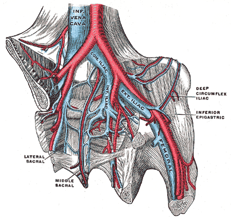
The inferior vena cava is a large vein that carries the deoxygenated blood from the lower and middle body into the right atrium of the heart. It is formed by the joining of the right and the left common iliac veins, usually at the level of the fifth lumbar vertebra. The inferior vena cava is the lower (" inferior") of the two venae cavae, the two large veins that carry deoxygenated blood from the body to the right atrium of the heart: the inferior vena cava carries blood from the lower half of the body whilst the superior vena cava carries blood from the upper half of the body. Together, the venae cavae (in addition to the coronary sinus, which carries blood from the muscle of the heart itself) form the venous counterparts of the aorta. It is a large retroperitoneal vein that lies posterior to the abdominal cavity and runs along the right side of the vertebral column. It enters the right auricle at the lower right, back side of the heart. The name derives from la, ve ... [...More Info...] [...Related Items...] OR: [Wikipedia] [Google] [Baidu] |
Median Cubital Vein
In human anatomy, the median cubital vein (or median basilic vein) is a superficial vein of the upper limb. It lies in the cubital fossa superficial to the bicipital aponeurosis. It connects the cephalic vein and the basilic vein. It becomes prominent when pressure is applied. It is routinely used for venipuncture (taking blood) and as a site for an intravenous cannula. This is due to its particularly wide lumen, and its tendency to remain stationary upon needle insertion. Structure The median cubital vein is a superficial vein of the upper limb. It lies in the cubital fossa superficial to the bicipital aponeurosis. It connects the cephalic vein and the basilic vein. It becomes prominent when pressure is applied to upstream veins, as venous blood builds up. Variations The median cubital vein shows a wide range of variations. More commonly, the vein forms an H-pattern with the cephalic and basilic veins making up the sides. Other forms include an M-pattern, where the vein bra ... [...More Info...] [...Related Items...] OR: [Wikipedia] [Google] [Baidu] |
Cephalic Vein
In human anatomy, the cephalic vein is a superficial vein in the arm. It originates from the radial end of the dorsal venous network of hand, and ascends along the radial (lateral) side of the arm before emptying into the axillary vein. At the elbow, it communicates with the basilic vein via the median cubital vein. Anatomy The cephalic vein is situated within the superficial fascia along the anterolateral surface of the biceps. Origin The cephalic vein forms over the anatomical snuffbox at the radial end of the dorsal venous network of hand. Course and relations From its origin, it ascends ascends up the lateral aspect of the radius. Near the shoulder, the cephalic vein passes between the deltoid and pectoralis major muscles (deltopectoral groove) and through the clavipectoral triangle, where it empties into the axillary vein. Anastomoses It communicates with the basilic vein via the median cubital vein at the elbow. Clinical significance The cephalic vein is of ... [...More Info...] [...Related Items...] OR: [Wikipedia] [Google] [Baidu] |
X-ray
An X-ray, or, much less commonly, X-radiation, is a penetrating form of high-energy electromagnetic radiation. Most X-rays have a wavelength ranging from 10 picometers to 10 nanometers, corresponding to frequencies in the range 30 petahertz to 30 exahertz ( to ) and energies in the range 145 eV to 124 keV. X-ray wavelengths are shorter than those of UV rays and typically longer than those of gamma rays. In many languages, X-radiation is referred to as Röntgen radiation, after the German scientist Wilhelm Conrad Röntgen, who discovered it on November 8, 1895. He named it ''X-radiation'' to signify an unknown type of radiation.Novelline, Robert (1997). ''Squire's Fundamentals of Radiology''. Harvard University Press. 5th edition. . Spellings of ''X-ray(s)'' in English include the variants ''x-ray(s)'', ''xray(s)'', and ''X ray(s)''. The most familiar use of X-rays is checking for fractures (broken bones), but X-rays are also used in other ways. ... [...More Info...] [...Related Items...] OR: [Wikipedia] [Google] [Baidu] |
Digital Subtraction Angiography
Digital subtraction angiography (DSA) is a fluoroscopy technique used in interventional radiology to clearly visualize blood vessels in a bony or dense soft tissue environment. Images are produced using contrast medium by subtracting a "pre-contrast image" or ''mask'' from subsequent images, once the contrast medium has been introduced into a structure. Hence the term "digital ''subtraction'' angiography. Subtraction angiography was first described in 1935 and in English sources in 1962 as a manual technique. Digital technology made DSA practical starting in the 1970s. Procedure DSA and fluoroscopy In traditional angiography, images are acquired by exposing an area of interest with time-controlled x-rays while injecting a contrast medium into the blood vessels. The image obtained includes the blood vessels, together with all overlying and underlying structures. The images are useful for determining anatomical position and variations, but unhelpful for visualizing blood vessels acc ... [...More Info...] [...Related Items...] OR: [Wikipedia] [Google] [Baidu] |
Vein
Veins are blood vessels in humans and most other animals that carry blood towards the heart. Most veins carry deoxygenated blood from the tissues back to the heart; exceptions are the pulmonary and umbilical veins, both of which carry oxygenated blood to the heart. In contrast to veins, arteries carry blood away from the heart. Veins are less muscular than arteries and are often closer to the skin. There are valves (called ''pocket valves'') in most veins to prevent backflow. Structure Veins are present throughout the body as tubes that carry blood back to the heart. Veins are classified in a number of ways, including superficial vs. deep, pulmonary vs. systemic, and large vs. small. * Superficial veins are those closer to the surface of the body, and have no corresponding arteries. * Deep veins are deeper in the body and have corresponding arteries. * Perforator veins drain from the superficial to the deep veins. These are usually referred to in the lower limbs and feet. * ... [...More Info...] [...Related Items...] OR: [Wikipedia] [Google] [Baidu] |


