|
Retinotopy
Retinotopy (from Greek τόπος, place) is the mapping of visual input from the retina to neurons, particularly those neurons within the visual stream. For clarity, 'retinotopy' can be replaced with 'retinal mapping', and 'retinotopic' with 'retinally mapped'. Visual field maps (retinotopic maps) are found in many amphibian and mammalian species, though the specific size, number, and spatial arrangement of these maps can differ considerably. Sensory topographies can be found throughout the brain and are critical to the understanding of one's external environment. Moreover, the study of sensory topographies and retinotopy in particular has furthered our understanding of how neurons encode and organize sensory signals. Retinal mapping of the visual field is maintained through various points of the visual pathway including but not limited to the retina, the dorsal lateral geniculate nucleus, the optic tectum, the primary visual cortex (V1), and higher visual areas (V2-V4). Retin ... [...More Info...] [...Related Items...] OR: [Wikipedia] [Google] [Baidu] |
Retinotopic English
Retinotopy (from Greek τόπος, place) is the mapping of visual input from the retina to neurons, particularly those neurons within the visual stream. For clarity, 'retinotopy' can be replaced with 'retinal mapping', and 'retinotopic' with 'retinally mapped'. Visual field maps (retinotopic maps) are found in many amphibian and mammalian species, though the specific size, number, and spatial arrangement of these maps can differ considerably. Sensory topographies can be found throughout the brain and are critical to the understanding of one's external environment. Moreover, the study of sensory topographies and retinotopy in particular has furthered our understanding of how neurons encode and organize sensory signals. Retinal mapping of the visual field is maintained through various points of the visual pathway including but not limited to the retina, the dorsal lateral geniculate nucleus, the optic tectum, the primary visual cortex (V1), and higher visual areas (V2-V4). Retino ... [...More Info...] [...Related Items...] OR: [Wikipedia] [Google] [Baidu] |
Dorsomedial Area
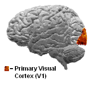
The visual cortex of the brain is the area of the cerebral cortex that processes visual information. It is located in the occipital lobe. Sensory input originating from the eyes travels through the lateral geniculate nucleus in the thalamus and then reaches the visual cortex. The area of the visual cortex that receives the sensory input from the lateral geniculate nucleus is the primary visual cortex, also known as visual area 1 ( V1), Brodmann area 17, or the striate cortex. The extrastriate areas consist of visual areas 2, 3, 4, and 5 (also known as V2, V3, V4, and V5, or Brodmann area 18 and all Brodmann area 19). Both hemispheres of the brain include a visual cortex; the visual cortex in the left hemisphere receives signals from the right visual field, and the visual cortex in the right hemisphere receives signals from the left visual field. Introduction The primary visual cortex (V1) is located in and around the calcarine fissure in the occipital lobe. Each hemisphere's V1 ... [...More Info...] [...Related Items...] OR: [Wikipedia] [Google] [Baidu] |
Visual Cortex
The visual cortex of the brain is the area of the cerebral cortex that processes visual information. It is located in the occipital lobe. Sensory input originating from the eyes travels through the lateral geniculate nucleus in the thalamus and then reaches the visual cortex. The area of the visual cortex that receives the sensory input from the lateral geniculate nucleus is the primary visual cortex, also known as visual area 1 ( V1), Brodmann area 17, or the striate cortex. The extrastriate areas consist of visual areas 2, 3, 4, and 5 (also known as V2, V3, V4, and V5, or Brodmann area 18 and all Brodmann area 19). Both hemispheres of the brain include a visual cortex; the visual cortex in the left hemisphere receives signals from the right visual field, and the visual cortex in the right hemisphere receives signals from the left visual field. Introduction The primary visual cortex (V1) is located in and around the calcarine fissure in the occipital lobe. Each hemisphere's V ... [...More Info...] [...Related Items...] OR: [Wikipedia] [Google] [Baidu] |
Superior Colliculus
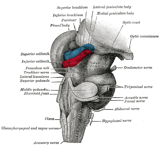
In neuroanatomy, the superior colliculus () is a structure lying on the roof of the mammalian midbrain. In non-mammalian vertebrates, the homologous structure is known as the optic tectum, or optic lobe. The adjective form '' tectal'' is commonly used for both structures. In mammals, the superior colliculus forms a major component of the midbrain. It is a paired structure and together with the paired inferior colliculi forms the corpora quadrigemina. The superior colliculus is a layered structure, with a pattern that is similar to all mammals. The layers can be grouped into the superficial layers ( stratum opticum and above) and the deeper remaining layers. Neurons in the superficial layers receive direct input from the retina and respond almost exclusively to visual stimuli. Many neurons in the deeper layers also respond to other modalities, and some respond to stimuli in multiple modalities. The deeper layers also contain a population of motor-related neurons, capable of acti ... [...More Info...] [...Related Items...] OR: [Wikipedia] [Google] [Baidu] |
Cerebral Cortex
The cerebral cortex, also known as the cerebral mantle, is the outer layer of neural tissue of the cerebrum of the brain in humans and other mammals. The cerebral cortex mostly consists of the six-layered neocortex, with just 10% consisting of allocortex. It is separated into two cortices, by the longitudinal fissure that divides the cerebrum into the left and right cerebral hemispheres. The two hemispheres are joined beneath the cortex by the corpus callosum. The cerebral cortex is the largest site of neural integration in the central nervous system. It plays a key role in attention, perception, awareness, thought, memory, language, and consciousness. The cerebral cortex is part of the brain responsible for cognition. In most mammals, apart from small mammals that have small brains, the cerebral cortex is folded, providing a greater surface area in the confined volume of the cranium. Apart from minimising brain and cranial volume, cortical folding is crucial f ... [...More Info...] [...Related Items...] OR: [Wikipedia] [Google] [Baidu] |
Meridian (perimetry, Visual Field)
Meridian (plural: "meridians") is used in perimetry and in specifying visual fields. According to IPS Perimetry Standards 1978 (2002): ''" Perimetry is the measurement of n observer'svisual functions ... at topographically defined loci in the visual field. The visual field is that portion of the external environment of the observer n which when he or she issteadily fixating ...e or shecan detect visual stimuli."'' In perimetry, the observer's eye is considered to be at the centre of an imaginary sphere. More precisely, the centre of the sphere is in the centre of the pupil of the observer's eye. The observer is looking at a point, the fixation point, on the interior of the sphere. The visual field can be considered to be all parts of the sphere for which the observer can see a particular test stimulus. If we consider this surface to be that on which an observer can see anything, then it is a section of the sphere somewhat larger than a hemisphere. In reality it is smaller than ... [...More Info...] [...Related Items...] OR: [Wikipedia] [Google] [Baidu] |
Superior Colliculus
In neuroanatomy, the superior colliculus () is a structure lying on the roof of the mammalian midbrain. In non-mammalian vertebrates, the homologous structure is known as the optic tectum, or optic lobe. The adjective form '' tectal'' is commonly used for both structures. In mammals, the superior colliculus forms a major component of the midbrain. It is a paired structure and together with the paired inferior colliculi forms the corpora quadrigemina. The superior colliculus is a layered structure, with a pattern that is similar to all mammals. The layers can be grouped into the superficial layers ( stratum opticum and above) and the deeper remaining layers. Neurons in the superficial layers receive direct input from the retina and respond almost exclusively to visual stimuli. Many neurons in the deeper layers also respond to other modalities, and some respond to stimuli in multiple modalities. The deeper layers also contain a population of motor-related neurons, capable of acti ... [...More Info...] [...Related Items...] OR: [Wikipedia] [Google] [Baidu] |
Gyri (neuroanatomy)
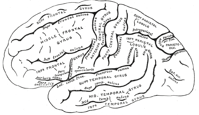
In neuroanatomy, a gyrus (pl. gyri) is a ridge on the cerebral cortex. It is generally surrounded by one or more sulci (depressions or furrows; sg. ''sulcus''). Gyri and sulci create the folded appearance of the brain in humans and other mammals. Structure The gyri are part of a system of folds and ridges that create a larger surface area for the human brain and other mammalian brains. Because the brain is confined to the skull, brain size is limited. Ridges and depressions create folds allowing a larger cortical surface area, and greater cognitive function, to exist in the confines of a smaller cranium. Development The human brain undergoes gyrification during fetal and neonatal development. In embryonic development, all mammalian brains begin as smooth structures derived from the neural tube. A cerebral cortex without surface convolutions is lissencephalic, meaning 'smooth-brained'. As development continues, gyri and sulci begin to take shape on the fetal brain, w ... [...More Info...] [...Related Items...] OR: [Wikipedia] [Google] [Baidu] |
Human
Humans (''Homo sapiens'') are the most abundant and widespread species of primate, characterized by bipedalism and exceptional cognitive skills due to a large and complex brain. This has enabled the development of advanced tools, culture, and language. Humans are highly social and tend to live in complex social structures composed of many cooperating and competing groups, from families and kinship networks to political states. Social interactions between humans have established a wide variety of values, social norms, and rituals, which bolster human society. Its intelligence and its desire to understand and influence the environment and to explain and manipulate phenomena have motivated humanity's development of science, philosophy, mythology, religion, and other fields of study. Although some scientists equate the term ''humans'' with all members of the genus '' Homo'', in common usage, it generally refers to ''Homo sapiens'', the only extant member. Anato ... [...More Info...] [...Related Items...] OR: [Wikipedia] [Google] [Baidu] |
Macaque
The macaques () constitute a genus (''Macaca'') of gregarious Old World monkeys of the subfamily Cercopithecinae. The 23 species of macaques inhabit ranges throughout Asia, North Africa, and (in one instance) Gibraltar. Macaques are principally frugivorous (preferring fruit), although their diet also includes seeds, leaves, flowers, and tree bark. Some species, such as the crab-eating macaque, subsist on a diet of invertebrates and occasionally small vertebrates. On average, southern pig-tailed macaques in Malaysia eat about 70 large rats each per year. All macaque social groups are matriarchal, arranged around dominant females. Macaques are found in a variety of habitats throughout the Asian continent and are highly adaptable. Certain species have learned to live with humans and have become invasive in some human-settled environments, such as the island of Mauritius and Silver Springs State Park in Florida. Macaques can be a threat to wildlife conservation as well as to hum ... [...More Info...] [...Related Items...] OR: [Wikipedia] [Google] [Baidu] |
Sulci (neuroanatomy)
In neuroanatomy, a sulcus (Latin: "furrow", pl. ''sulci'') is a depression or groove in the cerebral cortex. It surrounds a gyrus (pl. gyri), creating the characteristic folded appearance of the brain in humans and other mammals. The larger sulci are usually called fissures. Structure Sulci, the grooves, and gyri, the folds or ridges, make up the folded surface of the cerebral cortex. Larger or deeper sulci are termed fissures, and in many cases the two terms are interchangeable. The folded cortex creates a larger surface area for the brain in humans and other mammals. When looking at the human brain, two-thirds of the surface are hidden in the grooves. The sulci and fissures are both grooves in the cortex, but they are differentiated by size. A sulcus is a shallower groove that surrounds a gyrus. A fissure is a large furrow that divides the brain into lobes and also into the two hemispheres as the longitudinal fissure. Importance of expanded surface area As the surfa ... [...More Info...] [...Related Items...] OR: [Wikipedia] [Google] [Baidu] |


