|
Reflex
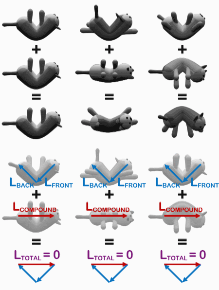
In biology, a reflex, or reflex action, is an involuntary, unplanned sequence or action and nearly instantaneous response to a stimulus. Reflexes are found with varying levels of complexity in organisms with a nervous system. A reflex occurs via neural pathways in the nervous system called reflex arcs. A stimulus initiates a neural signal, which is carried to a synapse. The signal is then transferred across the synapse to a motor neuron which evokes a target response. These neural signals do not always travel to the brain, so many reflexes are an automatic response to a stimulus that does not receive or need conscious thought. Many reflexes are fine-tuned to increase organism survival and self-defense. This is observed in reflexes such as the startle reflex, which provides an automatic response to an unexpected stimuli, and the feline righting reflex, which reorients a cat's body when falling to ensure safe landing. The simplest type of reflex, a short-latency reflex, has a ... [...More Info...] [...Related Items...] OR: [Wikipedia] [Google] [Baidu] |
Pupillary Light Reflex
The pupillary light reflex (PLR) or photopupillary reflex is a reflex that controls the diameter of the pupil, in response to the intensity (luminance) of light that falls on the retinal ganglion cells of the retina in the back of the eye, thereby assisting in adaptation of vision to various levels of lightness/darkness. A greater intensity of light causes the pupil to constrict ( miosis/myosis; thereby allowing less light in), whereas a lower intensity of light causes the pupil to dilate ( mydriasis, expansion; thereby allowing more light in). Thus, the pupillary light reflex regulates the intensity of light entering the eye. Light shone into one eye will cause both pupils to constrict. Terminology The pupil is the dark circular opening in the center of the iris and is where light enters the eye. By analogy with a camera, the pupil is equivalent to aperture, whereas the iris is equivalent to the diaphragm. It may be helpful to consider the ''Pupillary reflex'' as an Iris' ref ... [...More Info...] [...Related Items...] OR: [Wikipedia] [Google] [Baidu] |
Stretch Reflex
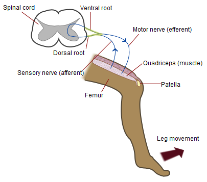
The stretch reflex (myotatic reflex), or more accurately "muscle stretch reflex", is a muscle contraction in response to stretching within the muscle. The reflex functions to maintain the muscle at a constant length. The term deep tendon reflex is often used by many health workers and students to refer to this reflex. "Tendons have little to do with the response, other than being responsible for mechanically transmitting the sudden stretch from the reflex hammer to the muscle spindle. In addition, some muscles with stretch reflexes have no tendons (e.g., "jaw jerk" of the masseter muscle)". As an example of a spinal reflex, it results in a fast response that involves an afferent signal into the spinal cord and an efferent signal out to the muscle. The stretch reflex can be a monosynaptic reflex which provides automatic regulation of skeletal muscle length, whereby the signal entering the spinal cord arises from a change in muscle length or velocity. It can also include a polysyna ... [...More Info...] [...Related Items...] OR: [Wikipedia] [Google] [Baidu] |
Righting Reflex
The righting reflex, also known as the labyrinthine righting reflex, is a reflex that corrects the orientation of the body when it is taken out of its normal upright position. It is initiated by the vestibular system, which detects that the body is not erect and causes the head to move back into position as the rest of the body follows. The perception of head movement involves the body sensing linear acceleration or the force of gravity through the otoliths, and angular acceleration through the semicircular canals. The reflex uses a combination of visual system inputs, vestibular inputs, and somatosensory inputs to make postural adjustments when the body becomes displaced from its normal vertical position. These inputs are used to create what is called an efference copy. This means that the brain makes comparisons in the cerebellum between expected posture and perceived posture, and corrects for the difference. The reflex takes 6 or 7 weeks to perfect, but can be affected by various t ... [...More Info...] [...Related Items...] OR: [Wikipedia] [Google] [Baidu] |
Golgi Tendon Reflex
The Golgi tendon reflex (also called inverse stretch reflex, autogenic inhibition, tendon reflex) is an inhibitory effect on the muscle resulting from the muscle tension stimulating Golgi tendon organs (GTO) of the muscle, and hence it is self-induced. The reflex arc is a negative feedback mechanism preventing too much tension on the muscle and tendon. When the tension is extreme, the inhibition can be so great it overcomes the excitatory effects on the muscle's alpha motoneurons causing the muscle to suddenly relax. This reflex is also called the inverse myotatic reflex, because it is the inverse of the stretch reflex. GTOs' inhibitory effects come from their reflex arcs: the Ib sensory fibers that are sent through the dorsal root into the spinal cord to synapse on Ib inhibitory interneurons that in turn terminate directly on the motor neurons that innervate the same muscle. The fibers also make direct excitatory synapses onto motoneurons that innervate the antagonist muscle. No ... [...More Info...] [...Related Items...] OR: [Wikipedia] [Google] [Baidu] |
Reflex Arc
A reflex arc is a neural pathway that controls a reflex. In vertebrates, most sensory neurons do not pass directly into the brain, but synapse in the spinal cord. This allows for faster reflex actions to occur by activating spinal motor neurons without the delay of routing signals through the brain. The brain will receive the input while the reflex is being carried out and the analysis of the signal takes place after the reflex action. There are two types: autonomic reflex arc (affecting inner organs) and somatic reflex arc (affecting muscles). Autonomic reflexes sometimes involve the spinal cord and some somatic reflexes are mediated more by the brain than the spinal cord. During a somatic reflex, nerve signals travel along the following pathway: # ''Somatic receptors'' in the skin, muscles and tendons # ''Afferent nerve fibers'' carry signals from the somatic receptors to the posterior horn of the spinal cord or to the brainstem # An ''integrating center'', the point at whic ... [...More Info...] [...Related Items...] OR: [Wikipedia] [Google] [Baidu] |
Tendon Reflex
Tendon reflex (or T-reflex) may refer to: *The stretch reflex or muscle stretch reflex (MSR), when the stretch is created by a blow upon a muscle tendon. This is the commonly used definition of the term. Albeit a misnomer, in this sense a common example is the standard patellar reflex or knee-jerk response. Stretch reflex tests are used to determine the integrity of the spinal cord and peripheral nervous system, and they can be used to determine the presence of a neuromuscular disease. ::Note that the term "deep tendon reflex", if it refers to the muscle stretch reflex, is a misnomer. "Tendons have little to do with the response, other than being responsible for mechanically transmitting the sudden stretch from the reflex hammer to the muscle spindle. In addition, some muscles with stretch reflexes have no tendons (e.g., "jaw jerk" of the masseter muscle)". *The Golgi tendon reflex, which is a reflex to extensive tension on a tendon; it functions to protect musculoskeletal inte ... [...More Info...] [...Related Items...] OR: [Wikipedia] [Google] [Baidu] |
Patellar Reflex
The patellar reflex, also called the knee reflex or knee-jerk, is a stretch reflex which tests the L2, L3, and L4 segments of the spinal cord. Mechanism Striking of the patellar tendon with a reflex hammer just below the patella stretches the muscle spindle in the quadriceps muscle. This produces a signal which travels back to the spinal cord and synapses (without interneurons) at the level of L3 or L4 in the spinal cord, completely independent of higher centres. From there, an alpha motor neuron conducts an efferent impulse back to the quadriceps femoris muscle, triggering contraction. This contraction, coordinated with the relaxation of the antagonistic flexor hamstring muscle causes the leg to kick. There is a latency of around 18 ms between stretch of the patellar tendon and the beginning of contraction of the quadriceps femoris muscle. This is a reflex of proprioception which helps maintain posture and balance, allowing to keep one's balance with little effort or cons ... [...More Info...] [...Related Items...] OR: [Wikipedia] [Google] [Baidu] |
Startle Response
In animals, including humans, the startle response is a largely unconscious defensive response to sudden or threatening stimuli, such as sudden noise or sharp movement, and is associated with negative affect.Rammirez-Moreno, David. "A computational model for the modulation of the prepulse inhibition of the acoustic startle reflex". ''Biological Cybernetics'', 2012, p. 169 Usually the onset of the startle response is a startle reflex reaction. The startle reflex is a brainstem reflectory reaction (reflex) that serves to protect vulnerable parts, such as the back of the neck (whole-body startle) and the eyes (eyeblink) and facilitates escape from sudden stimuli. It is found across many different species, throughout all stages of life. A variety of responses may occur depending on the affected individual's emotional state, body posture, preparation for execution of a motor task, or other activities. The startle response is implicated in the formation of specific phobias. Startl ... [...More Info...] [...Related Items...] OR: [Wikipedia] [Google] [Baidu] |
Triceps Reflex
The triceps reflex, a deep tendon reflex, is a reflex as it elicits involuntary contraction of the triceps brachii muscle. It is initiated by the Cervical (of the neck region) spinal nerve 7 nerve root (the small segment of the nerve that emerges from the spinal cord). The reflex is tested as part of the neurological examination to assess the sensory and motor pathways within the C7 and C8 spinal nerves. Testing The test can be performed by tapping the triceps tendonA tendon is a strip or sheet of connective tissue that transmits the force generated by the contraction of muscle to the bone by attaching with it. Thus, in simple words, a tendon attaches a muscle to a bone with the sharp end of a reflex hammer while the forearm is hanging loose at a right angle to the arm. A sudden contraction of the triceps muscle causes extension,A straightening at the elbow joint) of the forearm and indicates a normal reflex. Reflex arc The arc involves the stretch receptors in the tricep ... [...More Info...] [...Related Items...] OR: [Wikipedia] [Google] [Baidu] |
H-reflex
The H-reflex (or Hoffmann's reflex) is a reflectory reaction of muscles after electrical stimulation of sensory fibers (Ia afferents stemming from muscle spindles) in their innervating nerves (for example, those located behind the knee). The H-reflex test is performed using an electric stimulator, which gives usually a square-wave current of short duration and small amplitude (higher stimulations might involve alpha fibers, causing an F-wave, compromising the results), and an EMG set, to record the muscle response. That response is usually a clear wave, called H-wave, 28-35 ms after the stimulus, not to be confused with an F-wave. An M-wave, an early response, occurs 3-6 ms after the onset of stimulation. The H and F-waves are later responses. As the stimulus increases, the amplitude of the F-wave increases only slightly, and the H-wave decreases, and at supramaximal stimulus, the H-wave will disappear. The M-wave does the opposite of the H-wave. As the stimulus increases th ... [...More Info...] [...Related Items...] OR: [Wikipedia] [Google] [Baidu] |
Ankle Jerk Reflex
The ankle jerk reflex, also known as the Achilles reflex, occurs when the Achilles tendon is tapped while the foot is dorsiflexed. It is a type of stretch reflex that tests the function of the gastrocnemius muscle and the nerve that supplies it. A positive result would be the jerking of the foot towards its plantar surface. Being a deep tendon reflex, it is monosynaptic. It is also a stretch reflex. These are monosynaptic spinal segmental reflexes. When they are intact, integrity of the following is confirmed: cutaneous innervation, motor supply, and cortical input to the corresponding spinal segment. Root value This reflex is mediated by the S1 spinal segment of the spinal cord. Procedure and components Ankle of the patient is relaxed. It is helpful to support the ball of the foot at least somewhat to put some tension in the Achilles tendon, but don’t completely dorsiflex the ankle. A small strike is given on the Achilles tendon using a rubber hammer to elicit the respons ... [...More Info...] [...Related Items...] OR: [Wikipedia] [Google] [Baidu] |
Biceps Reflex
Biceps reflex is a reflex test that examines the function of the C5 reflex arc and the C6 reflex arc. The test is performed by using a tendon hammer to quickly depress the biceps brachii tendon as it passes through the cubital fossa. Specifically, the test activates the stretch receptors inside the biceps brachii muscle which communicates mainly with the C5 spinal nerve and partially with the C6 spinal nerve to induce a reflex contraction of the biceps muscle and jerk of the forearm. A strong contraction indicates a "brisk" reflex, and a weak or absent reflex is known as "diminished". Brisk or absent reflexes are used as clues to the location of neurological disease. Typically, brisk reflexes are found in lesions of upper motor neurons, and absent or reduced reflexes are found in lower motor neuron lesions. A change in the biceps reflex indicates pathology at the level of musculocutaneous nerve The musculocutaneous nerve arises from the lateral cord of the brachial plexus, o ... [...More Info...] [...Related Items...] OR: [Wikipedia] [Google] [Baidu] |


