|
Plexus
In neuroanatomy, a plexus (from the Latin term for "braid") is a branching network of vessels or nerves. The vessels may be blood vessels (veins, capillaries) or lymphatic vessels. The nerves are typically axons outside the central nervous system. The standard plural form in English is plexuses. Alternatively, the Latin plural plexūs may be used. Types Nerve plexuses The four primary nerve plexuses are the cervical plexus, brachial plexus, lumbar plexus, and the sacral plexus. Cardiac plexus Celiac plexus Renal plexus Venous plexus Choroid plexus The choroid plexus is a part of the central nervous system in the brain and consists of capillaries, brain ventricles, and ependymal cells. Invertebrates The plexus is the characteristic form of nervous system in the coelenterates and persists with modifications in the flatworms. The nerves of the radially symmetric echinoderm An echinoderm () is any member of the phylum Echinodermata (). The adults are recognis ... [...More Info...] [...Related Items...] OR: [Wikipedia] [Google] [Baidu] |
Brachial Plexus
The brachial plexus is a network () of nerves formed by the anterior rami of the lower four cervical nerves and first thoracic nerve ( C5, C6, C7, C8, and T1). This plexus extends from the spinal cord, through the cervicoaxillary canal in the neck, over the first rib, and into the armpit, it supplies afferent and efferent nerve fibers the to chest, shoulder, arm, forearm, and hand. Structure The brachial plexus is divided into five ''roots'', three ''trunks'', six ''divisions'' (three anterior and three posterior), three ''cords'', and five ''branches''. There are five "terminal" branches and numerous other "pre-terminal" or "collateral" branches, such as the subscapular nerve, the thoracodorsal nerve, and the long thoracic nerve, that leave the plexus at various points along its length. A common structure used to identify part of the brachial plexus in cadaver dissections is the M or W shape made by the musculocutaneous nerve, lateral cord, median nerve, medial cord, a ... [...More Info...] [...Related Items...] OR: [Wikipedia] [Google] [Baidu] |
Nerve Plexus
A nerve plexus is a plexus (branching network) of intersecting nerves. A nerve plexus is composed of afferent and efferent fibers that arise from the merging of the anterior rami of spinal nerves and blood vessels. There are five spinal nerve plexuses, except in the thoracic region, as well as other forms of autonomic plexuses, many of which are a part of the enteric nervous system. The nerves that arise from the plexuses have both sensory and motor functions. These functions include muscle contraction, the maintenance of body coordination and control, and the reaction to sensations such as heat, cold, pain, and pressure. There are several plexuses in the body, including: *Spinal Plexuses **Cervical plexus - serves the head, neck and shoulders **Brachial plexus - serves the chest, shoulders, arms and hands ** Lumbosacral plexus *** Lumbar plexus - serves the back, abdomen, groin, thighs, knees, and calves ****Subsartorial plexus - below the sartorius muscle of thigh *** Sacral ple ... [...More Info...] [...Related Items...] OR: [Wikipedia] [Google] [Baidu] |
Choroid Plexus
The choroid plexus, or plica choroidea, is a plexus of cells that arises from the tela choroidea in each of the ventricles of the brain. Regions of the choroid plexus produce and secrete most of the cerebrospinal fluid (CSF) of the central nervous system. The choroid plexus consists of modified ependymal cells surrounding a core of capillaries and loose connective tissue. Multiple cilia on the ependymal cells move to circulate the cerebrospinal fluid. Structure Location There is a choroid plexus in each of the four ventricles. In the lateral ventricles it is found in the body, and continued in an enlarged amount in the atrium. There is no choroid plexus in the anterior horn. In the third ventricle there is a small amount in the roof that is continuous with that in the body, via the interventricular foramina, the channels that connect the lateral ventricles with the third ventricle. A choroid plexus is in part of the roof of the fourth ventricle. Microanatomy T ... [...More Info...] [...Related Items...] OR: [Wikipedia] [Google] [Baidu] |
Lumbar Plexus
The lumbar plexus is a web of nerves (a nervous plexus) in the lumbar region of the body which forms part of the larger lumbosacral plexus. It is formed by the Ventral ramus of spinal nerve, divisions of the first four lumbar nerves (L1-L4) and from contributions of the subcostal nerve (T12), which is the last Thoracic nerves, thoracic nerve. Additionally, the ventral rami of the fourth lumbar nerve pass communicating branches, the lumbosacral trunk, to the sacral plexus. The nerves of the lumbar plexus pass in front of the hip joint and mainly support the anterior part of the thigh.''Thieme Atlas of anatomy'' (2006), pp 470-471 The plexus is formed lateral to the intervertebral foramina and passes through Psoas major muscle, psoas major. Its smaller motor branches are distributed directly to psoas major, while the larger branches leave the muscle at various sites to run obliquely down through the pelvis to leave under the inguinal ligament with the exception of the obturator n ... [...More Info...] [...Related Items...] OR: [Wikipedia] [Google] [Baidu] |
Sacral Plexus
In human anatomy, the sacral plexus is a nerve plexus which provides motor and sensory nerves for the posterior thigh, most of the lower leg and foot, and part of the pelvis. It is part of the lumbosacral plexus and emerges from the lumbar vertebrae and sacral vertebrae (L4-S4).''Thieme Atlas of Anatomy'' (2006), pp 470-471 A sacral plexopathy is a disorder affecting the nerves of the sacral plexus, usually caused by trauma, nerve compression, vascular disease, or infection. Symptoms may include pain, loss of motor control, and sensory deficits. Structure The sacral plexus is formed by: * the lumbosacral trunk * the anterior division of the first sacral nerve * portions of the anterior divisions of the second and third sacral nerves The nerves forming the sacral plexus converge toward the lower part of the greater sciatic foramen, and unite to form a flattened band, from the anterior and posterior surfaces of which several branches arise. The band itself is continued as th ... [...More Info...] [...Related Items...] OR: [Wikipedia] [Google] [Baidu] |
Cervical Plexus
The cervical plexus is a plexus of the anterior rami of the first four cervical spinal nerves which arise from C1 to C4 cervical segment in the neck. They are located laterally to the transverse processes between prevertebral muscles from the medial side and vertebral (m. scalenus, m. levator scapulae, m. splenius cervicis) from lateral side. There is anastomosis with accessory nerve, hypoglossal nerve and sympathetic trunk. It is located in the neck, deep to the sternocleidomastoid muscle. Nerves formed from the cervical plexus innervate the back of the head, as well as some neck muscles. The branches of the cervical plexus emerge from the posterior triangle at the nerve point, a point which lies midway on the posterior border of the sternocleidomastoid. Branches The cervical plexus has two types of branches: cutaneous and muscular. *Cutaneous (4 branches): ** Lesser occipital nerve - innervates the skin and the scalp posterosuperior to the auricle (C2) ** Great auricular ... [...More Info...] [...Related Items...] OR: [Wikipedia] [Google] [Baidu] |
Brain Ventricle
The ventricular system is a set of four interconnected cavities known as cerebral ventricles in the brain. Within each ventricle is a region of choroid plexus which produces the circulating cerebrospinal fluid (CSF). The ventricular system is continuous with the central canal of the spinal cord from the fourth ventricle, allowing for the flow of CSF to circulate. All of the ventricular system and the central canal of the spinal cord are lined with ependyma, a specialised form of epithelium connected by tight junctions that make up the blood–cerebrospinal fluid barrier. Structure The system comprises four ventricles: * lateral ventricles right and left (one for each hemisphere) * third ventricle * fourth ventricle There are several foramina, openings acting as channels, that connect the ventricles. The interventricular foramina (also called the foramina of Monro) connect the lateral ventricles to the third ventricle through which the cerebrospinal fluid can flow. Ventri ... [...More Info...] [...Related Items...] OR: [Wikipedia] [Google] [Baidu] |
Echinoderm
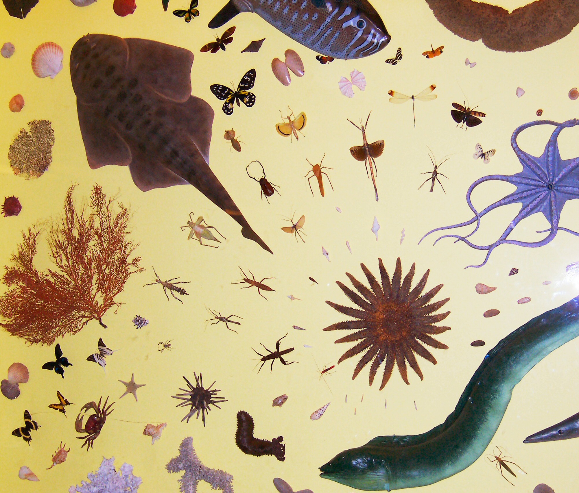
An echinoderm () is any member of the phylum Echinodermata (). The adults are recognisable by their (usually five-point) radial symmetry, and include starfish, brittle stars, sea urchins, sand dollars, and sea cucumbers, as well as the sea lilies or "stone lilies". Adult echinoderms are found on the sea bed at every ocean depth, from the intertidal zone to the abyssal zone. The phylum contains about 7,000 living species, making it the second-largest grouping of deuterostomes, after the chordates. Echinoderms are the largest entirely marine phylum. The first definitive echinoderms appeared near the start of the Cambrian. The echinoderms are important both ecologically and geologically. Ecologically, there are few other groupings so abundant in the biotic desert of the deep sea, as well as shallower oceans. Most echinoderms are able to reproduce asexually and regenerate tissue, organs, and limbs; in some cases, they can undergo complete regeneration from a single li ... [...More Info...] [...Related Items...] OR: [Wikipedia] [Google] [Baidu] |
Radial Symmetry
Symmetry in biology refers to the symmetry observed in organisms, including plants, animals, fungi, and bacteria. External symmetry can be easily seen by just looking at an organism. For example, take the face of a human being which has a plane of symmetry down its centre, or a pine cone with a clear symmetrical spiral pattern. Internal features can also show symmetry, for example the tubes in the human body (responsible for transporting gases, nutrients, and waste products) which are cylindrical and have several planes of symmetry. Biological symmetry can be thought of as a balanced distribution of duplicate body parts or shapes within the body of an organism. Importantly, unlike in mathematics, symmetry in biology is always approximate. For example, plant leaves – while considered symmetrical – rarely match up exactly when folded in half. Symmetry is one class of patterns in nature whereby there is near-repetition of the pattern element, either by reflection or rotation ... [...More Info...] [...Related Items...] OR: [Wikipedia] [Google] [Baidu] |
Flatworm
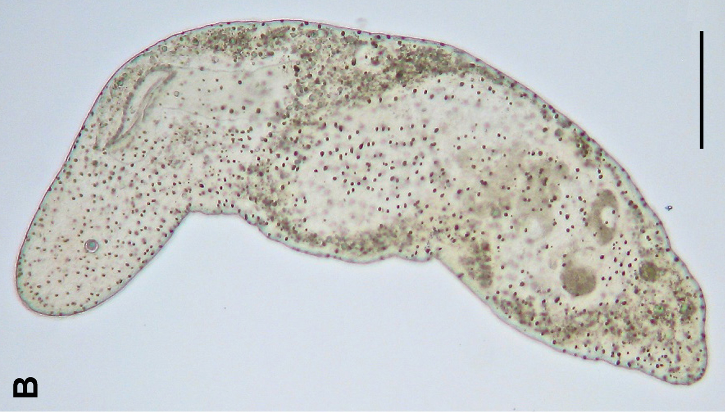
The flatworms, flat worms, Platyhelminthes, or platyhelminths (from the Greek πλατύ, ''platy'', meaning "flat" and ἕλμινς (root: ἑλμινθ-), ''helminth-'', meaning "worm") are a phylum of relatively simple bilaterian, unsegmented, soft-bodied invertebrates. Unlike other bilaterians, they are acoelomates (having no body cavity), and have no specialized circulatory and respiratory organs, which restricts them to having flattened shapes that allow oxygen and nutrients to pass through their bodies by diffusion. The digestive cavity has only one opening for both ingestion (intake of nutrients) and egestion (removal of undigested wastes); as a result, the food cannot be processed continuously. In traditional medicinal texts, Platyhelminthes are divided into Turbellaria, which are mostly non- parasitic animals such as planarians, and three entirely parasitic groups: Cestoda, Trematoda and Monogenea; however, since the turbellarians have since been proven not to be ... [...More Info...] [...Related Items...] OR: [Wikipedia] [Google] [Baidu] |
Coelenterate
Radiata or Radiates is a historical taxonomic rank that was used to classify animals with radially symmetric body plans. The term Radiata is no longer accepted, as it united several different groupings of animals that do not form a monophyletic group under current views of animal phylogeny. The similarities once offered in justification of the taxon, such as radial symmetry, are now taken to be the result of either incorrect evaluations by early researchers or convergent evolution, rather than an indication of a common ancestor. Because of this, the term is used mostly in a historical context. In the early 19th century, Georges Cuvier united Ctenophora and Cnidaria in the Radiata ('' Zoophytes''). Thomas Cavalier-Smith, in 1983, redefined Radiata as a subkingdom consisting of Myxozoa, Placozoa, Cnidaria and Ctenophora. Lynn Margulis and K. V. Schwartz later redefined Radiata in their '' Five Kingdom'' classification, this time including only Cnidaria and Ctenophora. This de ... [...More Info...] [...Related Items...] OR: [Wikipedia] [Google] [Baidu] |
Ependymal Cells
The ependyma is the thin neuroepithelial ( simple columnar ciliated epithelium) lining of the ventricular system of the brain and the central canal of the spinal cord. The ependyma is one of the four types of neuroglia in the central nervous system (CNS). It is involved in the production of cerebrospinal fluid (CSF), and is shown to serve as a reservoir for neuroregeneration. Structure The ependyma is made up of ependymal cells called ependymocytes, a type of glial cell. These cells line the ventricles in the brain and the central canal of the spinal cord, which become filled with cerebrospinal fluid. These are nervous tissue cells with simple columnar shape, much like that of some mucosal epithelial cells. Early monociliated ependymal cells are differentiated to multiciliated ependymal cells for their function in circulating cerebrospinal fluid. The basal membranes of these cells are characterized by tentacle-like extensions that attach to astrocytes. The apical side is cov ... [...More Info...] [...Related Items...] OR: [Wikipedia] [Google] [Baidu] |


