|
Metaphysis
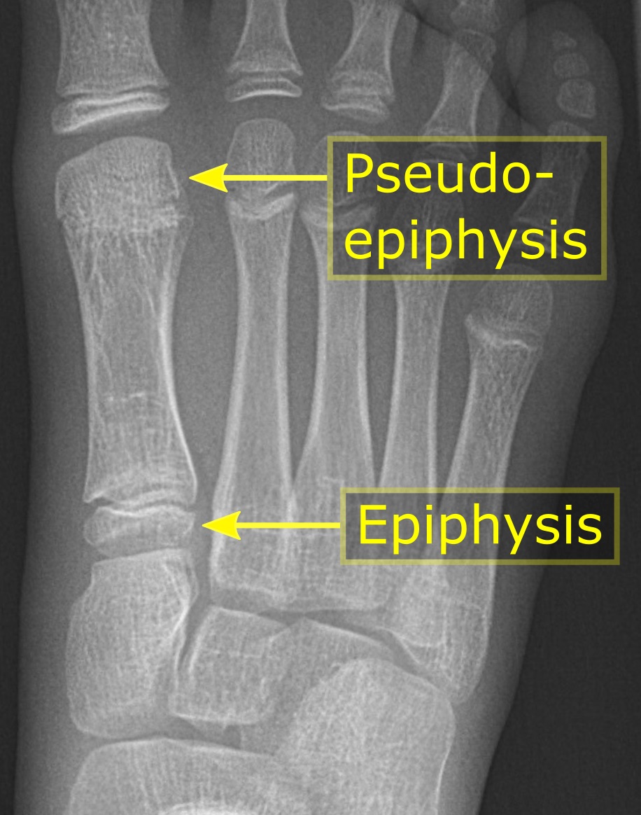
The metaphysis is the neck portion of a long bone between the epiphysis and the diaphysis. It contains the growth plate, the part of the bone that grows during childhood, and as it grows it ossifies near the diaphysis and the epiphyses. The metaphysis contains a diverse population of cells including mesenchymal stem cells, which give rise to bone and fat cells, as well as hematopoietic stem cells which give rise to a variety of blood cells as well as bone-destroying cells called osteoclasts. Thus the metaphysis contains a highly metabolic set of tissues including trabecular (spongy) bone, blood vessels , as well as Marrow Adipose Tissue (MAT). The metaphysis may be divided anatomically into three components based on tissue content: a cartilaginous component (epiphyseal plate), a bony component (metaphysis) and a fibrous component surrounding the periphery of the plate. The growth plate synchronizes chondrogenesis with osteogenesis or interstitial cartilage growth with both a ... [...More Info...] [...Related Items...] OR: [Wikipedia] [Google] [Baidu] |
Epiphysis
The epiphysis () is the rounded end of a long bone, at its joint with adjacent bone(s). Between the epiphysis and diaphysis (the long midsection of the long bone) lies the metaphysis, including the epiphyseal plate (growth plate). At the joint, the epiphysis is covered with articular cartilage; below that covering is a zone similar to the epiphyseal plate, known as subchondral bone. The epiphysis is filled with red bone marrow, which produces erythrocytes (red blood cells). Structure There are four types of epiphysis: # Pressure epiphysis: The region of the long bone that forms the joint is a pressure epiphysis (e.g. the head of the femur, part of the hip joint complex). Pressure epiphyses assist in transmitting the weight of the human body and are the regions of the bone that are under pressure during movement or locomotion. Another example of a pressure epiphysis is the head of the humerus which is part of the shoulder complex. condyles of femur and tibia also comes un ... [...More Info...] [...Related Items...] OR: [Wikipedia] [Google] [Baidu] |
Diaphysis
The diaphysis is the main or midsection (shaft) of a long bone. It is made up of cortical bone and usually contains bone marrow and adipose tissue (fat). It is a middle tubular part composed of compact bone which surrounds a central marrow cavity which contains red or yellow marrow. In diaphysis, primary ossification occurs. Ewing sarcoma tends to occur at the diaphysis.Physical Medicine and Rehabilitation Board Review, Cuccurullo Additional images Illu long bone.jpg File:EpiMetaDiaphyse.jpg, Long bone See also *Epiphysis The epiphysis () is the rounded end of a long bone, at its joint with adjacent bone(s). Between the epiphysis and diaphysis (the long midsection of the long bone) lies the metaphysis, including the epiphyseal plate (growth plate). At the jo ... * Metaphysis References Skeletal system Long bones {{musculoskeletal-stub ... [...More Info...] [...Related Items...] OR: [Wikipedia] [Google] [Baidu] |
Long Bone
The long bones are those that are longer than they are wide. They are one of five types of bones: long, short, flat, irregular and sesamoid. Long bones, especially the femur and tibia, are subjected to most of the load during daily activities and they are crucial for skeletal mobility. They grow primarily by elongation of the diaphysis, with an epiphysis at each end of the growing bone. The ends of epiphyses are covered with hyaline cartilage ("articular cartilage"). The longitudinal growth of long bones is a result of endochondral ossification at the epiphyseal plate. Bone growth in length is stimulated by the production of growth hormone (GH), a secretion of the anterior lobe of the pituitary gland. The long bone category includes the femora, tibiae, and fibulae of the legs; the humeri, radii, and ulnae of the arms; metacarpals and metatarsals of the hands and feet, the phalanges of the fingers and toes, and the clavicles or collar bones. The long bones of the hum ... [...More Info...] [...Related Items...] OR: [Wikipedia] [Google] [Baidu] |
Chondrogenesis
Chondrogenesis is the process by which cartilage is developed. Cartilage in fetal development In embryogenesis, the skeletal system is derived from the mesoderm germ layer. Chondrification (also known as chondrogenesis) is the process by which cartilage is formed from condensed mesenchyme tissue, which differentiates into chondrocytes and begins secreting the molecules that form the extracellular matrix. Early in fetal development, the greater part of the skeleton is cartilaginous. This ''temporary'' cartilage is gradually replaced by bone ( Endochondral ossification), a process that ends at puberty. In contrast, the cartilage in the joints remains unossified during the whole of life and is, therefore, ''permanent''. Mineralization Adult hyaline articular cartilage is progressively mineralized at the junction between cartilage and bone. It is then termed ''articular calcified cartilage''. A mineralization front advances through the base of the hyaline articular cartilage at ... [...More Info...] [...Related Items...] OR: [Wikipedia] [Google] [Baidu] |
Non-ossifying Fibroma
A non-ossifying fibroma (NOF) is a benign bone tumor of the osteoclastic giant cell-rich tumor type. It generally occurs in the metaphysis of long bones in children and adolescents. Typically, there are no symptoms unless there is a fracture. It can occur as part of a syndrome such as when multiple non-ossifying fibromas occur in neurofibromatosis, or Jaffe-Campanacci syndrome in combination with cafe-au-lait spots, mental retardation, hypogonadism, eye and cardiovascular abnormalities. Diagnosis is by X-ray or MRI, usually when investigating a person for something else. Medical imaging typically shows a well defined radiolucent lesion, with a distinct multilocular appearance, sometimes looking like bubbles. It is usually around 1-2cm in size, but be as large as 7cm. They consist of foci consist of collagen rich connective tissue, fibroblasts, histiocytes and osteoclasts. Usually no treatment is required. Surgical curettage and bone grafting may be required if it is large. ... [...More Info...] [...Related Items...] OR: [Wikipedia] [Google] [Baidu] |
Aneurysmal Bone Cyst
Aneurysmal bone cyst (ABC) is a non-cancerous bone tumor composed of multiple varying sizes of spaces in a bone which are filled with blood. The term is a misnomer, as the lesion is neither an aneurysm nor a cyst. It generally presents with pain and swelling in the affected bone. Pressure on neighbouring tissues may cause compression effects such as neurological symptoms. The cause is unknown. Diagnosis involves medical imaging. CT scan and X-ray show lytic expansion lesions with clear borders. MRI reveals fluid levels. Treatment is usually by curettage, bone grafting or surgically removing the part of bone. 20–30% may recur, usually in the first couple of years after treatment, particularly in children. It is rare. The incidence is around 0.15 cases per one million per year. Aneurysmal bone cyst was first described by Jaffe and Lichtenstein in 1942. Signs and symptoms The afflicted may have relatively small amounts of pain that will quickly increase in severity over ... [...More Info...] [...Related Items...] OR: [Wikipedia] [Google] [Baidu] |
Simple Bone Cyst
A unicameral bone cyst, also known as a simple bone cyst, is a cavity filled with a yellow-colored fluid. It is considered to be benign since it does not spread beyond the bone. Unicameral bone cysts can be classified into two categories: active and latent. An active cyst is adjacent to the epiphyseal plate and tends to grow until it fills the entire diaphysis, the shaft, of the bone; depending on the invasiveness of the cyst, it can cause a pathological fracture or even destroy the epiphyseal plate leading to the permanent shortening of the bone. A latent cyst is located away from the epiphyseal plate and is more likely to heal with treatment. It is typically diagnosed in under 20 year olds. Although unicameral bone cysts can form in any bone structure, it is predominantly found in the proximal humerus and proximal femur; additionally, it affects males twice as often as females. Signs and symptoms Most unicameral bone cysts do not cause any symptoms and are discovered as acciden ... [...More Info...] [...Related Items...] OR: [Wikipedia] [Google] [Baidu] |
Fibrous Dysplasia
Fibrous dysplasia is a disorder where normal bone and marrow is replaced with fibrous tissue, resulting in formation of bone that is weak and prone to expansion. As a result, most complications result from fracture, deformity, functional impairment, and pain. Disease occurs along a broad clinical spectrum ranging from asymptomatic, incidental lesions, to severe disabling disease. Disease can affect one bone ( monostotic), multiple ( polyostotic), or all bones (panostotic) and may occur in isolation or in combination with café au lait skin macules and hyperfunctioning endocrinopathies, termed McCune–Albright syndrome. More rarely, fibrous dysplasia may be associated with intramuscular myxomas, termed Mazabraud's syndrome. Fibrous dysplasia is very rare, and there is no known cure. Fibrous dysplasia is not a form of cancer. Presentation Fibrous dysplasia is a mosaic disease that can involve any part or combination of the craniofacial, axillary, and/or appendicular skeleton. ... [...More Info...] [...Related Items...] OR: [Wikipedia] [Google] [Baidu] |
Enchondroma
Enchondroma is a type of benign bone tumor belonging to the group of cartilage tumors. There may be no symptoms, or it may present typically in the short tubular bones of the hands with a swelling, pain or pathological fracture. Diagnosis is by X-ray, CT scan and sometimes MRI. Most occur as a less than three centimetre size single tumor. When several occur in one long bone or several bones, the syndrome is called enchondromatosis. Where there are no symptoms, treatment is often not needed. If treatment is required, curettage may be performed. Less than 1% become malignant, unless part of a syndrome. They comprise around 30% of cartilage tumors. 90% of tumors in the hand are enchondromas. Symptoms and signs Individuals with an enchondroma often have no symptoms at all. The following are the most common symptoms of an enchondroma. However, each individual may experience symptoms differently. Symptoms may include: * Pain that may occur at the site of the tumor if the tumor ... [...More Info...] [...Related Items...] OR: [Wikipedia] [Google] [Baidu] |
Osteoblastoma
Osteoblastoma is an uncommon osteoid tissue-forming primary neoplasm of the bone. It has clinical and histologic manifestations similar to those of osteoid osteoma; therefore, some consider the two tumors to be variants of the same disease, with osteoblastoma representing a giant osteoid osteoma. However, an aggressive type of osteoblastoma has been recognized, making the relationship less clear. Although similar to osteoid osteoma, it is larger (between 2 and 6 cm). Signs and symptoms Patients with osteoblastoma usually present with pain of several months' duration. In contrast to the pain associated with osteoid osteoma, the pain of osteoblastoma usually is less intense, usually not worse at night, and not relieved readily with salicylates (aspirin and related compounds). If the lesion is superficial, the patient may have localized swelling and tenderness. Spinal lesions can cause painful scoliosis, although this is less common with osteoblastoma than with osteoid osteom ... [...More Info...] [...Related Items...] OR: [Wikipedia] [Google] [Baidu] |
Fibrosarcoma
Fibrosarcoma (fibroblastic sarcoma) is a malignant mesenchymal tumour derived from fibrous connective tissue and characterized by the presence of immature proliferating fibroblasts or undifferentiated anaplastic spindle cells in a storiform pattern. Fibrosarcomas mainly arise in people between the ages of 25–79 It originates in fibrous tissues of the bone and invades long or flat bones such as the femur, tibia, and mandible. It also involves the periosteum and overlying muscle. Presentation Adult-type Individuals presenting with fibrosarcoma are usually adults thirty to fifty-five years old, often presenting with pain. Among adults, fibrosarcomas develop equally in men and women. Infantile-type In infants, fibrosarcoma (often termed congenital infantile fibrosarcoma) is usually congenital. Infants presenting with this fibrosarcoma usually do so in the first two years of their life. Cytogenetically, congenital infantile fibrosarcoma is characterized by the majority of cas ... [...More Info...] [...Related Items...] OR: [Wikipedia] [Google] [Baidu] |
Chondrosarcoma
Chondrosarcoma is a bone sarcoma, a primary cancer composed of cells derived from transformed cells that produce cartilage. A chondrosarcoma is a member of a category of tumors of bone and soft tissue known as sarcomas. About 30% of bone sarcomas are chondrosarcomas. It is resistant to chemotherapy and radiotherapy. Unlike other primary bone sarcomas that mainly affect children and adolescents, a chondrosarcoma can present at any age. It more often affects the axial skeleton than the appendicular skeleton. Types Symptoms and signs * Back or thigh pain * Sciatica * Bladder Symptoms * Unilateral edema Causes The cause is unknown. There may be a history of enchondroma or osteochondroma. A small minority of secondary chondrosarcomas occur in people with Maffucci syndrome and Ollier disease. It has been associated with faulty isocitrate dehydrogenase 1 and 2 enzymes, which are also associated with gliomas and leukemias. Diagnosis Imaging studies – including radiographs ("x-ray ... [...More Info...] [...Related Items...] OR: [Wikipedia] [Google] [Baidu] |


