|
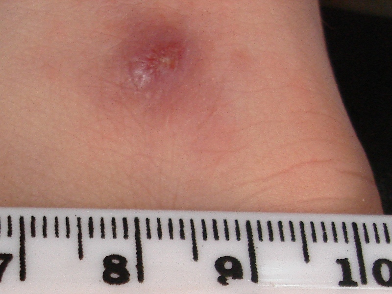
Koebner Phenomenon
The Koebner phenomenon or Köbner phenomenon (, ), also called the Koebner response or the isomorphic response, attributed to Heinrich Köbner, is the appearance of skin lesions on lines of trauma. The Koebner phenomenon may result from either a linear exposure or irritation. Conditions demonstrating linear lesions after a linear exposure to a causative agent include: molluscum contagiosum, warts and toxicodendron dermatitis (a dermatitis caused by a genus of plants including poison ivy). Warts and molluscum contagiosum lesions can be spread in linear patterns by self-scratching (" auto-inoculation"). Toxicodendron dermatitis lesions are often linear from brushing up against the plant. Causes of the Koebner phenomenon that are secondary to scratching rather than an infective or chemical cause include vitiligo, psoriasis, lichen planus, lichen nitidus, pityriasis rubra pilaris, and keratosis follicularis (Darier disease). Definition The Koebner phenomenon describes skin l ... [...More Info...] [...Related Items...] OR: [Wikipedia] [Google] [Baidu] |
Heinrich Köbner
Heinrich Koebner (German spelling Köbner); (2 December 1838 – 3 September 1904) was a German-Jewish dermatologist born in Breslau. He studied medicine in Berlin, earning his doctorate in 1859 at Breslau. Afterwards he performed hospital duties in Vienna under Ferdinand von Hebra (1816–1880) and in Paris with Alfred Hardy (1811–1893). In 1876 he became director of the policlinic for syphilis and diseases of the skin at the University of Breslau. In 1884 he established a new policlinic in Berlin, where he provided classes for physicians. Koebner phenomenon Koebner was a renowned dermatologist known for his research of psoriasis, epidermolysis bullosa simplex and various fungal disorders. He is best known for the eponymous "Koebner phenomenon", also known as isomorphic phenomenon. The Koebner phenomenon is the development of isomorphic pathologic lesions in the traumatized "uninvolved skin" of persons who have cutaneous diseases such as psoriasis. In other ... [...More Info...] [...Related Items...] OR: [Wikipedia] [Google] [Baidu] |
Flat Warts
Flat warts, technically known as Verruca plana, are reddish-brown or flesh-colored, slightly raised, flat-surfaced, well-demarcated papule of 2 to 5 mm in diameter. Upon close inspection, these lesions have a surface that is "finely verrucous".Lookingbill, Donald, et al. ''Principles of Dermatology''. Saunders. 2000. Pages 68-69. . Most often, these lesions affect the hands, legs, or face, and a linear arrangement is not uncommon. At histopathology, flat warts have cells with prominent perinuclear vacuolization around pyknotic, basophilic Basophilic is a technical term used by pathologists. It describes the appearance of cells, tissues and cellular structures as seen through the microscope after a histological section has been stained with a basic dye. The most common such dye i ..., centrally located nuclei that may be located in the granular layer. Last Update: May 13, 2019. These are referred to as "owl's eye cells." Additional images References External l ... [...More Info...] [...Related Items...] OR: [Wikipedia] [Google] [Baidu] |
Pyoderma Gangrenosum
Pyoderma gangrenosum is a rare, inflammatory skin disease where painful pustules or nodules become ulcers that progressively grow. Pyoderma gangrenosum is not infectious. Treatments may include corticosteroids, ciclosporin, infliximab, or canakinumab. The disease was identified in 1930. It affects approximately 1 person in 100,000 in the population. Though it can affect people of any age, it mostly affects people in their 40s and 50s. Types There are two main types of pyoderma gangrenosum: * the 'typical' ulcerative form, which occurs in the legs * an 'atypical' form that is more superficial and occurs in the hands and other parts of the body Other variations are: * Peristomal pyoderma gangrenosum comprises 15% of all cases of pyoderma * Bullous pyoderma gangrenosum * Pustular pyoderma gangrenosum * Vegetative pyoderma gangrenosum Presentation Associations The following are conditions commonly associated with pyoderma gangrenosum: * Inflammatory bowel disease: ** Ulcerati ... [...More Info...] [...Related Items...] OR: [Wikipedia] [Google] [Baidu] |
Post Kala Azar Dermal Leishmaniasis
Leishmaniasis is a wide array of clinical manifestations caused by parasites of the trypanosome genus '' Leishmania''. It is generally spread through the bite of phlebotomine sandflies, '' Phlebotomus'' and '' Lutzomyia'', and occurs most frequently in the tropics and sub-tropics of Africa, Asia, the Americas, and southern Europe. The disease can present in three main ways: cutaneous, mucocutaneous, or visceral. The cutaneous form presents with skin ulcers, while the mucocutaneous form presents with ulcers of the skin, mouth, and nose. The visceral form starts with skin ulcers and later presents with fever, low red blood cell count, and enlarged spleen and liver. Infections in humans are caused by more than 20 species of ''Leishmania''. Risk factors include poverty, malnutrition, deforestation, and urbanization. All three types can be diagnosed by seeing the parasites under microscopy. Additionally, visceral disease can be diagnosed by blood tests. Leishmaniasis can be ... [...More Info...] [...Related Items...] OR: [Wikipedia] [Google] [Baidu] |
Cutaneous Leishmaniasis
Cutaneous leishmaniasis is the most common form of leishmaniasis affecting humans. It is a skin infection caused by a single-celled parasite that is transmitted by the bite of a phlebotomine sand fly. There are about thirty species of '' Leishmania'' that may cause cutaneous leishmaniasis. This disease is considered to be a zoonosis (an infectious disease that is naturally transmissible from animals to humans), with the exception of ''Leishmania tropica'' — which is often an anthroponotic disease (an infectious disease that is naturally transmissible from humans to vertebrate animals). Signs and symptoms Post kala-azar dermal leishmaniasis Post-kala-azar dermal leishmaniasis (PKDL) is a recurrence of kala-azar that may appear on the skin of affected individuals months and up to 20 years after being partially treated, untreated or even in those considered adequately treated. In Sudan, they can be demonstrated in up to 60% of treated cases. They manifest as hypopigmented ... [...More Info...] [...Related Items...] OR: [Wikipedia] [Google] [Baidu] |
Systemic-onset Juvenile Idiopathic Arthritis
Systemic-onset juvenile idiopathic arthritis (or the juvenile onset form of Still's disease) is a type of juvenile idiopathic arthritis (JIA) with extra-articular manifestations like fever and rash apart from arthritis. It was originally called systemic-onset juvenile rheumatoid arthritis or Still's disease. Predominantly extra-articular manifestations like high fevers, rheumatic rash, enlargement of the liver and spleen, enlargement of the lymph nodes, and anemia. Other manifestations include inflammation of the pleura, inflammation of the pericardium, inflammation of the heart's muscular tissue, and inflammation of the peritoneum are also seen. It is sometimes called "juvenile-onset Still's disease" to distinguish it from adult-onset Still's disease. However, there is some evidence that the main difference between two conditions is the age of onset. Presentation Systemic JIA is characterized by arthritis, fever, which typically is higher than the low-grade fever associat ... [...More Info...] [...Related Items...] OR: [Wikipedia] [Google] [Baidu] |
Juvenile Idiopathic Arthritis
Juvenile idiopathic arthritis (JIA) is the most common, chronic rheumatic disease of childhood, affecting approximately one per 1,000 children. ''Juvenile'', in this context, refers to disease onset before 16 years of age, while ''idiopathic'' refers to a condition with no defined cause, and ''arthritis'' is inflammation within the joint. JIA is an autoimmune, noninfective, inflammatory joint disease, the cause of which remains poorly understood. It is characterised by chronic joint inflammation. JIA is a subset of childhood arthritis, but unlike other, more transient forms of childhood arthritis, JIA persists for at least six weeks, and in some children is a lifelong condition. It differs significantly from forms of arthritis commonly seen in adults (osteoarthritis, rheumatoid arthritis), in terms of cause, disease associations, and prognosis. The prognosis for children with JIA has improved dramatically over recent decades, particularly with the introduction of biological ther ... [...More Info...] [...Related Items...] OR: [Wikipedia] [Google] [Baidu] |
Lupus
Lupus, technically known as systemic lupus erythematosus (SLE), is an autoimmune disease in which the body's immune system mistakenly attacks healthy tissue in many parts of the body. Symptoms vary among people and may be mild to severe. Common symptoms include painful and swollen joints, fever, chest pain, hair loss, mouth ulcers, swollen lymph nodes, feeling tired, and a red rash which is most commonly on the face. Often there are periods of illness, called flares, and periods of remission during which there are few symptoms. The cause of SLE is not clear. It is thought to involve a mixture of genetics combined with environmental factors. Among identical twins, if one is affected there is a 24% chance the other one will also develop the disease. Female sex hormones, sunlight, smoking, vitamin D deficiency, and certain infections are also believed to increase a person's risk. The mechanism involves an immune response by autoantibodies against a person's own tissues ... [...More Info...] [...Related Items...] OR: [Wikipedia] [Google] [Baidu] |
Necrobiosis Lipoidica
Necrobiosis lipoidica is a necrotising skin condition that usually occurs in patients with diabetes mellitus but can also be associated with rheumatoid arthritis. In the former case it may be called necrobiosis lipoidica diabeticorum (NLD). NLD occurs in approximately 0.3% of the diabetic population, with the majority of those affected are women (approximately 3:1 females to males affected). The severity or control of diabetes in an individual does not affect who will or will not get NLD. Better maintenance of diabetes after being diagnosed with NLD will not change how quickly the NLD will resolve. Signs and symptoms NL/NLD most frequently appears on the patient's shins, often on both legs, although it may also occur on forearms, hands, trunk, and, rarely, nipple, penis, and surgical sites. The lesions are often asymptomatic but may become tender and ulcerate when injured. The first symptom of NL is often a "bruised" appearance (erythema) that is not necessarily associated with ... [...More Info...] [...Related Items...] OR: [Wikipedia] [Google] [Baidu] |
Kaposi Sarcoma
Kaposi's sarcoma (KS) is a type of cancer that can form masses in the skin, in lymph nodes, in the mouth, or in other organs. The skin lesions are usually painless, purple and may be flat or raised. Lesions can occur singly, multiply in a limited area, or may be widespread. Depending on the sub-type of disease and level of immune suppression, KS may worsen either gradually or quickly. Except for Classical KS where there is generally no immune suppression, KS is caused by a combination of immune suppression (such as due to HIV/AIDS) and infection by Human herpesvirus 8 (HHV8 – also called KS-associated herpesvirus (KSHV)). Four sub-types are described: classic, endemic, immunosuppression therapy-related (also called iatrogenic), and epidemic (also called AIDS-related). Classic KS tends to affect older men in regions where KSHV is highly prevalent (Mediterranean, Eastern Europe, Middle East), is usually slow-growing, and most often affects only the legs. Endemic KS is most comm ... [...More Info...] [...Related Items...] OR: [Wikipedia] [Google] [Baidu] |
Elastosis Perforans Serpiginosa
Elastosis perforans serpiginosa is a unique perforating disorder characterized by transepidermal elimination of elastic fibers and distinctive clinical lesions, which are serpiginous in distribution and can be associated with specific diseases.Freedberg, et al. (2003). ''Fitzpatrick's Dermatology in General Medicine''. (6th ed.). Page 1041. McGraw-Hill. . File:Histopathology of elastosis perforans serpiginosa.jpg, Histopathology of elastosis perforans serpiginosa: Degenerated elastic fibers and transepidermal perforating canals (arrow points at one of them) File:Autosomal dominant - en.svg, This condition is inherited in an autosomal dominant manner. See also * List of cutaneous conditions * Poikiloderma vasculare atrophicans Poikiloderma vasculare atrophicans (PVA), is a cutaneous condition (skin disease) characterized by hypo- or hyperpigmentation (diminished or heightened skin pigmentation, respectively), telangiectasia and skin atrophy. Other names for the conditio ... ... [...More Info...] [...Related Items...] OR: [Wikipedia] [Google] [Baidu] |


