|
Kidney
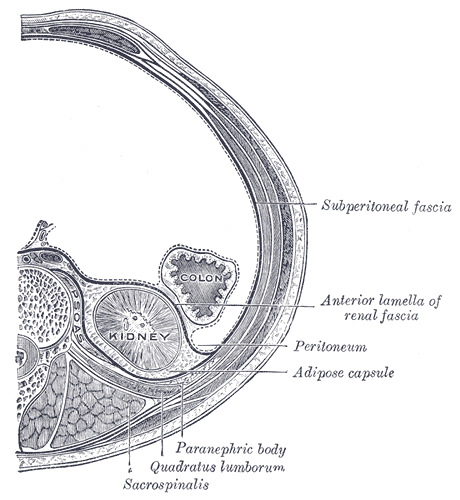
The kidneys are two reddish-brown bean-shaped organs found in vertebrates. They are located on the left and right in the retroperitoneal space, and in adult humans are about in length. They receive blood from the paired renal arteries; blood exits into the paired renal veins. Each kidney is attached to a ureter, a tube that carries excreted urine to the bladder. The kidney participates in the control of the volume of various body fluids, fluid osmolality, acid–base balance, various electrolyte concentrations, and removal of toxins. Filtration occurs in the glomerulus: one-fifth of the blood volume that enters the kidneys is filtered. Examples of substances reabsorbed are solute-free water, sodium, bicarbonate, glucose, and amino acids. Examples of substances secreted are hydrogen, ammonium, potassium and uric acid. The nephron is the structural and functional unit of the kidney. Each adult human kidney contains around 1 million nephrons, while a mouse kidney co ... [...More Info...] [...Related Items...] OR: [Wikipedia] [Google] [Baidu] |
Nephron
The nephron is the minute or microscopic structural and functional unit of the kidney. It is composed of a renal corpuscle and a renal tubule. The renal corpuscle consists of a tuft of capillaries called a glomerulus and a cup-shaped structure called Bowman's capsule. The renal tubule extends from the capsule. The capsule and tubule are connected and are composed of epithelial cells with a lumen. A healthy adult has 1 to 1.5 million nephrons in each kidney. Blood is filtered as it passes through three layers: the endothelial cells of the capillary wall, its basement membrane, and between the foot processes of the podocytes of the lining of the capsule. The tubule has adjacent peritubular capillaries that run between the descending and ascending portions of the tubule. As the fluid from the capsule flows down into the tubule, it is processed by the epithelial cells lining the tubule: water is reabsorbed and substances are exchanged (some are added, others are removed); first ... [...More Info...] [...Related Items...] OR: [Wikipedia] [Google] [Baidu] |
Ureter
The ureters are tubes made of smooth muscle that propel urine from the kidneys to the urinary bladder. In a human adult, the ureters are usually long and around in diameter. The ureter is lined by urothelial cells, a type of transitional epithelium, and has an additional smooth muscle layer that assists with peristalsis in its lowest third. The ureters can be affected by a number of diseases, including urinary tract infections and kidney stone. is when a ureter is narrowed, due to for example chronic inflammation. Congenital abnormalities that affect the ureters can include the development of two ureters on the same side or abnormally placed ureters. Additionally, reflux of urine from the bladder back up the ureters is a condition commonly seen in children. The ureters have been identified for at least two thousand years, with the word "ureter" stemming from the stem relating to urinating and seen in written records since at least the time of Hippocrates. It is, however, ... [...More Info...] [...Related Items...] OR: [Wikipedia] [Google] [Baidu] |
Renal Vein
The renal veins are large-calibre veins that drain blood filtered by the kidneys into the inferior vena cava. There is one renal vein draining each kidney. Because the inferior vena cava is on the right half of the body, the left renal vein is longer than the right one. Structure One renal vein drains each kidney. A renal vein is situated anterior to its corresponding accompanying renal artery. The renal veins empty into the inferior vena cava, entering it at nearly a 90° angle. Due to the right-ward displacement of the inferior vena cava from the midline, the left renal vein is some 3 times longer than the right one (~7.5 cm and ~2.5 cm, respectively). The renal vein divides into 4 divisions upon entering the kidney: * the anterior branch which receives blood from the anterior portion of the kidney and, * the posterior branch which receives blood from the posterior portion. Tributaries Because the tributaries of the inferior vena cava are not bilaterally symmetrical, the l ... [...More Info...] [...Related Items...] OR: [Wikipedia] [Google] [Baidu] |
Urinary System
The urinary system, also known as the urinary tract or renal system, consists of the kidneys, ureters, bladder, and the urethra. The purpose of the urinary system is to eliminate waste from the body, regulate blood volume and blood pressure, control levels of electrolytes and metabolites, and regulate blood pH. The urinary tract is the body's drainage system for the eventual removal of urine. The kidneys have an extensive blood supply via the renal arteries which leave the kidneys via the renal vein. Each kidney consists of functional units called nephrons. Following filtration of blood and further processing, wastes (in the form of urine) exit the kidney via the ureters, tubes made of smooth muscle fibres that propel urine towards the urinary bladder, where it is stored and subsequently expelled from the body by urination ( voiding). The female and male urinary system are very similar, differing only in the length of the urethra. Urine is formed in the kidneys through a ... [...More Info...] [...Related Items...] OR: [Wikipedia] [Google] [Baidu] |
Potassium
Potassium is the chemical element with the symbol K (from Neo-Latin '' kalium'') and atomic number19. Potassium is a silvery-white metal that is soft enough to be cut with a knife with little force. Potassium metal reacts rapidly with atmospheric oxygen to form flaky white potassium peroxide in only seconds of exposure. It was first isolated from potash, the ashes of plants, from which its name derives. In the periodic table, potassium is one of the alkali metals, all of which have a single valence electron in the outer electron shell, that is easily removed to create an ion with a positive charge – a cation, that combines with anions to form salts. Potassium in nature occurs only in ionic salts. Elemental potassium reacts vigorously with water, generating sufficient heat to ignite hydrogen emitted in the reaction, and burning with a lilac- colored flame. It is found dissolved in sea water (which is 0.04% potassium by weight), and occurs in many minerals such as ... [...More Info...] [...Related Items...] OR: [Wikipedia] [Google] [Baidu] |
Renal Vein
The renal veins are large-calibre veins that drain blood filtered by the kidneys into the inferior vena cava. There is one renal vein draining each kidney. Because the inferior vena cava is on the right half of the body, the left renal vein is longer than the right one. Structure One renal vein drains each kidney. A renal vein is situated anterior to its corresponding accompanying renal artery. The renal veins empty into the inferior vena cava, entering it at nearly a 90° angle. Due to the right-ward displacement of the inferior vena cava from the midline, the left renal vein is some 3 times longer than the right one (~7.5 cm and ~2.5 cm, respectively). The renal vein divides into 4 divisions upon entering the kidney: * the anterior branch which receives blood from the anterior portion of the kidney and, * the posterior branch which receives blood from the posterior portion. Tributaries Because the tributaries of the inferior vena cava are not bilaterally symmetrical, the l ... [...More Info...] [...Related Items...] OR: [Wikipedia] [Google] [Baidu] |
Glomerulus (kidney)
The glomerulus (plural glomeruli) is a network of small blood vessels (capillaries) known as a ''tuft'', located at the beginning of a nephron in the kidney. Each of the two kidneys contains about one million nephrons. The tuft is structurally supported by the mesangium (the space between the blood vessels), composed of intraglomerular mesangial cells. The blood is filtered across the capillary walls of this tuft through the glomerular filtration barrier, which yields its filtrate of water and soluble substances to a cup-like sac known as Bowman's capsule. The filtrate then enters the renal tubule of the nephron. The glomerulus receives its blood supply from an afferent arteriole of the renal arterial circulation. Unlike most capillary beds, the glomerular capillaries exit into efferent arterioles rather than venules. The resistance of the efferent arterioles causes sufficient hydrostatic pressure within the glomerulus to provide the force for ultrafiltration. The glomeru ... [...More Info...] [...Related Items...] OR: [Wikipedia] [Google] [Baidu] |
Uric Acid
Uric acid is a heterocyclic compound of carbon, nitrogen, oxygen, and hydrogen with the formula C5H4N4O3. It forms ions and salts known as urates and acid urates, such as ammonium acid urate. Uric acid is a product of the metabolic breakdown of purine nucleotides, and it is a normal component of urine. High blood concentrations of uric acid can lead to gout and are associated with other medical conditions, including diabetes and the formation of ammonium acid urate kidney stones. Chemistry Uric acid was first isolated from kidney stones in 1776 by Swedish chemist Carl Wilhelm Scheele. In 1882, the Ukrainian chemist Ivan Horbaczewski first synthesized uric acid by melting urea with glycine. Uric acid displays lactam–lactim tautomerism (also often described as keto–enol tautomerism). Although the lactim form is expected to possess some degree of aromaticity, uric acid crystallizes in the lactam form, with computational chemistry also indicating that tautomer to be ... [...More Info...] [...Related Items...] OR: [Wikipedia] [Google] [Baidu] |
Retroperitoneal Space
The retroperitoneal space (retroperitoneum) is the anatomical space (sometimes a potential space) behind (''retro'') the peritoneum. It has no specific delineating anatomical structures. Organs are retroperitoneal if they have peritoneum on their anterior side only. Structures that are not suspended by mesentery in the abdominal cavity and that lie between the parietal peritoneum and abdominal wall are classified as retroperitoneal. This is different from organs that are not retroperitoneal, which have peritoneum on their posterior side and are suspended by mesentery in the abdominal cavity. The retroperitoneum can be further subdivided into the following: *Perirenal (or perinephric) space *Anterior pararenal (or paranephric) space *Posterior pararenal (or paranephric) space Retroperitoneal structures Structures that lie behind the peritoneum are termed "retroperitoneal". Organs that were once suspended within the abdominal cavity by mesentery but migrated posterior to the p ... [...More Info...] [...Related Items...] OR: [Wikipedia] [Google] [Baidu] |
Renal Artery
The renal arteries are paired arteries that supply the kidneys with blood. Each is directed across the crus of the diaphragm, so as to form nearly a right angle. The renal arteries carry a large portion of total blood flow to the kidneys. Up to a third of total cardiac output can pass through the renal arteries to be filtered by the kidneys. Structure The renal arteries normally arise at a 90° angle off of the left interior side of the abdominal aorta, immediately below the superior mesenteric artery. They have a radius of approximately 0.25 cm, 0.26 cm at the root. The measured mean diameter can differ depending on the imaging method used. For example, the diameter was found to be 5.04 ± 0.74 mm using ultrasound but 5.68 ± 1.19 mm using angiography. Due to the anatomical position of the aorta, the inferior vena cava, and the kidneys, the right renal artery is normally longer than the left renal artery. * The right passes behind the inferior vena cava, ... [...More Info...] [...Related Items...] OR: [Wikipedia] [Google] [Baidu] |
Vitamin D
Vitamin D is a group of Lipophilicity, fat-soluble secosteroids responsible for increasing intestinal absorption of calcium, magnesium, and phosphate, and many other biological effects. In humans, the most important compounds in this group are vitamin D3 (cholecalciferol) and vitamin D2 (ergocalciferol). The major natural source of the vitamin is Chemical synthesis, synthesis of cholecalciferol in the Epidermis#Layers, lower layers of epidermis of the skin through a chemical reaction that is dependent on Health effects of sunlight exposure, sun exposure (specifically Ultraviolet#Subtypes, UVB radiation). Cholecalciferol and ergocalciferol can be ingested from the diet and dietary supplement, supplements. Only a few foods, such as the flesh of fatty fish, naturally contain significant amounts of vitamin D. In the U.S. and other countries, cow's milk and plant-derived milk substitutes are fortified with vitamin D, as are many breakfast cereals. Mushrooms exposed to ultraviolet l ... [...More Info...] [...Related Items...] OR: [Wikipedia] [Google] [Baidu] |
Renal Artery
The renal arteries are paired arteries that supply the kidneys with blood. Each is directed across the crus of the diaphragm, so as to form nearly a right angle. The renal arteries carry a large portion of total blood flow to the kidneys. Up to a third of total cardiac output can pass through the renal arteries to be filtered by the kidneys. Structure The renal arteries normally arise at a 90° angle off of the left interior side of the abdominal aorta, immediately below the superior mesenteric artery. They have a radius of approximately 0.25 cm, 0.26 cm at the root. The measured mean diameter can differ depending on the imaging method used. For example, the diameter was found to be 5.04 ± 0.74 mm using ultrasound but 5.68 ± 1.19 mm using angiography. Due to the anatomical position of the aorta, the inferior vena cava, and the kidneys, the right renal artery is normally longer than the left renal artery. * The right passes behind the inferior vena cava, ... [...More Info...] [...Related Items...] OR: [Wikipedia] [Google] [Baidu] |


