|
Decorin
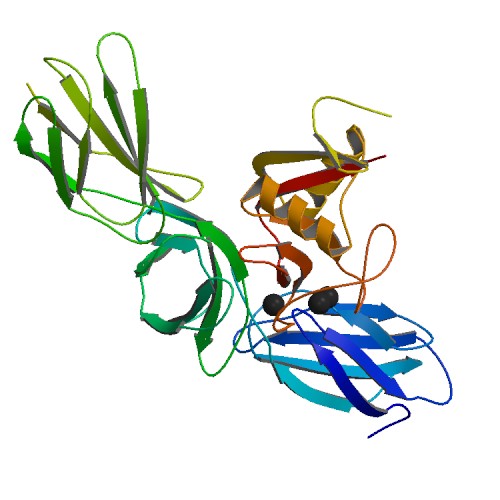
Decorin is a protein that in humans is encoded by the ''DCN'' gene. Decorin is a proteoglycan that is on average 90 - 140 kilodaltons (kDa) in molecular weight. It belongs to the small leucine-rich proteoglycan (SLRP) family and consists of a protein core containing leucine repeats with a glycosaminoglycan (GAG) chain consisting of either chondroitin sulfate (CS) or dermatan sulfate (DS). Decorin is a small cellular or pericellular matrix proteoglycan and is closely related in structure to biglycan protein. Decorin and biglycan are thought to be the result of a gene duplication. This protein is a component of connective tissue, binds to type I collagen fibrils, and plays a role in matrix assembly. Naming Decorin's name is a derivative of both the fact that it "decorates" collagen type I, and that it interacts with the "d" and "e" bands of fibrils of this collagen. Function Decorin appears to influence fibrillogenesis, and also interacts with fibronectin, thromb ... [...More Info...] [...Related Items...] OR: [Wikipedia] [Google] [Baidu] |
Myokine
A myokine is one of several hundred cytokines or other small proteins (~5–20 kDa) and proteoglycan peptides that are produced and released by skeletal muscle cells (muscle fibers) in response to muscular contractions. They have autocrine, paracrine and/or endocrine effects; their systemic effects occur at picomolar concentrations. Receptors for myokines are found on muscle, fat, liver, pancreas, bone, heart, immune, and brain cells. The location of these receptors reflects the fact that myokines have multiple functions. Foremost, they are involved in exercise-associated metabolic changes, as well as in the metabolic changes following training adaptation. They also participate in tissue regeneration and repair, maintenance of healthy bodily functioning, immunomodulation; and cell signaling, expression and differentiation. History The definition and use of the term myokine first occurred in 2003. In 2008, the first myokine, myostatin, was identified. The gp130 receptor cytokine ... [...More Info...] [...Related Items...] OR: [Wikipedia] [Google] [Baidu] |
Biglycan
Biglycan is a small leucine-rich repeat proteoglycan (SLRP) which is found in a variety of extracellular matrix tissues, including bone, cartilage and tendon. In humans, biglycan is encoded by the ''BGN'' gene which is located on the X chromosome. The name "biglycan" was proposed in an article by Fisher, Termine and Young in an article in the Journal of Biological Chemistry in 1989 because the proteoglycan contained two GAG chains; formerly it was known as proteoglycan-I (PG-I). Structure Biglycan consists of a protein core containing leucine-rich repeat regions and two glycosaminoglycan (GAG) chains consisting of either chondroitin sulfate (CS) or dermatan sulfate (DS), with DS being more abundant in most connective tissues. The CS/DS chains are attached at amino acids 5 and 10 in human biglycan. The composition of the GAG chains has been reported as varying according to tissue of origin. Non-glycanated forms of biglycan (no GAG chains) increase with age in human artic ... [...More Info...] [...Related Items...] OR: [Wikipedia] [Google] [Baidu] |
Proteoglycan
Proteoglycans are proteins that are heavily glycosylated. The basic proteoglycan unit consists of a "core protein" with one or more covalently attached glycosaminoglycan (GAG) chain(s). The point of attachment is a serine (Ser) residue to which the glycosaminoglycan is joined through a tetrasaccharide bridge (e.g. chondroitin sulfate-GlcA- Gal-Gal- Xyl-PROTEIN). The Ser residue is generally in the sequence -Ser- Gly-X-Gly- (where X can be any amino acid residue but proline), although not every protein with this sequence has an attached glycosaminoglycan. The chains are long, linear carbohydrate polymers that are negatively charged under physiological conditions due to the occurrence of sulfate and uronic acid groups. Proteoglycans occur in connective tissue. Types Proteoglycans are categorized by their relative size (large and small) and the nature of their glycosaminoglycan chains. Types include: Certain members are considered members of the "small leucine-rich proteogly ... [...More Info...] [...Related Items...] OR: [Wikipedia] [Google] [Baidu] |
Leucine-rich Repeat
A leucine-rich repeat (LRR) is a protein structural motif that forms an α/β horseshoe fold. It is composed of repeating 20–30 amino acid stretches that are unusually rich in the hydrophobic amino acid leucine. These tandem repeats commonly fold together to form a solenoid protein domain, termed leucine-rich repeat domain. Typically, each repeat unit has beta strand- turn-alpha helix structure, and the assembled domain, composed of many such repeats, has a horseshoe shape with an interior parallel beta sheet and an exterior array of helices. One face of the beta sheet and one side of the helix array are exposed to solvent and are therefore dominated by hydrophilic residues. The region between the helices and sheets is the protein's hydrophobic core and is tightly sterically packed with leucine residues. Leucine-rich repeats are frequently involved in the formation of protein–protein interactions. Examples Leucine-rich repeat motifs have been identified in a large n ... [...More Info...] [...Related Items...] OR: [Wikipedia] [Google] [Baidu] |
Endothelial Cell
The endothelium is a single layer of squamous endothelial cells that line the interior surface of blood vessels and lymphatic vessels. The endothelium forms an interface between circulating blood or lymph in the lumen and the rest of the vessel wall. Endothelial cells form the barrier between vessels and tissue and control the flow of substances and fluid into and out of a tissue. Endothelial cells in direct contact with blood are called vascular endothelial cells whereas those in direct contact with lymph are known as lymphatic endothelial cells. Vascular endothelial cells line the entire circulatory system, from the heart to the smallest capillaries. These cells have unique functions that include fluid filtration, such as in the glomerulus of the kidney, blood vessel tone, hemostasis, neutrophil recruitment, and hormone trafficking. Endothelium of the interior surfaces of the heart chambers is called endocardium. An impaired function can lead to serious health issues thr ... [...More Info...] [...Related Items...] OR: [Wikipedia] [Google] [Baidu] |
TGF-beta
Transforming growth factor beta (TGF-β) is a multifunctional cytokine belonging to the transforming growth factor superfamily that includes three different mammalian isoforms (TGF-β 1 to 3, HGNC symbols TGFB1, TGFB2, TGFB3) and many other signaling proteins. TGFB proteins are produced by all white blood cell lineages. Activated TGF-β complexes with other factors to form a serine/threonine kinase complex that binds to TGF-β receptors. TGF-β receptors are composed of both type 1 and type 2 receptor subunits. After the binding of TGF-β, the type 2 receptor kinase phosphorylates and activates the type 1 receptor kinase that activates a signaling cascade. This leads to the activation of different downstream substrates and regulatory proteins, inducing transcription of different target genes that function in differentiation, chemotaxis, proliferation, and activation of many immune cells. TGF-β is secreted by many cell types, including macrophages, in a latent form in whic ... [...More Info...] [...Related Items...] OR: [Wikipedia] [Google] [Baidu] |
TGF-beta 1
Transforming growth factor beta (TGF-β) is a multifunctional cytokine belonging to the transforming growth factor superfamily that includes three different mammalian isoforms (TGF-β 1 to 3, HGNC symbols TGFB1, TGFB2, TGFB3) and many other signaling proteins. TGFB proteins are produced by all white blood cell lineages. Activated TGF-β complexes with other factors to form a serine/threonine kinase complex that binds to TGF-β receptors. TGF-β receptors are composed of both type 1 and type 2 receptor subunits. After the binding of TGF-β, the type 2 receptor kinase phosphorylates and activates the type 1 receptor kinase that activates a signaling cascade. This leads to the activation of different downstream substrates and regulatory proteins, inducing transcription of different target genes that function in differentiation, chemotaxis, proliferation, and activation of many immune cells. TGF-β is secreted by many cell types, including macrophages, in a latent form in which ... [...More Info...] [...Related Items...] OR: [Wikipedia] [Google] [Baidu] |
Cell Cycle
The cell cycle, or cell-division cycle, is the series of events that take place in a cell that cause it to divide into two daughter cells. These events include the duplication of its DNA (DNA replication) and some of its organelles, and subsequently the partitioning of its cytoplasm, chromosomes and other components into two daughter cells in a process called cell division. In cells with nuclei ( eukaryotes, i.e., animal, plant, fungal, and protist cells), the cell cycle is divided into two main stages: interphase and the mitotic (M) phase (including mitosis and cytokinesis). During interphase, the cell grows, accumulating nutrients needed for mitosis, and replicates its DNA and some of its organelles. During the mitotic phase, the replicated chromosomes, organelles, and cytoplasm separate into two new daughter cells. To ensure the proper replication of cellular components and division, there are control mechanisms known as cell cycle checkpoints after each of the key s ... [...More Info...] [...Related Items...] OR: [Wikipedia] [Google] [Baidu] |
Autophagy
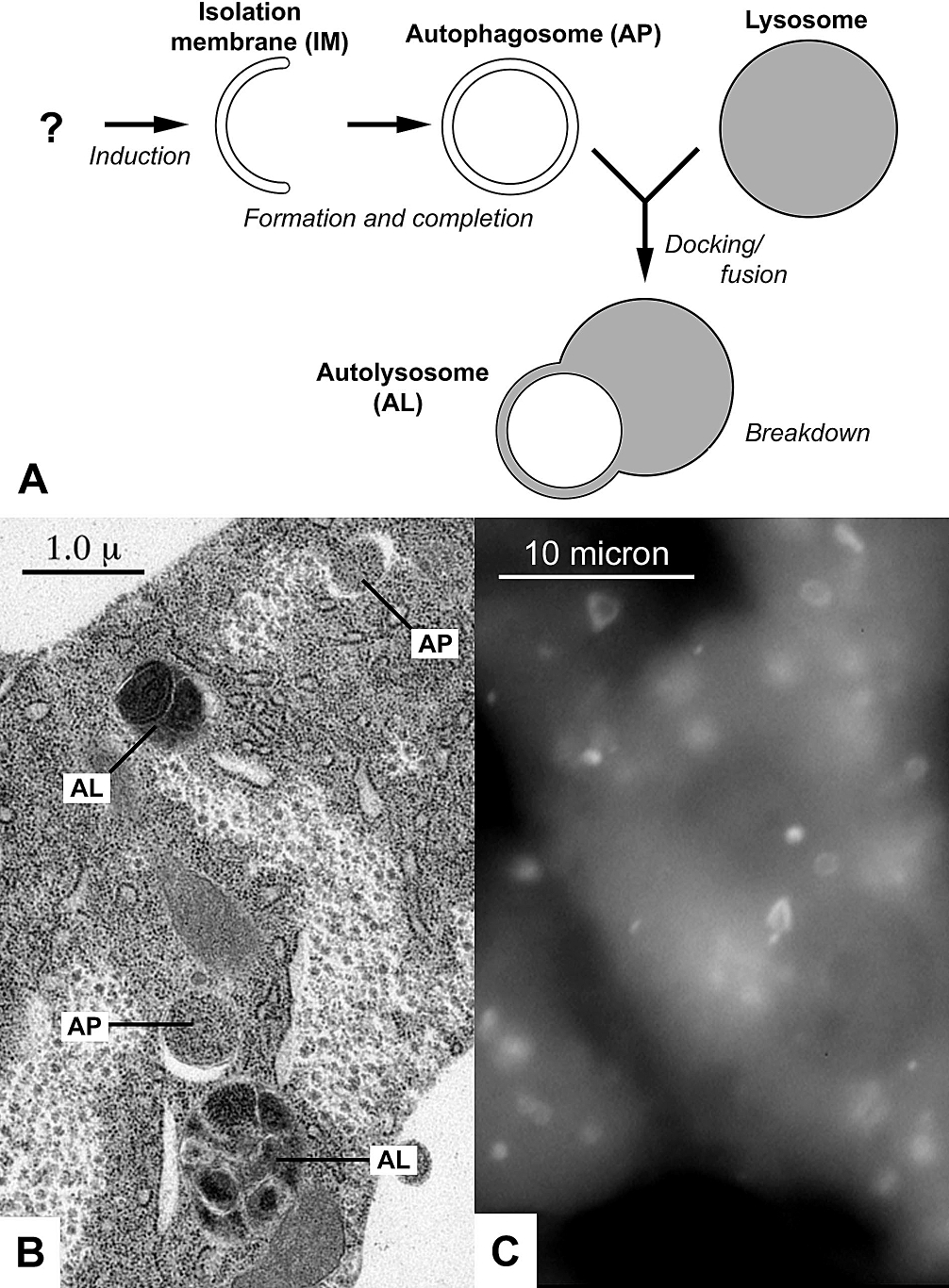
Autophagy (or autophagocytosis; from the Ancient Greek , , meaning "self-devouring" and , , meaning "hollow") is the natural, conserved degradation of the cell that removes unnecessary or dysfunctional components through a lysosome-dependent regulated mechanism. It allows the orderly degradation and recycling of cellular components. Although initially characterized as a primordial degradation pathway induced to protect against starvation, it has become increasingly clear that autophagy also plays a major role in the homeostasis of non-starved cells. Defects in autophagy have been linked to various human diseases, including neurodegeneration and cancer, and interest in modulating autophagy as a potential treatment for these diseases has grown rapidly. Four forms of autophagy have been identified: macroautophagy, microautophagy, chaperone-mediated autophagy (CMA), and crinophagy. In macroautophagy (the most thoroughly researched form of autophagy), cytoplasmic components (like m ... [...More Info...] [...Related Items...] OR: [Wikipedia] [Google] [Baidu] |
Protein
Proteins are large biomolecules and macromolecules that comprise one or more long chains of amino acid residues. Proteins perform a vast array of functions within organisms, including catalysing metabolic reactions, DNA replication, responding to stimuli, providing structure to cells and organisms, and transporting molecules from one location to another. Proteins differ from one another primarily in their sequence of amino acids, which is dictated by the nucleotide sequence of their genes, and which usually results in protein folding into a specific 3D structure that determines its activity. A linear chain of amino acid residues is called a polypeptide. A protein contains at least one long polypeptide. Short polypeptides, containing less than 20–30 residues, are rarely considered to be proteins and are commonly called peptides. The individual amino acid residues are bonded together by peptide bonds and adjacent amino acid residues. The sequence of amino acid ... [...More Info...] [...Related Items...] OR: [Wikipedia] [Google] [Baidu] |
Angiogenesis
Angiogenesis is the physiological process through which new blood vessels form from pre-existing vessels, formed in the earlier stage of vasculogenesis. Angiogenesis continues the growth of the vasculature by processes of sprouting and splitting. Vasculogenesis is the embryonic formation of endothelial cells from mesoderm cell precursors, and from neovascularization, although discussions are not always precise (especially in older texts). The first vessels in the developing embryo form through vasculogenesis, after which angiogenesis is responsible for most, if not all, blood vessel growth during development and in disease. Angiogenesis is a normal and vital process in growth and development, as well as in wound healing and in the formation of granulation tissue. However, it is also a fundamental step in the transition of tumors from a benign state to a malignant one, leading to the use of angiogenesis inhibitors in the treatment of cancer. The essential role of angiogenes ... [...More Info...] [...Related Items...] OR: [Wikipedia] [Google] [Baidu] |
VEGFR2
Kinase insert domain receptor (KDR, a type IV receptor tyrosine kinase) also known as vascular endothelial growth factor receptor 2 (VEGFR-2) is a VEGF receptor. ''KDR'' is the human gene encoding it. KDR has also been designated as CD309 (cluster of differentiation 309). KDR is also known as Flk1 (Fetal Liver Kinase 1). The Q472H germline ''KDR'' genetic variant affects VEGFR-2 phosphorylation and has been found to associate with microvessel density in NSCLC. Interactions Kinase insert domain receptor has been shown to interact with SHC2, Annexin A5 and SHC1. See also * Cluster of differentiation * VEGF receptors VEGF receptors are receptors for vascular endothelial growth factor (VEGF). There are three main subtypes of VEGFR, numbered 1, 2 and 3. Also, they may be membrane-bound (mbVEGFR) or soluble (sVEGFR), depending on alternative splicing. Inhi ... References Further reading * * * * * * * * External links * * Clusters of diffe ... [...More Info...] [...Related Items...] OR: [Wikipedia] [Google] [Baidu] |


