|
Cystography
In radiology and urology, a cystography (also known as cystogram) is a procedure used to visualise the urinary bladder. Using a urinary catheter, radiocontrast is instilled in the bladder, and X-ray imaging is performed. Cystography can be used to evaluate bladder cancer, vesicoureteral reflux, bladder polyps, and hydronephrosis. It requires less radiation than pelvic CT, although it is less sensitive and specific than MRI or CT. In adult cases, the patient is typically instructed to void three times, after which a post voiding image is obtained to see how much urine is left within the bladder (residual urine), which is useful to evaluate bladder contraction dysfunction. A final radiograph of the kidneys after the procedure is finished is performed to evaluate for occult vesicoureteral reflux that was not seen during the procedure itself. CT cystography CT cystography is performed by filling up the urinary bladder using diluted iodinated contrast to visualise any bladder inj ... [...More Info...] [...Related Items...] OR: [Wikipedia] [Google] [Baidu] |
Voiding Cystourethrogram
In urology, voiding cystourethrography (VCUG) is a frequently performed technique for visualizing a person's urethra and urinary bladder while the person urinates (voids). It is used in the diagnosis of vesicoureteral reflux (kidney reflux), among other disorders. The technique consists of catheterizing the person in order to fill the bladder with a radiocontrast agent, typically diatrizoic acid. Under fluoroscopy (real time x-rays) the radiologist watches the contrast enter the bladder and looks at the anatomy of the patient. If the contrast moves into the ureters and back into the kidneys, the radiologist makes the diagnosis of vesicoureteral reflux, and gives the degree of severity a score. The exam ends when the person voids while the radiologist is watching under fluoroscopy. Consumption of fluid promotes excretion of contrast media after the procedure. It is important to watch the contrast during voiding, because this is when the bladder has the most pressure, and it ... [...More Info...] [...Related Items...] OR: [Wikipedia] [Google] [Baidu] |
Vesicoureteral Reflux
Vesicoureteral reflux (VUR), also known as vesicoureteric reflux, is a condition in which urine flows retrograde, or backward, from the bladder into one or both ureters and then to the renal calyx or kidneys. Urine normally travels in one direction (forward, or anterograde) from the kidneys to the bladder via the ureters, with a 1-way valve at the vesicoureteral (ureteral-bladder) junction preventing backflow. The valve is formed by oblique tunneling of the distal ureter through the wall of the bladder, creating a short length of ureter (1–2 cm) that can be compressed as the bladder fills. Reflux occurs if the ureter enters the bladder without sufficient tunneling, i.e., too "end-on". Signs and symptoms Most children with vesicoureteral reflux are asymptomatic. Vesicoureteral reflux may be diagnosed as a result of further evaluation of dilation of the kidney or ureters draining urine from the kidney while in utero as well as when a sibling has VUR (though routine testin ... [...More Info...] [...Related Items...] OR: [Wikipedia] [Google] [Baidu] |
Ureter
The ureters are tubes made of smooth muscle that propel urine from the kidneys to the urinary bladder. In a human adult, the ureters are usually long and around in diameter. The ureter is lined by urothelial cells, a type of transitional epithelium, and has an additional smooth muscle layer that assists with peristalsis in its lowest third. The ureters can be affected by a number of diseases, including urinary tract infections and kidney stone. is when a ureter is narrowed, due to for example chronic inflammation. Congenital abnormalities that affect the ureters can include the development of two ureters on the same side or abnormally placed ureters. Additionally, reflux of urine from the bladder back up the ureters is a condition commonly seen in children. The ureters have been identified for at least two thousand years, with the word "ureter" stemming from the stem relating to urinating and seen in written records since at least the time of Hippocrates. It is, however, ... [...More Info...] [...Related Items...] OR: [Wikipedia] [Google] [Baidu] |
Iodinated Contrast
Iodinated contrast is a form of intravenous radiocontrast agent containing iodine, which enhances the visibility of vascular structures and organs during radiographic procedures. Some pathologies, such as cancer, have particularly improved visibility with iodinated contrast. The radiodensity of iodinated contrast is 25–30 Hounsfield units (HU) per milligram of iodine per milliliter at a tube voltage of 100–120 kVp. Types Iodine-based contrast media are usually classified as ionic or nonionic. Both types are used most commonly in radiology due to their relatively harmless interaction with the body and its solubility. Contrast media are primarily used to visualize vessels and changes in tissues on radiography and CT (computerized tomography). Contrast media can also be used for tests of the urinary tract, uterus and fallopian tubes. It may cause the patient to feel as if they have had urinary incontinence. It also puts a metallic taste in the mouth of the patient. The iodin ... [...More Info...] [...Related Items...] OR: [Wikipedia] [Google] [Baidu] |
Cystoscopy
Cystoscopy is endoscopy of the urinary bladder via the urethra. It is carried out with a cystoscope. The urethra is the tube that carries urine from the bladder to the outside of the body. The cystoscope has lenses like a telescope or microscope. These lenses let the physician focus on the inner surfaces of the urinary tract. Some cystoscopes use optical fibres (flexible glass fibres) that carry an image from the tip of the instrument to a viewing piece at the other end. Cystoscopes range from pediatric to adult and from the thickness of a pencil up to approximately 9 mm and have a light at the tip. Many cystoscopes have extra tubes to guide other instruments for surgical procedures to treat urinary problems. There are two main types of cystoscopy—flexible and rigid—differing in the flexibility of the cystoscope. Flexible cystoscopy is carried out with local anaesthesia on both sexes. Typically, a topical anesthetic, most often xylocaine gel (common brand names are Ane ... [...More Info...] [...Related Items...] OR: [Wikipedia] [Google] [Baidu] |
Cholecystography
Oral cholecystography is a radiological procedure used to visualize the gallbladder and biliary channels, developed in 1924 by American surgeons Evarts Ambrose Graham Evarts Ambrose Graham (1883–1957) was an American academic, physician, and surgeon. Early years and military service Born in Chicago, Illinois to a surgeon, Dr. David Wilson Graham, and Ida Ansbach Barned Graham, Evarts attended college at Prin ... and Warren Henry Cole. It is usually indicated in cases of suspected gallbladder disease, and can also be used to determine or rule out the presence of intermittent obstruction of the bile ducts or recurrent biliary disease after biliary surgery. A radiopaque cholegraphic (contrast) agent, usually iopanoic acid (Telepaque) or its sodium or calcium salt, is orally administered, which is absorbed by the intestine. This excreted material will collect in the gallbladder, where reabsorption of water concentrates the excreted contrast. Since only 10% of gallstones are ra ... [...More Info...] [...Related Items...] OR: [Wikipedia] [Google] [Baidu] |
Computed Tomography Of The Abdomen And Pelvis
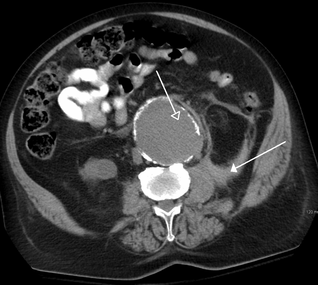
Computed tomography of the abdomen and pelvis is an application of computed tomography (CT) and is a sensitive method for diagnosis of abdominal diseases. It is used frequently to determine stage of cancer and to follow progress. It is also a useful test to investigate acute abdominal pain (especially of the lower quadrants, whereas ultrasound is the preferred first line investigation for right upper quadrant pain). Renal stones, appendicitis, pancreatitis, diverticulitis, abdominal aortic aneurysm, and bowel obstruction are conditions that are readily diagnosed and assessed with CT. CT is also the first line for detecting solid organ injury after trauma. Advantages Multidetector CT (MDCT) can clearly delineate anatomic structures in the abdomen, which is critical in the diagnosis of internal diaphragmatic and other nonpalpable or unsuspected hernias. MDCT also offers clear detail of the abdominal wall allowing wall hernias to be identified accurately. Contrast administra ... [...More Info...] [...Related Items...] OR: [Wikipedia] [Google] [Baidu] |
Haematuria
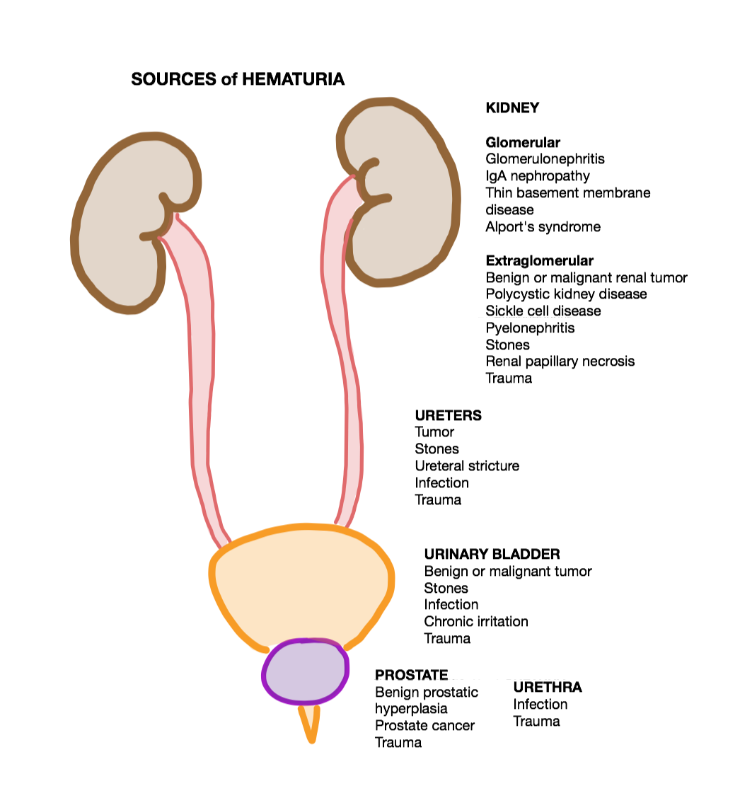
Hematuria or haematuria is defined as the presence of blood or red blood cells in the urine. “Gross hematuria” occurs when urine appears red, brown, or tea-colored due to the presence of blood. Hematuria may also be subtle and only detectable with a microscope or laboratory test. Blood that enters and mixes with the urine can come from any location within the urinary system, including the kidney, ureter, urinary bladder, urethra, and in men, the prostate. Common causes of hematuria include urinary tract infection (UTI), kidney stones, viral illness, trauma, bladder cancer, and exercise. These causes are grouped into glomerular and non-glomerular causes, depending on the involvement of the glomerulus of the kidney. But not all red urine is hematuria. Other substances such as certain medications and foods (e.g. blackberries, beets, food dyes) can cause urine to appear red. Menstruation in women may also cause the appearance of hematuria and may result in a positive urine dipstick ... [...More Info...] [...Related Items...] OR: [Wikipedia] [Google] [Baidu] |
Voiding
Urination, also known as micturition, is the release of urine from the urinary bladder through the urethra to the outside of the body. It is the urinary system's form of excretion. It is also known medically as micturition, voiding, uresis, or, rarely, emiction, and known colloquially by various names including peeing, weeing, and pissing. In healthy humans (and many other animals), the process of urination is under voluntary control. In infants, some elderly individuals, and those with neurological injury, urination may occur as a reflex. It is normal for adult humans to urinate up to seven times during the day. In some animals, in addition to expelling waste material, urination can mark territory or express submissiveness. Physiologically, urination involves coordination between the central, autonomic, and somatic nervous systems. Brain centres that regulate urination include the pontine micturition center, periaqueductal gray, and the cerebral cortex. In placental mam ... [...More Info...] [...Related Items...] OR: [Wikipedia] [Google] [Baidu] |
American Society Of Radiologic Technologists
The American Society of Radiologic Technologists (ASRT), located in Albuquerque, New Mexico, is a professional membership association for medical imaging technologists, radiation therapists, and radiologic science students. ASRT members may specialize in a specific area of radiologic technology, such as computed tomography, mammography, magnetic resonance imaging or nuclear medicine. ASRT provides members with continuing educational opportunities, promotes radiologic technology as a career, and monitors state and federal legislation that affects the profession. It also works with other organizations to establish standards of practice for the profession and developing educational curricula. The ASRT is governed by an elected Board of Directors and a House of Delegates and has affiliate relationships with 54 state or local societies. Local affiliated societies operate independently of the national organization, but ASRT provides them with assistance and guidance upon request. The ... [...More Info...] [...Related Items...] OR: [Wikipedia] [Google] [Baidu] |
Radiology
Radiology ( ) is the medical discipline that uses medical imaging to diagnose diseases and guide their treatment, within the bodies of humans and other animals. It began with radiography (which is why its name has a root referring to radiation), but today it includes all imaging modalities, including those that use no electromagnetic radiation (such as ultrasonography and magnetic resonance imaging), as well as others that do, such as computed tomography (CT), fluoroscopy, and nuclear medicine including positron emission tomography (PET). Interventional radiology is the performance of usually minimally invasive medical procedures with the guidance of imaging technologies such as those mentioned above. The modern practice of radiology involves several different healthcare professions working as a team. The radiologist is a medical doctor who has completed the appropriate post-graduate training and interprets medical images, communicates these findings to other physicians ... [...More Info...] [...Related Items...] OR: [Wikipedia] [Google] [Baidu] |
Magnetic Resonance Imaging
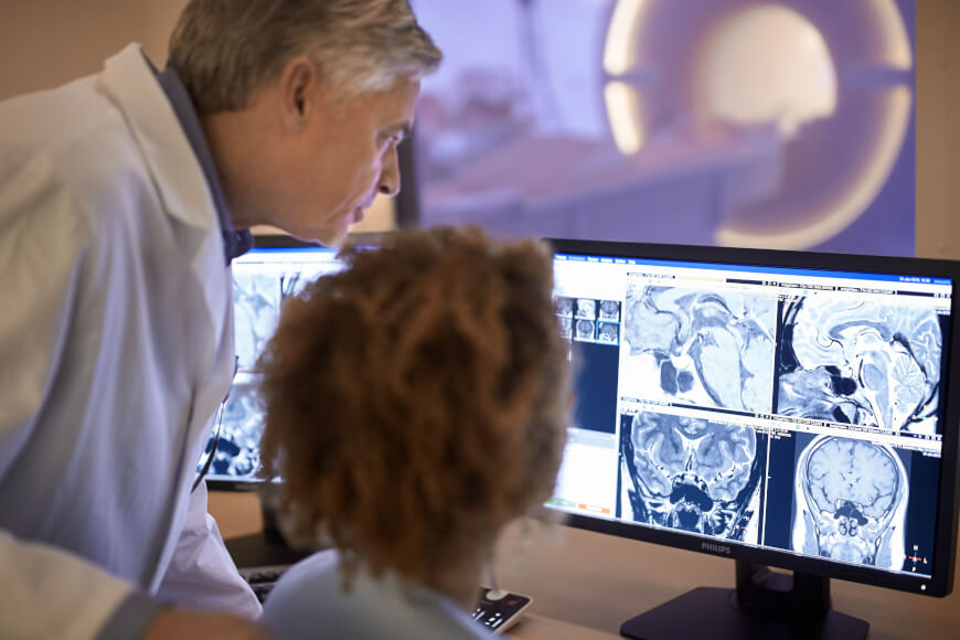
Magnetic resonance imaging (MRI) is a medical imaging technique used in radiology to form pictures of the anatomy and the physiological processes of the body. MRI scanners use strong magnetic fields, magnetic field gradients, and radio waves to generate images of the organs in the body. MRI does not involve X-rays or the use of ionizing radiation, which distinguishes it from CT and PET scans. MRI is a medical application of nuclear magnetic resonance (NMR) which can also be used for imaging in other NMR applications, such as NMR spectroscopy. MRI is widely used in hospitals and clinics for medical diagnosis, staging and follow-up of disease. Compared to CT, MRI provides better contrast in images of soft-tissues, e.g. in the brain or abdomen. However, it may be perceived as less comfortable by patients, due to the usually longer and louder measurements with the subject in a long, confining tube, though "Open" MRI designs mostly relieve this. Additionally, implants and ... [...More Info...] [...Related Items...] OR: [Wikipedia] [Google] [Baidu] |


