|
Bronchus
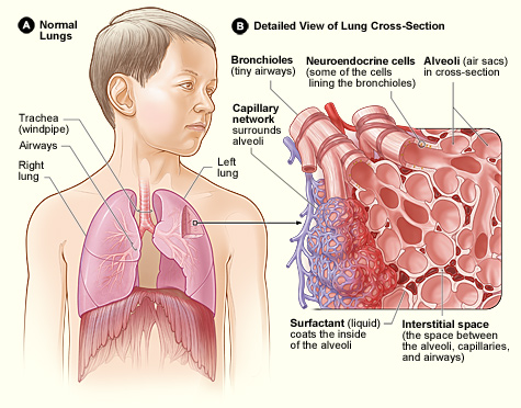
A bronchus is a passage or airway in the lower respiratory tract that conducts air into the lungs. The first or primary bronchi pronounced (BRAN-KAI) to branch from the trachea at the carina are the right main bronchus and the left main bronchus. These are the widest bronchi, and enter the right lung, and the left lung at each hilum. The main bronchi branch into narrower secondary bronchi or lobar bronchi, and these branch into narrower tertiary bronchi or segmental bronchi. Further divisions of the segmental bronchi are known as 4th order, 5th order, and 6th order segmental bronchi, or grouped together as subsegmental bronchi. The bronchi, when too narrow to be supported by cartilage, are known as bronchioles. No gas exchange takes place in the bronchi. Structure The trachea (windpipe) divides at the carina into two main or primary bronchi, the left bronchus and the right bronchus. The carina of the trachea is located at the level of the sternal angle and the fifth thoracic ... [...More Info...] [...Related Items...] OR: [Wikipedia] [Google] [Baidu] |
Lower Respiratory Tract
The respiratory tract is the subdivision of the respiratory system involved with the process of Respiration (physiology), respiration in mammals. The respiratory tract is lined with respiratory epithelium as respiratory mucosa. Air is breathed in through the nose to the nasal cavity, where a layer of nasal mucosa acts as a filter and traps pollutants and other harmful substances found in the air. Next, air moves into the pharynx, a passage that contains the intersection between the esophagus, oesophagus and the larynx. The opening of the larynx has a special flap of cartilage, the epiglottis, that opens to allow air to pass through but closes to prevent food from moving into the airway. From the larynx, air moves into the trachea and down to the intersection known as the carina of trachea, carina that branches to form the right and left primary (main) bronchus, bronchi. Each of these bronchi branches into a Bronchus, secondary (lobar) bronchus that branches into Bronchus, terti ... [...More Info...] [...Related Items...] OR: [Wikipedia] [Google] [Baidu] |
Respiratory System
The respiratory system (also respiratory apparatus, ventilatory system) is a biological system consisting of specific organs and structures used for gas exchange in animals and plants. The anatomy and physiology that make this happen varies greatly, depending on the size of the organism, the environment in which it lives and its evolutionary history. In land animals the respiratory surface is internalized as linings of the lungs. Gas exchange in the lungs occurs in millions of small air sacs; in mammals and reptiles these are called alveoli, and in birds they are known as atria. These microscopic air sacs have a very rich blood supply, thus bringing the air into close contact with the blood. These air sacs communicate with the external environment via a system of airways, or hollow tubes, of which the largest is the trachea, which branches in the middle of the chest into the two main bronchi. These enter the lungs where they branch into progressively narrower secondary and ... [...More Info...] [...Related Items...] OR: [Wikipedia] [Google] [Baidu] |
Bronchiole
The bronchioles or bronchioli (pronounced ''bron-kee-oh-lee'') are the smaller branches of the bronchial airways in the lower respiratory tract. They include the terminal bronchioles, and finally the respiratory bronchioles that mark the start of the respiratory zone delivering air to the gas exchanging units of the alveoli. The bronchioles no longer contain the cartilage that is found in the bronchi, or glands in their submucosa. Structure The pulmonary lobule is the portion of the lung ventilated by one bronchiole. Bronchioles are approximately 1 mm or less in diameter and their walls consist of ciliated cuboidal epithelium and a layer of smooth muscle. Bronchioles divide into even smaller bronchioles, called ''terminal'', which are 0.5 mm or less in diameter. Terminal bronchioles in turn divide into smaller respiratory bronchioles which divide into alveolar ducts. Terminal bronchioles mark the end of the conducting division of air flow in the respiratory sy ... [...More Info...] [...Related Items...] OR: [Wikipedia] [Google] [Baidu] |
Respiratory Bronchiole
The bronchioles or bronchioli (pronounced ''bron-kee-oh-lee'') are the smaller branches of the bronchial airways in the lower respiratory tract. They include the terminal bronchioles, and finally the respiratory bronchioles that mark the start of the respiratory zone delivering air to the gas exchanging units of the alveoli. The bronchioles no longer contain the cartilage that is found in the bronchi, or glands in their submucosa. Structure The pulmonary lobule is the portion of the lung ventilated by one bronchiole. Bronchioles are approximately 1 mm or less in diameter and their walls consist of ciliated cuboidal epithelium and a layer of smooth muscle. Bronchioles divide into even smaller bronchioles, called ''terminal'', which are 0.5 mm or less in diameter. Terminal bronchioles in turn divide into smaller respiratory bronchioles which divide into alveolar ducts. Terminal bronchioles mark the end of the conducting division of air flow in the respiratory syste ... [...More Info...] [...Related Items...] OR: [Wikipedia] [Google] [Baidu] |
Terminal Bronchiole
The bronchioles or bronchioli (pronounced ''bron-kee-oh-lee'') are the smaller branches of the bronchial airways in the lower respiratory tract. They include the terminal bronchioles, and finally the respiratory bronchioles that mark the start of the respiratory zone delivering air to the gas exchanging units of the alveoli. The bronchioles no longer contain the cartilage that is found in the bronchi, or glands in their submucosa. Structure The pulmonary lobule is the portion of the lung ventilated by one bronchiole. Bronchioles are approximately 1 mm or less in diameter and their walls consist of ciliated cuboidal epithelium and a layer of smooth muscle. Bronchioles divide into even smaller bronchioles, called ''terminal'', which are 0.5 mm or less in diameter. Terminal bronchioles in turn divide into smaller respiratory bronchioles which divide into alveolar ducts. Terminal bronchioles mark the end of the conducting division of air flow in the respiratory syste ... [...More Info...] [...Related Items...] OR: [Wikipedia] [Google] [Baidu] |
Eparterial Branch
The eparterial bronchus (right superior lobar bronchus) is a branch of the right main bronchus given off about 2.5 cm from the bifurcation of the trachea. This branch supplies the superior lobe of the right lung and is the most superior of all secondary bronchi. It arises above the level of the right pulmonary artery, and for this reason is named the eparterial bronchus. All other distributions falling below the pulmonary artery are termed ''hyparterial''. The eparterial bronchus is the only secondary bronchus with a specific name apart from the name of its corresponding lobe. Name The classification of ''eparterial'' and ''hyparterial'' is attributed to Swiss people, Swiss anatomist and anthropologist Christoph Theodor Aeby, and is central to his model of the anatomical lung. He presented this model in a monograph titled, "Der Bronchialbaum der Säugethiere und des Menschen, nebst Bemerkungen über den Bronchialbaum der Vögel und Reptilien". References External links< ... [...More Info...] [...Related Items...] OR: [Wikipedia] [Google] [Baidu] |
Eparterial Bronchus
The eparterial bronchus (right superior lobar bronchus) is a branch of the right main bronchus given off about 2.5 cm from the bifurcation of the trachea. This branch supplies the superior lobe of the right lung and is the most superior of all secondary bronchi. It arises above the level of the right pulmonary artery, and for this reason is named the eparterial bronchus. All other distributions falling below the pulmonary artery are termed ''hyparterial''. The eparterial bronchus is the only secondary bronchus with a specific name apart from the name of its corresponding lobe. Name The classification of ''eparterial'' and ''hyparterial'' is attributed to Swiss anatomist and anthropologist An anthropologist is a person engaged in the practice of anthropology. Anthropology is the study of aspects of humans within past and present societies. Social anthropology, cultural anthropology and philosophical anthropology study the norms an ... Christoph Theodor Aeby, and is cen ... [...More Info...] [...Related Items...] OR: [Wikipedia] [Google] [Baidu] |
Right Lung
The lungs are the primary organs of the respiratory system in humans and most other animals, including some snails and a small number of fish. In mammals and most other vertebrates, two lungs are located near the backbone on either side of the heart. Their function in the respiratory system is to extract oxygen from the air and transfer it into the bloodstream, and to release carbon dioxide from the bloodstream into the atmosphere, in a process of gas exchange. Respiration is driven by different muscular systems in different species. Mammals, reptiles and birds use their different muscles to support and foster breathing. In earlier tetrapods, air was driven into the lungs by the pharyngeal muscles via buccal pumping, a mechanism still seen in amphibians. In humans, the main muscle of respiration that drives breathing is the diaphragm. The lungs also provide airflow that makes vocal sounds including human speech possible. Humans have two lungs, one on the left and one on th ... [...More Info...] [...Related Items...] OR: [Wikipedia] [Google] [Baidu] |
Human Lung
The lungs are the primary organs of the respiratory system in humans and most other animals, including some snails and a small number of fish. In mammals and most other vertebrates, two lungs are located near the backbone on either side of the heart. Their function in the respiratory system is to extract oxygen from the air and transfer it into the bloodstream, and to release carbon dioxide from the bloodstream into the atmosphere, in a process of gas exchange. Respiration is driven by different muscular systems in different species. Mammals, reptiles and birds use their different muscles to support and foster breathing. In earlier tetrapods, air was driven into the lungs by the pharyngeal muscles via buccal pumping, a mechanism still seen in amphibians. In humans, the main muscle of respiration that drives breathing is the diaphragm. The lungs also provide airflow that makes vocal sounds including human speech possible. Humans have two lungs, one on the left and one on ... [...More Info...] [...Related Items...] OR: [Wikipedia] [Google] [Baidu] |
Lung
The lungs are the primary organs of the respiratory system in humans and most other animals, including some snails and a small number of fish. In mammals and most other vertebrates, two lungs are located near the backbone on either side of the heart. Their function in the respiratory system is to extract oxygen from the air and transfer it into the bloodstream, and to release carbon dioxide from the bloodstream into the atmosphere, in a process of gas exchange. Respiration is driven by different muscular systems in different species. Mammals, reptiles and birds use their different muscles to support and foster breathing. In earlier tetrapods, air was driven into the lungs by the pharyngeal muscles via buccal pumping, a mechanism still seen in amphibians. In humans, the main muscle of respiration that drives breathing is the diaphragm. The lungs also provide airflow that makes vocal sounds including human speech possible. Humans have two lungs, one on the left an ... [...More Info...] [...Related Items...] OR: [Wikipedia] [Google] [Baidu] |
Root Of The Lung
The root of the lung is a group of structures that emerge at the hilum of each lung, just above the middle of the mediastinal surface and behind the cardiac impression of the lung. It is nearer to the back (posterior border) than the front (anterior border). The root of the lung is connected by the structures that form it to the heart and the trachea. The rib cage is separated from the lung by a two-layered membranous coating, the pleura. The hilum is the large triangular depression where the connection between the parietal pleura (covering the rib cage) and the visceral pleura (covering the lung) is made, and this marks the meeting point between the mediastinum and the pleural cavities. Location The root of the right lung lies behind the superior vena cava and part of the right atrium, and below the azygos vein. That of the left lung passes beneath the aortic arch and in front of the descending aorta; the phrenic nerve, pericardiacophrenic artery and vein, and the anterior p ... [...More Info...] [...Related Items...] OR: [Wikipedia] [Google] [Baidu] |


