|
Bronchomalacia
Bronchomalacia is a term for weak cartilage in the walls of the bronchial tubes, often occurring in children under a day. Bronchomalacia means 'floppiness' of some part of the bronchi. Patients present with noisy breathing and/or wheezing. There is collapse of a main stem bronchus on exhalation. If the trachea is also involved the term tracheobronchomalacia (TBM) is used. If only the upper airway the trachea is involved it is called tracheomalacia (TM). There are two types of bronchomalacia. Primary bronchomalacia is due to a deficiency in the cartilaginous rings. Secondary bronchomalacia may occur by extrinsic compression from an enlarged vessel, a vascular ring or a bronchogenic cyst. Though uncommon, idiopathic (of unknown cause) tracheobronchomalacia has been described in older adults. Cause Bronchomalacia can best be described as a birth defect of the bronchus in the respiratory tract. Congenital malacia of the large airways is one of the few causes of irreversible airways ob ... [...More Info...] [...Related Items...] OR: [Wikipedia] [Google] [Baidu] |
Tracheomalacia
Tracheomalacia is a condition or incident where the cartilage that keeps the airway (trachea) open is soft such that the trachea partly collapses especially during increased airflow. This condition is most commonly seen in infants and young children. The usual symptom is stridor when a person breathes out. This is usually known as a collapsed windpipe. The trachea normally opens slightly during breathing in and narrows slightly during breathing out. These processes are exaggerated in tracheomalacia, leading to airway collapse on breathing out. If the condition extends further to the large airways (bronchi) (if there is also bronchomalacia), it is termed tracheobronchomalacia. The same condition can also affect the larynx, which is called laryngomalacia. The term is from ''trachea'' and the Greek μαλακία, ''softening'' Signs and symptoms Tracheomalacia occurs when the walls of the trachea collapse. This can happen because the walls of the windpipe are weak, or it can ... [...More Info...] [...Related Items...] OR: [Wikipedia] [Google] [Baidu] |
Vertebrate Trachea
The trachea, also known as the windpipe, is a cartilaginous tube that connects the larynx to the bronchi of the lungs, allowing the passage of air, and so is present in almost all air-breathing animals with lungs. The trachea extends from the larynx and branches into the two primary bronchi. At the top of the trachea the cricoid cartilage attaches it to the larynx. The trachea is formed by a number of horseshoe-shaped rings, joined together vertically by overlying ligaments, and by the trachealis muscle at their ends. The epiglottis closes the opening to the larynx during swallowing. The trachea begins to form in the second month of embryo development, becoming longer and more fixed in its position over time. It is epithelium lined with column-shaped cells that have hair-like extensions called cilia, with scattered goblet cells that produce protective mucins. The trachea can be affected by inflammation or infection, usually as a result of a viral illness affecting other part ... [...More Info...] [...Related Items...] OR: [Wikipedia] [Google] [Baidu] |
Bronchiectasis
Bronchiectasis is a disease in which there is permanent enlargement of parts of the airways of the lung. Symptoms typically include a chronic cough with mucus production. Other symptoms include shortness of breath, coughing up blood, and chest pain. Wheezing and nail clubbing may also occur. Those with the disease often get lung infections. Bronchiectasis may result from a number of infectious and acquired causes, including measles, pneumonia, tuberculosis, immune system problems, as well as the genetic disorder cystic fibrosis. Cystic fibrosis eventually results in severe bronchiectasis in nearly all cases. The cause in 10–50% of those without cystic fibrosis is unknown. The mechanism of disease is breakdown of the airways due to an excessive inflammatory response. Involved airways (bronchi) become enlarged and thus less able to clear secretions. These secretions increase the amount of bacteria in the lungs, resulting in airway blockage and further breakdown of the airways. ... [...More Info...] [...Related Items...] OR: [Wikipedia] [Google] [Baidu] |
Respirology
Pulmonology (, , from Latin ''pulmō, -ōnis'' "lung" and the Greek suffix "study of"), pneumology (, built on Greek πνεύμων "lung") or pneumonology () is a medical specialty that deals with diseases involving the respiratory tract.ACP: Pulmonology: Internal Medicine Subspecialty . Acponline.org. Retrieved on 2011-09-30. It is also known as respirology, respiratory medicine, or chest medicine in some countries and areas. Pulmonology is considered a branch of internal medicine, and is related to intensive care medicine ...
[...More Info...] [...Related Items...] OR: [Wikipedia] [Google] [Baidu] |
Larynx
The larynx (), commonly called the voice box, is an organ in the top of the neck involved in breathing, producing sound and protecting the trachea against food aspiration. The opening of larynx into pharynx known as the laryngeal inlet is about 4–5 centimeters in diameter. The larynx houses the vocal cords, and manipulates pitch and volume, which is essential for phonation. It is situated just below where the tract of the pharynx splits into the trachea and the esophagus. The word ʻlarynxʼ (plural ʻlaryngesʼ) comes from the Ancient Greek word ''lárunx'' ʻlarynx, gullet, throat.ʼ Structure The triangle-shaped larynx consists largely of cartilages that are attached to one another, and to surrounding structures, by muscles or by fibrous and elastic tissue components. The larynx is lined by a ciliated columnar epithelium except for the vocal folds. The cavity of the larynx extends from its triangle-shaped inlet, to the epiglottis, and to the circular outlet at the ... [...More Info...] [...Related Items...] OR: [Wikipedia] [Google] [Baidu] |
Respiratory Pathology
The respiratory system (also respiratory apparatus, ventilatory system) is a biological system consisting of specific organs and structures used for gas exchange in animals and plants. The anatomy and physiology that make this happen varies greatly, depending on the size of the organism, the environment in which it lives and its evolutionary history. In land animals the respiratory surface is internalized as linings of the lungs. Gas exchange in the lungs occurs in millions of small air sacs; in mammals and reptiles these are called alveoli, and in birds they are known as atria. These microscopic air sacs have a very rich blood supply, thus bringing the air into close contact with the blood. These air sacs communicate with the external environment via a system of airways, or hollow tubes, of which the largest is the trachea, which branches in the middle of the chest into the two main bronchi. These enter the lungs where they branch into progressively narrower secondary and ter ... [...More Info...] [...Related Items...] OR: [Wikipedia] [Google] [Baidu] |
Congenital Cystic Adenomatoid Malformation
Congenital pulmonary airway malformation (CPAM), formerly known as congenital cystic adenomatoid malformation (CCAM), is a congenital disorder of the lung similar to bronchopulmonary sequestration. In CPAM, usually an entire lobe of lung is replaced by a non-working cystic piece of abnormal lung tissue. This abnormal tissue will never function as normal lung tissue. The underlying cause for CPAM is unknown. It occurs in approximately 1 in every 30,000 pregnancies. In most cases the outcome of a fetus with CPAM is very good. In rare cases, the cystic mass grows so large as to limit the growth of the surrounding lung and cause pressure against the heart. In these situations, the CPAM can be life-threatening for the fetus. CPAM can be separated into five types, based on clinical and pathologic features. CPAM type 1 is the most common, with large cysts and a good prognosis. CPAM type 2 (with medium-sized cysts) often has a poor prognosis, owing to its frequent association with other si ... [...More Info...] [...Related Items...] OR: [Wikipedia] [Google] [Baidu] |
Pulmonary Sequestration
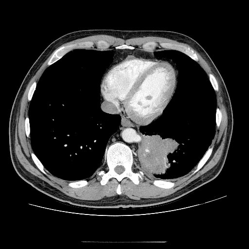
A pulmonary sequestration is a medical condition wherein a piece of tissue that ultimately develops into lung tissue is not attached to the pulmonary arterial blood supply, as is the case in normally developing lung. This sequestered tissue is therefore not connected to the normal bronchial airway architecture, and fails to function in, and contribute to, respiration of the organism. This condition is usually diagnosed in children and is generally thought to be congenital in nature. More and more, these lesions are diagnosed ''in utero'' by prenatal ultrasound. Presentation Symptoms can vary greatly, but they include a persistent dry cough. Complications Failure to have a pulmonary sequestration removed can lead to a number of complications. These include: * Potentially fatal hemorrhage * The creation of a left-right shunt, where blood flows in a shortcut through the feed off the aorta * Chronic infection with diseases such as ** Bronchiectasis ** Tuberculosis ** Aspergillo ... [...More Info...] [...Related Items...] OR: [Wikipedia] [Google] [Baidu] |
Lung
The lungs are the primary organs of the respiratory system in humans and most other animals, including some snails and a small number of fish. In mammals and most other vertebrates, two lungs are located near the backbone on either side of the heart. Their function in the respiratory system is to extract oxygen from the air and transfer it into the bloodstream, and to release carbon dioxide from the bloodstream into the atmosphere, in a process of gas exchange. Respiration is driven by different muscular systems in different species. Mammals, reptiles and birds use their different muscles to support and foster breathing. In earlier tetrapods, air was driven into the lungs by the pharyngeal muscles via buccal pumping, a mechanism still seen in amphibians. In humans, the main muscle of respiration that drives breathing is the diaphragm. The lungs also provide airflow that makes vocal sounds including human speech possible. Humans have two lungs, one on the left an ... [...More Info...] [...Related Items...] OR: [Wikipedia] [Google] [Baidu] |
Laryngomalacia
Laryngomalacia (literally, "soft larynx") is the most common cause of chronic stridor in infancy, in which the soft, immature cartilage of the upper larynx collapses inward during inhalation, causing airway obstruction. It can also be seen in older patients, especially those with neuromuscular conditions resulting in weakness of the muscles of the throat. However, the infantile form is much more common. Laryngomalacia is one of the most common laryngeal congenital disease in infancy and public education about the signs and symptoms of the disease is lacking. Signs and symptoms In infantile laryngomalacia, the supraglottic larynx (the part above the vocal cords) is tightly curled, with a short band holding the cartilage shield in the front (the epiglottis) tightly to the mobile cartilage in the back of the larynx (the arytenoids). These bands are known as the aryepiglottic folds. The shortened aryepiglottic folds cause the epiglottis to be curled on itself. This is the well ... [...More Info...] [...Related Items...] OR: [Wikipedia] [Google] [Baidu] |
Laryngocele
A laryngocele is a congenital anomalous air sac Air sacs are spaces within an organism where there is the constant presence of air. Among modern animals, birds possess the most air sacs (9–11), with their extinct dinosaurian relatives showing a great increase in the pneumatization (presence ... communicating with the cavity of the larynx, which may bulge outward on the neck. It may also be acquired, as seen in glassblowers, due to continual forced expiration producing increased pressures in the larynx which leads to dilatation of the laryngeal ventricle ( sinus of Morgagni). It is also seen in people with chronic obstructive airway disease. Additional images References External links {{Congenital malformations and deformations of respiratory system Human head and neck Congenital disorders of respiratory system ... [...More Info...] [...Related Items...] OR: [Wikipedia] [Google] [Baidu] |


