wrist osteoarthritis on:
[Wikipedia]
[Google]
[Amazon]
Wrist osteoarthritis is gradual loss of articular cartilage and hypertrophic bone changes (osteophytes). While in many joints this is part of normal aging (senescence), in the wrist osteoarthritis usually occurs over years to decades after scapholunate interosseous ligament rupture or an unhealed fracture of the scaphoid. Characteristic symptoms including pain, deformity and stiffness. Pain intensity and incapability (limited function) are notably variable and do not correspond with arthritis severity on radiographs.
Osteoarthritis of the wrist can be
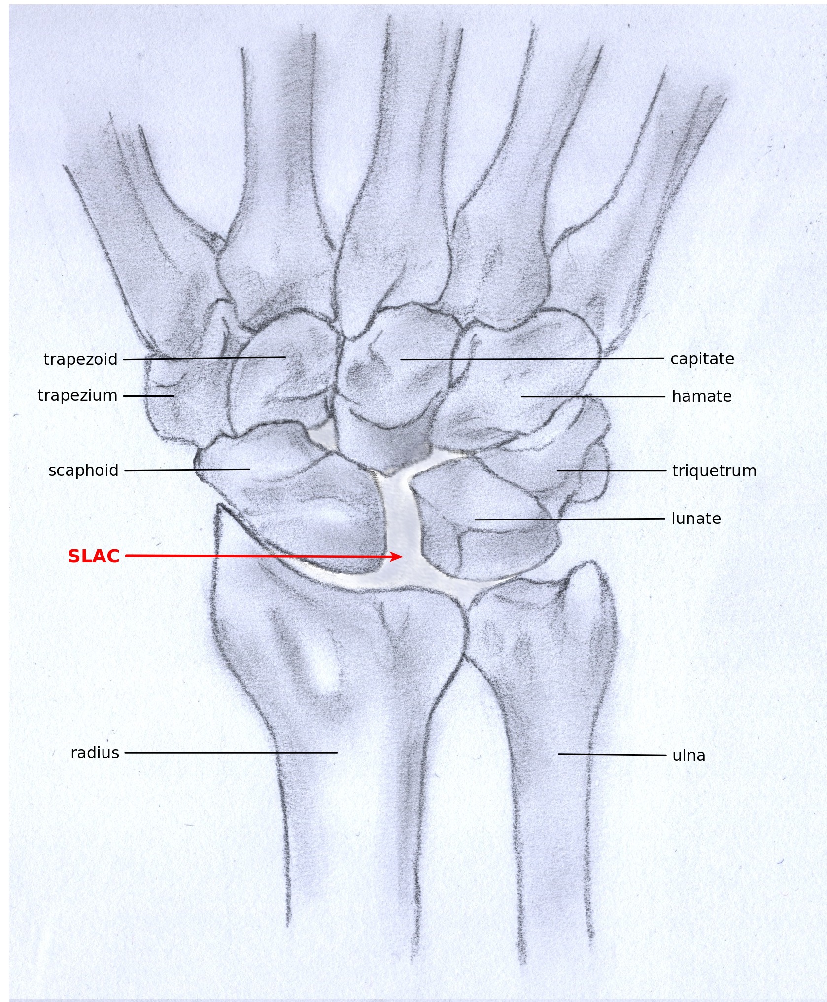
 SLAC and SNAC are two patterns of wrist osteoarthritis, following predictable patterns depending on the type of underlying injury. SLAC is caused by scapholunate ligament rupture, and SNAC is caused by a
 Scaphoid fracture
Image:Stadium1osteoarthritis.JPG, Stage I
Image:Stadium2osteoarthritis.JPG, Stage II
Image:Stadium3osteoathritis.JPG, Stage III
Image:Stadium4osteoarhtritis.JPG, Stage IV

 In order to understand the cause of post-traumatic wrist osteoarthritis it is important to know and understand the anatomy of the wrist. The hand is subdivided into three parts:
*
In the absence of gout, chondrocalcinosis, rheumatoid arthritis, or prior distal radius fracture, a person with gradual onset limited motion and pain in the wrist likely has wrist osteoarthritis.
idiopathic
An idiopathic disease is any disease with an unknown cause or mechanism of apparent spontaneous origin.
For some medical conditions, one or more causes are somewhat understood, but in a certain percentage of people with the condition, the cause ...
, but it is mostly seen as a post-traumatic condition. There are different types of post-traumatic osteoarthritis. Scapholunate advanced collapse ( SLAC) is the most common form, followed by scaphoid non-union advanced collapse (SNAC). Other post-traumatic causes such as intra-articular
A joint or articulation (or articular surface) is the connection made between bones, ossicles, or other hard structures in the body which link an animal's skeletal system into a functional whole.Saladin, Ken. Anatomy & Physiology. 7th ed. McGraw- ...
fractures
Fracture is the appearance of a crack or complete separation of an object or material into two or more pieces under the action of stress (mechanics), stress. The fracture of a solid usually occurs due to the development of certain displacemen ...
of the distal radius
In classical geometry, a radius (: radii or radiuses) of a circle or sphere is any of the line segments from its Centre (geometry), center to its perimeter, and in more modern usage, it is also their length. The radius of a regular polygon is th ...
or ulna
The ulna or ulnar bone (: ulnae or ulnas) is a long bone in the forearm stretching from the elbow to the wrist. It is on the same side of the forearm as the little finger, running parallel to the Radius (bone), radius, the forearm's other long ...
can also lead to wrist osteoarthritis, but are less common.
Types
 SLAC and SNAC are two patterns of wrist osteoarthritis, following predictable patterns depending on the type of underlying injury. SLAC is caused by scapholunate ligament rupture, and SNAC is caused by a
 scaphoid fracture
A scaphoid fracture is a bone fracture, break of the scaphoid bone in the wrist. Symptoms generally includes pain at the base of the thumb which is worse with use of the hand. The Anatomical snuff box, anatomic snuffbox is generally tender and sw ...
which does not heal non-union
Nonunion is permanent failure of healing following a broken bone unless intervention (such as surgery) is performed. A fracture with nonunion generally forms a structural resemblance to a fibrous joint, and is therefore often called a "false jo ...
.
SLAC is more common than SNAC; 55% of the patients with wrist osteoarthritis have a SLAC wrist.
SLAC
Scapholunate advanced collapse (SLAC) is a predictable pattern of wrist osteoarthritis that results from untreated long-standingscapholunate ligament
The scapholunate ligament is a ligament of the wrist.
Rupture of the scapholunate ligament causes scapholunate instability, which, if untreated, will eventually cause a predictable pattern of wrist osteoarthritis called scapholunate advanced co ...
rupture and the associated carpal malalignment. The misalignment is described as dorsal intercalated segment instability (DISI) which is where the lunate angulates towards the dorsal side of the hand.
SNAC
 Scaphoid fracture
 non-union
Nonunion is permanent failure of healing following a broken bone unless intervention (such as surgery) is performed. A fracture with nonunion generally forms a structural resemblance to a fibrous joint, and is therefore often called a "false jo ...
changes the shape of the scaphoid bone and results in DISI malalignment.
Scaphoid Non-union Advanced collapse (SNAC) is the pattern of osteoarthritis that develops in relation to the malalignment.
Stages
Post-traumatic osteoarthritis can be classified into four stages. These stages are similar between SLAC and SNAC wrists. Each stage has a different treatment. * Stage I: the osteoarthritis is only localized in the distal scaphoid and radial styloid. * Stage II: the osteoarthritis is localized in the entire radioscaphoid joint. * Stage III: the osteoarthritis is localized in the entire radioscaphoid joint with involvement of the capitolunate joint. * Stage IV: the osteoarthritis is located in the entire radiocarpal joint and in the intercarpal joints. It also may involve the distal radio-ulnar joint (DRUJ).Signs and symptoms
The most common initial presenting symptom of wrist osteoarthritis isjoint pain
Arthralgia () literally means 'joint pain'. Specifically, arthralgia is a symptom of injury, infection, illness (in particular arthritis), or an allergic reaction to medication
Medication (also called medicament, medicine, pharmaceutic ...
. Other signs and symptoms, as with any joint affected by osteoarthritis, include:
* Loss of motion stiffness, which can be worse after a period of rest, such as when one awakes in the morning.
* Deformity of the wrist. There is a characteristic dorsal radial fullness related to osteophytes and joint effusion.
* Crepitus (crackling), which is felt when the wrist is moved passively.
These symptoms can lead to loss of function and less daily activity.
Mechanism

 In order to understand the cause of post-traumatic wrist osteoarthritis it is important to know and understand the anatomy of the wrist. The hand is subdivided into three parts:
*
 Wrist
In human anatomy, the wrist is variously defined as (1) the carpus or carpal bones, the complex of eight bones forming the proximal skeletal segment of the hand; "The wrist contains eight bones, roughly aligned in two rows, known as the carpal ...
* Metacarpus
In human anatomy, the metacarpal bones or metacarpus, also known as the "palm bones", are the appendicular skeleton, appendicular bones that form the intermediate part of the hand between the phalanges (fingers) and the carpal bones (wrist, wris ...
* Digits
The wrist consists of eight small carpal bones
The carpal bones are the eight small bones that make up the wrist (carpus) that connects the hand to the forearm. The terms "carpus" and "carpal" are derived from the Latin wikt:carpus#Latin, carpus and the Greek language, Greek wikt:καρπός ...
. Each of these carpal bones has a different size and shape. They contribute towards the stability of the wrist and are ranked in two rows, each consisting of four bones.
Proximal row
Fromlateral
Lateral is a geometric term of location which may also refer to:
Biology and healthcare
* Lateral (anatomy), a term of location meaning "towards the side"
* Lateral cricoarytenoid muscle, an intrinsic muscle of the larynx
* Lateral release ( ...
to medial and when viewed from anterior
Standard anatomical terms of location are used to describe unambiguously the anatomy of humans and other animals. The terms, typically derived from Latin or Greek roots, describe something in its standard anatomical position. This position pro ...
, the proximal
Standard anatomical terms of location are used to describe unambiguously the anatomy of humans and other animals. The terms, typically derived from Latin or Greek roots, describe something in its standard anatomical position. This position prov ...
row is formed by the:
* Scaphoid
The scaphoid bone is one of the carpal bones of the wrist. It is situated between the hand and forearm on the thumb side of the wrist (also called the lateral or radial side). It forms the radial border of the carpal tunnel. The scaphoid bone ...
* Lunate
Lunate is a crescent or moon-shaped microlith. In the specialized terminology of lithic reduction, a lunate flake is a small, crescent-shaped lithic flake, flake removed from a stone tool during the process of pressure flaking.
In the Natufian cu ...
* Triquetral
The triquetral bone (; also called triquetrum, pyramidal, three-faced, and formerly cuneiform bone) is located in the wrist on the medial side of the proximal row of the carpus between the lunate and pisiform bones. It is on the ulnar side of the ...
* Pisiform
The pisiform bone ( or ), also spelled pisiforme (from the Latin ''pisiformis'', pea-shaped), is a small knobbly, sesamoid bone that is found in the wrist. It forms the ulnar border of the carpal tunnel.
Structure
The pisiform is a sesamoid bone, ...
Distal row
Fromlateral
Lateral is a geometric term of location which may also refer to:
Biology and healthcare
* Lateral (anatomy), a term of location meaning "towards the side"
* Lateral cricoarytenoid muscle, an intrinsic muscle of the larynx
* Lateral release ( ...
to medial and when viewed from anterior
Standard anatomical terms of location are used to describe unambiguously the anatomy of humans and other animals. The terms, typically derived from Latin or Greek roots, describe something in its standard anatomical position. This position pro ...
, the distal
Standard anatomical terms of location are used to describe unambiguously the anatomy of humans and other animals. The terms, typically derived from Latin or Greek roots, describe something in its standard anatomical position. This position provi ...
row is formed by the:
* Trapezium
* Trapezoid
In geometry, a trapezoid () in North American English, or trapezium () in British English, is a quadrilateral that has at least one pair of parallel sides.
The parallel sides are called the ''bases'' of the trapezoid. The other two sides are ...
* Capitate
* Hamate
The hamate bone (from Latin language, Latin wiktionary:hamatus, hamatus, "hooked"), or unciform bone (from Latin language, Latin ''wikt:uncus, uncus'', "hook"), Latin os hamatum and occasionally abbreviated as just hamatum, is a bone in the huma ...
Diagnosis
Osteoarthritis of the wrist is predominantly a clinical diagnosis, and thus is primarily based on the patients medical history, physical examination and wrist X-rays.Medical history
The person may or may not recall an old wrist injury.Physical examination
Examination may identify limited passive wrist motion, pain at the extremes of wrist motion, tenderness at the radioscaphoid joint, and dorsal radial prominence. Activities that use forceful wrist extension such as rising from a chair or push-ups may be painful.In the absence of gout, chondrocalcinosis, rheumatoid arthritis, or prior distal radius fracture, a person with gradual onset limited motion and pain in the wrist likely has wrist osteoarthritis.
X-rays
Radiographs can confirm the diagnosis of wrist osteoarthritis. The earliest sign is narrowing of the joint space between theradius
In classical geometry, a radius (: radii or radiuses) of a circle or sphere is any of the line segments from its Centre (geometry), center to its perimeter, and in more modern usage, it is also their length. The radius of a regular polygon is th ...
and the scaphoid and an osteophyte off the tip of the radial styloid.
SLAC
Because SLAC results from scapholunate ligament rupture, there is a larger space between the two bones, also known as the Terry Thomas sign. Scaphoid instability due to the ligament rupture can be stactic or dynamic. When the X-ray is diagnostic and there is a convincing Terry Thomas sign it is a static scaphoid instability. When the scaphoid is made unstable by either the patient or by manipulation by the examining physician it is a dynamic instability.
SNAC
In order to diagnose a SNAC wrist you need a PA view X-ray and a lateral view X-ray. As in SLAC, the lateral view X-ray is performed to see if there is a DISI.
Computed tomography
A computed tomography scan (CT scan), formerly called computed axial tomography scan (CAT scan), is a medical imaging technique used to obtain detailed internal images of the body. The personnel that perform CT scans are called radiographers or ...
(CT) or Magnetic Resonance Imaging
Magnetic resonance imaging (MRI) is a medical imaging technique used in radiology to generate pictures of the anatomy and the physiological processes inside the body. MRI scanners use strong magnetic fields, magnetic field gradients, and ...
(MRI) are rarely used to diagnose SNAC or SLAC wrist osteoarthritis because there is no additional value. Also, these techniques are much more expensive than a standard X-ray. CT or MRI may be used if there is a strong suspicion for another underlying pathology or disease.
Treatment
Post-traumatic wrist osteoarthritis can be accommodated. A wrist splint, ice, acetaminophen, and NSAIDs may alleviate symptoms. Surgery to change the wrist anatomy to attempt to alleviate pain is an option.Stage I
For stage I, normally, nonsurgical treatment is sufficient. Injections of corticosteroid may be considered. Keep in mind that corticosteroids provide, at best, temporary alleviation of discomfort. And corticosteroid injection harms cartilage. Since people with Stage 1 arthritis have good cartilage, one might be cautious with corticosteroid injection. Surgical options for mild arthritis may include neurectomy of the anterior and posterior interosseous nerves, or radial styloidectomy, in which the radial styloid is surgically removed from the distal radius.Stage II
The surgical options for stage II and III wrist osteoarthritis are excision of some of the bones with or without fusion (arthrodesis) of the others. The idea is to try to alleviate pain while maintaining some wrist motion. One technique is to remove one row of carpal bones. The bones closer to the forearm (proximal) are removed: scaphoid, lunate, and triquetrum. It is important that the radioscaphocapitate ligament is left intact, because if the ligament is not preserved the capitate bone will translate to the ulnar side of the wrist and move away from the distal radius. The new articulation of the capitate with the lunate fossa of the distal radius is not as congruent as the former scaphoid-lunate-radius joint. This and other issues contribute to potential to develop arthritis over the years. In part based on these concerns, some surgeon prefer to maintain the lunate in patients younger than 40 years proximal row carpectomy. A surgery called four-corner arthrodesis is an option. The capitate, lunate, hamate and triquetrum are fused together in this procedure and the scaphoid is excised. Before the arthrodesis is executed, the lunate must be reduced out of DISI position. Because the radiolunate joint is typically preserved in stage II SLAC and SNAC wrists, this joint can be the only remaining joint of the proximal wrist. Both procedures are often combined with wrist denervation, as described in the text of treatment stage I.Stage III
In Stage III wrist osteoarthritis, some surgeons offer patients proximal row carpectomy and interpose some of the wrist capsule to account for the arthritis in the capitate. Four-corner arthrodesis, as described above in stage II, is also an option.Stage IV
In this stage there are two surgical treatment options; total wristarthroplasty
Arthroplasty (literally " e-orming of joint") is an orthopedic surgical procedure where the articular surface of a musculoskeletal joint is replaced, remodeled, or realigned by osteotomy or some other procedure. It is an elective procedure that ...
and total wrist arthrodesis
Arthrodesis, also known as artificial ankylosis or syndesis, is the artificial induction of joint ossification between two bones by surgery. This is done to relieve intractable pain in a joint which cannot be managed by pain medication, splin ...
.
Total wrist arthrodesis is the standard surgical treatment for patients with stage IV wrist osteoarthritis. During this procedure the carpal bones are all fused together and are then fastened to the distal radius. This procedure eliminates all wrist motion, but heavy labor is still possible.
An option for people who want to maintain some motion, and are willing to avoid using force with the hand, is total wrist arthroplasty. There is some evidence that patients with a total wrist arthrodesis on one side and a total wrist arthroplasty on the other, prefer the total wrist arthroplasty. The procedure exists of a couple of elements. First, the proximal row is removed and the distal row is fastened to the metacarpals. Then, one side of the arthroplasty is placed upon the distal row and the other side on the distal radius. Additionally, the head of the ulna is removed. Arthroplasty can have problems that may lead to another surgery.
References
{{Commons category, Wrist osteoarthritis Arthritis Skeletal disorders