X-ray Generator on:
[Wikipedia]
[Google]
[Amazon]
An X-ray generator is a device that produces

 An X-ray generator generally contains an X-ray tube to produce the X-rays. Possibly,
, NDT Resource Center. Page fetched April 21, 2011.
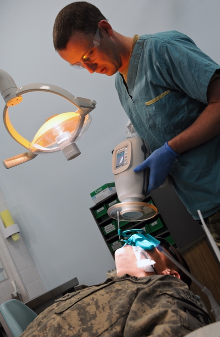
 In medical imaging applications, an X-ray machine has a control console that is used by a radiologic technologist to select X-ray techniques suitable for the specific exam, a power supply that creates and produces the desired kVp (peak kilovoltage), mA (milliamperes, sometimes referred to as mAs which is actually mA multiplied by the desired exposure length) for the X-ray tube, and the X-ray tube itself.
 A film of
X-rays
An X-ray, or, much less commonly, X-radiation, is a penetrating form of high-energy electromagnetic radiation. Most X-rays have a wavelength ranging from 10 picometers to 10 nanometers, corresponding to frequencies in the range 30&nbs ...
. Together with an X-ray detector
X-ray detectors are devices used to measure the flux, spatial distribution, spectrum, and/or other properties of X-rays.
Detectors can be divided into two major categories: imaging detectors (such as photographic plates and X-ray film (photograp ...
, it is commonly used in a variety of applications including medicine
Medicine is the science and practice of caring for a patient, managing the diagnosis, prognosis, prevention, treatment, palliation of their injury or disease, and promoting their health. Medicine encompasses a variety of health care pr ...
, X-ray fluorescence, electronic assembly inspection, and measurement of material thickness in manufacturing operations. In medical applications, X-ray generators are used by radiographer
Radiographers, also known as radiologic technologists, diagnostic radiographers and medical radiation technologists are healthcare professionals who specialize in the imaging of human anatomy for the diagnosis and treatment of pathology. Rad ...
s to acquire x-ray images of the internal structures (e.g., bones) of living organisms, and also in sterilization
Sterilization may refer to:
* Sterilization (microbiology), killing or inactivation of micro-organisms
* Soil steam sterilization, a farming technique that sterilizes soil with steam in open fields or greenhouses
* Sterilization (medicine) rende ...
.
Structure

 An X-ray generator generally contains an X-ray tube to produce the X-rays. Possibly,
 radioisotope
A radionuclide (radioactive nuclide, radioisotope or radioactive isotope) is a nuclide that has excess nuclear energy, making it unstable. This excess energy can be used in one of three ways: emitted from the nucleus as gamma radiation; transferr ...
s can also be used to generate X-rays.
An X-ray tube is a simple vacuum tube
A vacuum tube, electron tube, valve (British usage), or tube (North America), is a device that controls electric current flow in a high vacuum between electrodes to which an electric potential difference has been applied.
The type known as ...
that contains a cathode
A cathode is the electrode from which a conventional current leaves a polarized electrical device. This definition can be recalled by using the mnemonic ''CCD'' for ''Cathode Current Departs''. A conventional current describes the direction in whi ...
, which directs a stream of electrons into a vacuum, and an anode
An anode is an electrode of a polarized electrical device through which conventional current enters the device. This contrasts with a cathode, an electrode of the device through which conventional current leaves the device. A common mnemonic is ...
, which collects the electrons and is made of tungsten to evacuate the heat generated by the collision. When the electrons collide with the target, about 1% of the resulting energy is emitted as X-ray
An X-ray, or, much less commonly, X-radiation, is a penetrating form of high-energy electromagnetic radiation. Most X-rays have a wavelength ranging from 10 picometers to 10 nanometers, corresponding to frequencies in the range 30&nb ...
s, with the remaining 99% released as heat. Due to the high energy of the electrons that reach relativistic speeds the target is usually made of tungsten
Tungsten, or wolfram, is a chemical element with the symbol W and atomic number 74. Tungsten is a rare metal found naturally on Earth almost exclusively as compounds with other elements. It was identified as a new element in 1781 and first isol ...
even if other material can be used particularly in XRF applications.
An X-ray generator also needs to contain a cooling system to cool the anode; many X-ray generators use water or oil recirculating systems."X-ray Generators", NDT Resource Center. Page fetched April 21, 2011.
Medical imaging
 In medical imaging applications, an X-ray machine has a control console that is used by a radiologic technologist to select X-ray techniques suitable for the specific exam, a power supply that creates and produces the desired kVp (peak kilovoltage), mA (milliamperes, sometimes referred to as mAs which is actually mA multiplied by the desired exposure length) for the X-ray tube, and the X-ray tube itself.
History
The discovery of X-rays came from experimenting with Crookes tubes, an early experimental electrical discharge tube invented by English physicist William Crookes around 1869–1875. In 1895,Wilhelm Röntgen
Wilhelm Conrad Röntgen (; ; 27 March 184510 February 1923) was a German mechanical engineer and physicist, who, on 8 November 1895, produced and detected electromagnetic radiation in a wavelength range known as X-rays or Röntgen rays, an achie ...
discovered X-rays emanating from Crookes tubes and the many uses for X-rays were immediately apparent. One of the first X-ray photographs was made of the hand of Röntgen's wife. The image displayed both her wedding ring and bones. On January 18, 1896 an ''X-ray machine'' was formally displayed by Henry Louis Smith. A fully functioning unit was introduced to the public at the 1904 World's Fair by Clarence Dally. The technology developed quickly: In 1909 Mónico Sánchez Moreno had produced the first portable medical device and during World War I Marie Curie
Marie Salomea Skłodowska–Curie ( , , ; born Maria Salomea Skłodowska, ; 7 November 1867 – 4 July 1934) was a Polish and naturalized-French physicist and chemist who conducted pioneering research on radioactivity. She was the fir ...
led the development of X-ray machines mounted in "radiological cars" to provide mobile X-ray services for military field hospitals.
In the 1940s and 1950s, X-ray machines were used in stores to help sell footwear. These were known as Shoe-fitting fluoroscopes. However, as the harmful effects of X-ray
An X-ray, or, much less commonly, X-radiation, is a penetrating form of high-energy electromagnetic radiation. Most X-rays have a wavelength ranging from 10 picometers to 10 nanometers, corresponding to frequencies in the range 30&nb ...
radiation
In physics, radiation is the emission or transmission of energy in the form of waves or particles through space or through a material medium. This includes:
* ''electromagnetic radiation'', such as radio waves, microwaves, infrared, visi ...
were properly considered, they finally fell out of use. Shoe-fitting use of the device was first banned by the state of Pennsylvania
Pennsylvania (; ( Pennsylvania Dutch: )), officially the Commonwealth of Pennsylvania, is a state spanning the Mid-Atlantic, Northeastern, Appalachian, and Great Lakes regions of the United States. It borders Delaware to its southeast, ...
in 1957. (They were more a clever marketing tool to attract customers, rather than a fitting aid.) Together with Robert J. Van de Graaff, John G. Trump developed one of the first million-volt X-ray generators.
Overview
An X-ray imaging system consists of a generator control console where the operator selects desired techniques to obtain a quality readable image(kVp, mA and exposure time), an x-ray generator which controls the x-ray tube current, x-ray tube kilovoltage and x-ray emitting exposure time, an X-ray tube that converts the kilovoltage and mA into actual x-rays and an image detection system which can be either a film (analog technology) or a digital capture system and a PACS.Applications
X-ray machines are used inhealth care
Health care or healthcare is the improvement of health via the prevention, diagnosis, treatment, amelioration or cure of disease, illness, injury, and other physical and mental impairments in people. Health care is delivered by health pr ...
for visualising bone structures, during surgeries (especially orthopedic) to assist surgeons in reattaching broken bones with screws or structural plates, assisting cardiologists in locating blocked arteries and guiding stent placements or performing angioplasties and for other dense tissues such as tumour
A neoplasm () is a type of abnormal and excessive growth of tissue. The process that occurs to form or produce a neoplasm is called neoplasia. The growth of a neoplasm is uncoordinated with that of the normal surrounding tissue, and persists ...
s. Non-medicinal applications include security" \n\n\nsecurity.txt is a proposed standard for websites' security information that is meant to allow security researchers to easily report security vulnerabilities. The standard prescribes a text file called \"security.txt\" in the well known locat ...
and material analysis.
Medicine
The main fields in which x-ray machines are used in medicine areradiography
Radiography is an imaging technique using X-rays, gamma rays, or similar ionizing radiation and non-ionizing radiation to view the internal form of an object. Applications of radiography include medical radiography ("diagnostic" and "therapeu ...
, radiotherapy, and fluoroscopic-type procedures. Radiography is generally used for fast, highly penetrating images, and is usually used in areas with a high bone content but can also be used to look for tumors such as with mammography imaging. Some forms of radiography include:
* orthopantomogram — a panoramic x-ray of the jaw showing all the teeth at once
*mammography
Mammography (also called mastography) is the process of using low-energy X-rays (usually around 30 kVp) to examine the human breast for diagnosis and screening. The goal of mammography is the early detection of breast cancer, typically through ...
— x-rays of breast tissue
The breast is one of two prominences located on the upper ventral region of a primate's torso. Both females and males develop breasts from the same embryological tissues.
In females, it serves as the mammary gland, which produces and secret ...
*tomography
Tomography is imaging by sections or sectioning that uses any kind of penetrating wave. The method is used in radiology, archaeology, biology, atmospheric science, geophysics, oceanography, plasma physics, materials science, astrophysics, ...
— x-ray imaging in sections
In fluoroscopy, imaging of the digestive tract is done with the help of a radiocontrast agent such as barium sulfate
Barium sulfate (or sulphate) is the inorganic compound with the chemical formula Ba SO4. It is a white crystalline solid that is odorless and insoluble in water. It occurs as the mineral barite, which is the main commercial source of barium and ...
, which is opaque to X-rays.
Radiotherapy
Radiation therapy or radiotherapy, often abbreviated RT, RTx, or XRT, is a therapy using ionizing radiation, generally provided as part of cancer treatment to control or kill malignant cells and normally delivered by a linear accelerator. Rad ...
— the use of x-ray radiation to treat malignant and benign cancer cells, a non-imaging application
Fluoroscopy
Fluoroscopy () is an imaging technique that uses X-rays to obtain real-time moving images of the interior of an object. In its primary application of medical imaging, a fluoroscope () allows a physician to see the internal structure and function ...
is used in cases where real-time visualization is necessary (and is most commonly encountered in everyday life at airport security). Some medical applications of fluoroscopy include:
*angiography
Angiography or arteriography is a medical imaging technique used to visualize the inside, or lumen, of blood vessels and organs of the body, with particular interest in the arteries, veins, and the heart chambers. Modern angiography is perfor ...
— used to examine blood vessel
The blood vessels are the components of the circulatory system that transport blood throughout the human body. These vessels transport blood cells, nutrients, and oxygen to the tissues of the body. They also take waste and carbon dioxide awa ...
s in real time along with the placement of stents and other procedures to repair blocked arteries.
* barium enema — a procedure used to examine problems of the colon and lower gastrointestinal tract
The gastrointestinal tract (GI tract, digestive tract, alimentary canal) is the tract or passageway of the digestive system that leads from the mouth to the anus. The GI tract contains all the major organs of the digestive system, in humans and ...
*barium swallow
An upper gastrointestinal series, also called a barium swallow, barium study, or barium meal, is a series of radiographs used to examine the gastrointestinal tract for abnormalities. A contrast medium, usually a radiocontrast agent such as bar ...
— similar to a barium enema, but used to examine the upper gastrointestinal tract
*biopsy
A biopsy is a medical test commonly performed by a surgeon, interventional radiologist, or an interventional cardiologist. The process involves extraction of sample cells or tissues for examination to determine the presence or extent of a dise ...
— the removal of tissue for examination
* Pain Management - used to visually see and guide needles for administering/injecting pain medications, steroids or pain blocking medications throughout the spinal region.
* Orthopedic procedures - used to guide placement and removal of bone structure reinforcement plates, rods and fastening hardware used to aide the healing process and alignment of bone structures healing properly together.
X-rays are highly penetrating, ionizing radiation
Ionizing radiation (or ionising radiation), including nuclear radiation, consists of subatomic particles or electromagnetic waves that have sufficient energy to ionize atoms or molecules by detaching electrons from them. Some particles can travel ...
, therefore X-ray machines are used to take pictures of dense tissues such as bones and teeth. This is because bones absorb the radiation more than the less dense soft tissue. X-rays from a source pass through the body and onto a photographic cassette. Areas where radiation is absorbed show up as lighter shades of grey (closer to white). This can be used to diagnose broken or fractured bones.
In 2012, European Commission of Radiation Protection set leakage radiation limit from X-ray generators such as X-ray tubes and CT machines as one mGy/hour at one metre distance from the machine.
Security
X-ray machines are used to screen objects non-invasively. Luggage atairport
An airport is an aerodrome with extended facilities, mostly for commercial air transport. Airports usually consists of a landing area, which comprises an aerially accessible open space including at least one operationally active surfa ...
s and student baggage at some school
A school is an educational institution designed to provide learning spaces and learning environments for the teaching of students under the direction of teachers. Most countries have systems of formal education, which is sometimes co ...
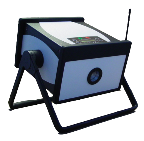
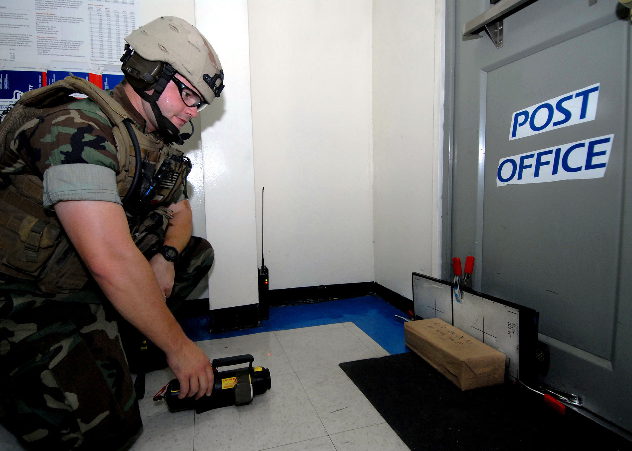
s are examined for possible weapons, including bombs. Prices of these Luggage X-rays vary from $50,000 to $300,000. The main parts of an X-ray Baggage Inspection System are the generator used to generate x-rays, the detector to detect radiation after passing through the baggage, signal processor unit (usually a PC) to process the incoming signal from the detector, and a conveyor system for moving baggage into the system. Portable pulsed X-ray Battery Powered X-ray Generator used in Security as shown in the figure provides EOD responders safer analysis of any possible target hazard.
Operation
When baggage is placed on the conveyor, it is moved into the machine by the operator. There is aninfrared
Infrared (IR), sometimes called infrared light, is electromagnetic radiation (EMR) with wavelengths longer than those of Light, visible light. It is therefore invisible to the human eye. IR is generally understood to encompass wavelengths from ...
transmitter and receiver assembly to detect the baggage when it enters the tunnel. This assembly gives the signal to switch on the generator and signal processing system. The signal processing system processes incoming signals from the detector and reproduce an image based upon the type of material and material density inside the baggage. This image is then sent to the display unit.
Color classification
The colour of the image displayed depends upon the material and material density : organic material such as paper, clothes and most explosives are displayed in orange. Mixed materials such as aluminum are displayed in green. Inorganic materials such as copper are displayed in blue and non-penetrable items are displayed in black (some machines display this as a yellowish green or red). The darkness of the color depends upon the density or thickness of the material. The material density determination is achieved by two-layer detector. The layers of the detector pixels are separated with a strip of metal. The metal absorbs soft rays, letting the shorter, more penetrating wavelengths through to the bottom layer of detectors, turning the detector to a crude two-band spectrometer.Advances in X-ray technology
 A film of
 carbon nanotube
A scanning tunneling microscopy image of a single-walled carbon nanotube
Rotating single-walled zigzag carbon nanotube
A carbon nanotube (CNT) is a tube made of carbon with diameters typically measured in nanometers.
''Single-wall carbon na ...
s (as a cathode) that emits electrons at room temperature when exposed to an electrical field has been fashioned into an X-ray device. An array of these emitters can be placed around a target item to be scanned and the images from each emitter can be assembled by computer software to provide a 3-dimensional image of the target in a fraction of the time it takes using a conventional X-ray device. The system also allows rapid, precise control, enabling prospective physiological gated imaging.
Engineers at the University of Missouri
The University of Missouri (Mizzou, MU, or Missouri) is a public land-grant research university in Columbia, Missouri. It is Missouri's largest university and the flagship of the four-campus University of Missouri System. MU was founded in ...
(MU), Columbia
Columbia may refer to:
* Columbia (personification), the historical female national personification of the United States, and a poetic name for America
Places North America Natural features
* Columbia Plateau, a geologic and geographic region i ...
, have invented a compact source of x-rays and other forms of radiation.
The radiation source is the size of a stick of gum and could be used to create portable x-ray scanners. A prototype handheld x-ray scanner using the source could be manufactured in as soon as three years.
See also
* Fluoroscope * Backscatter X-ray e.g., for security scanning passengers (rather than baggage) *X-ray crystallography
X-ray crystallography is the experimental science determining the atomic and molecular structure of a crystal, in which the crystalline structure causes a beam of incident X-rays to diffract into many specific directions. By measuring the angles ...
*Radiography
Radiography is an imaging technique using X-rays, gamma rays, or similar ionizing radiation and non-ionizing radiation to view the internal form of an object. Applications of radiography include medical radiography ("diagnostic" and "therapeu ...
* X-ray fluorescence
* X-ray astronomy (detectors)
Notes
References
# {{DEFAULTSORT:X-Ray Generator Generator Radiography Aviation security Explosive detection