Flexor Retinaculum Of The Hand on:
[Wikipedia]
[Google]
[Amazon]
The flexor retinaculum (transverse carpal ligament, or anterior annular ligament) is a
 In
In
fibrous
Fiber or fibre (from la, fibra, links=no) is a natural or artificial substance that is significantly longer than it is wide. Fibers are often used in the manufacture of other materials. The strongest engineering materials often incorporat ...
band on the palmar side of the hand near the wrist. It arches over the carpal bones
The carpal bones are the eight small bones that make up the wrist (or carpus) that connects the hand to the forearm. The term "carpus" is derived from the Latin carpus and the Greek καρπός (karpós), meaning "wrist". In human anatomy, t ...
of the hands, covering them and forming the carpal tunnel
In the human body, the carpal tunnel or carpal canal is the passageway on the palmar side of the wrist that connects the forearm to the hand.
The tunnel is bounded by the bones of the wrist and flexor retinaculum from connective tissue. Normall ...
.
Structure
The flexor retinaculum is a strong, fibrous band that covers the carpal bones on the palmar side of the hand near the wrist. It attaches to the bones near theradius
In classical geometry, a radius (plural, : radii) of a circle or sphere is any of the line segments from its Centre (geometry), center to its perimeter, and in more modern usage, it is also their length. The name comes from the latin ''radius'', ...
and ulna
The ulna (''pl''. ulnae or ulnas) is a long bone found in the forearm that stretches from the elbow to the smallest finger, and when in anatomical position, is found on the medial side of the forearm. That is, the ulna is on the same side of t ...
. On the ulnar side, the flexor retinaculum attaches to the pisiform bone
The pisiform bone ( or ), also spelled pisiforme (from the Latin ''pisifomis'', pea-shaped), is a small knobbly, sesamoid bone that is found in the wrist. It forms the ulnar border of the carpal tunnel.
Structure
The pisiform is a sesamoid bone, ...
and the hook of the hamate bone. On the radial side, it attaches to the tubercle of the scaphoid bone, and to the medial part of the palmar surface and the ridge of the trapezium bone
The trapezium bone (greater multangular bone) is a carpal bone in the hand. It forms the radial border of the carpal tunnel.
Structure
The trapezium is distinguished by a deep groove on its anterior surface. It is situated at the radial side o ...
.
The flexor retinaculum is continuous with the palmar carpal ligament, and deeper with the palmar aponeurosis. The ulnar artery and ulnar nerve
In human anatomy, the ulnar nerve is a nerve that runs near the ulna bone. The ulnar collateral ligament of elbow joint is in relation with the ulnar nerve. The nerve is the largest in the human body unprotected by muscle or bone, so injury is ...
, and the cutaneous branches of the median and ulnar nerve
In human anatomy, the ulnar nerve is a nerve that runs near the ulna bone. The ulnar collateral ligament of elbow joint is in relation with the ulnar nerve. The nerve is the largest in the human body unprotected by muscle or bone, so injury is ...
s, pass on top of the flexor retinaculum. On the radial side of the retinaculum is the tendon of the flexor carpi radialis, which lies in the groove on the greater multangular between the attachments of the ligament to the bone.
The tendons of the palmaris longus and flexor carpi ulnaris
The flexor carpi ulnaris (FCU) is a muscle of the forearm that flexes and adducts at the wrist joint.
Structure Origin
The flexor carpi ulnaris has two heads; a humeral head and ulnar head. The humeral head originates from the medial epicondyle of ...
are partly attached to the surface of the retinaculum; below, the short muscles of the thumb and little finger originate from the flexor retinaculum.
Function
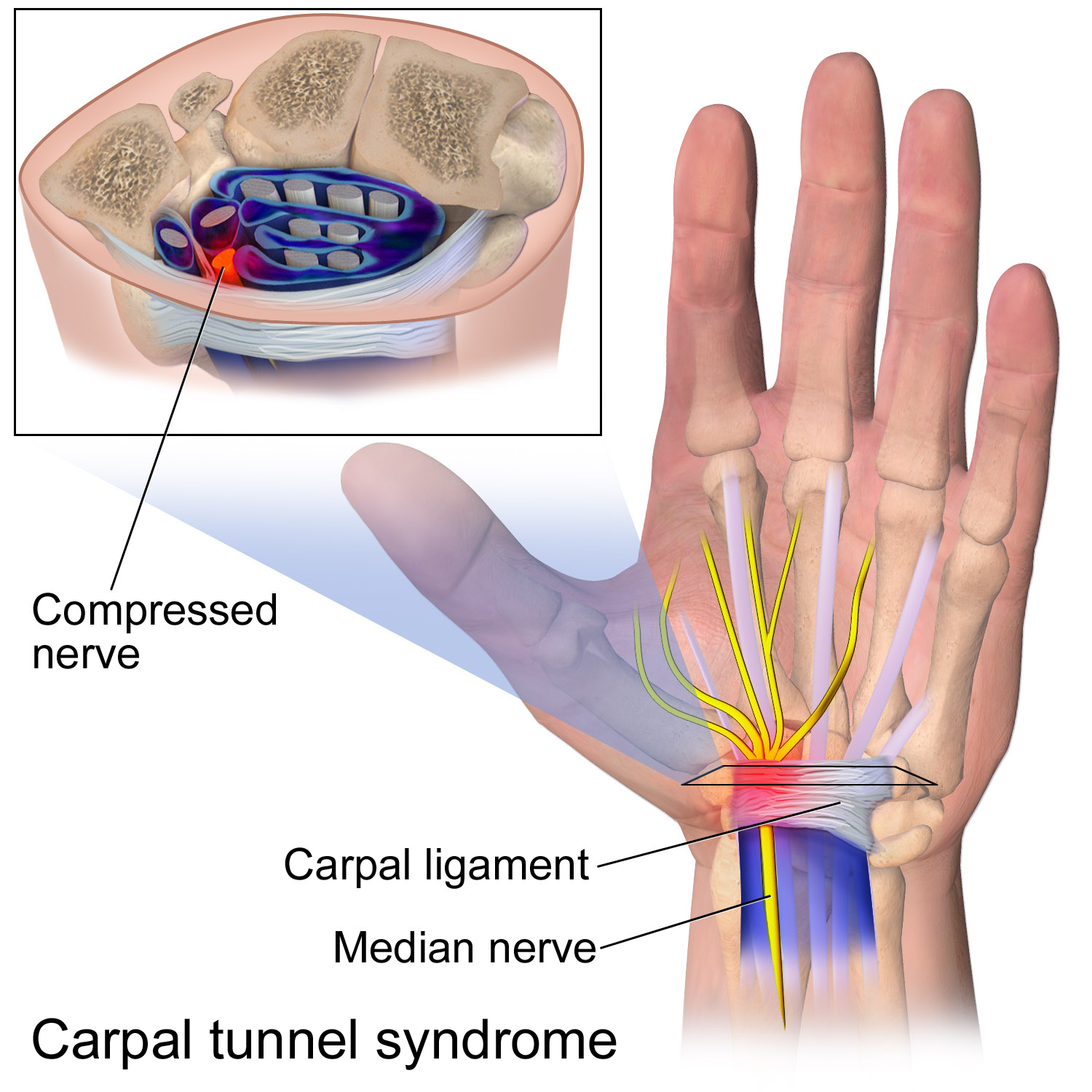
The flexor retinaculum is the roof of thecarpal tunnel
In the human body, the carpal tunnel or carpal canal is the passageway on the palmar side of the wrist that connects the forearm to the hand.
The tunnel is bounded by the bones of the wrist and flexor retinaculum from connective tissue. Normall ...
, through which the median nerve
The median nerve is a nerve in humans and other animals in the upper limb. It is one of the five main nerves originating from the brachial plexus.
The median nerve originates from the lateral and medial cords of the brachial plexus, and has cont ...
and tendons of muscles which flex the hand pass.
Clinical significance
 In
In carpal tunnel syndrome
Carpal tunnel syndrome (CTS) is the collection of symptoms and signs associated with median neuropathy at the carpal tunnel. Most CTS is related to idiopathic compression of the median nerve as it travels through the wrist at the carpal tunn ...
, one of the tendons or tissues in the carpal tunnel is inflamed, swollen, or fibrotic and puts pressure on the other structures in the tunnel, including the median nerve
The median nerve is a nerve in humans and other animals in the upper limb. It is one of the five main nerves originating from the brachial plexus.
The median nerve originates from the lateral and medial cords of the brachial plexus, and has cont ...
. Carpal tunnel syndrome is the most commonly reported nerve entrapment syndrome. It is often associated with repetitive motions of the wrist and fingers. It is because of this that pianists, meat cutters, and people with jobs involving extensive typing are at particularly high risk. The tough flexor retinaculum along with the rest of the carpal tunnel cannot expand, putting pressure on the median nerve running through the carpal tunnel with the flexor tendons of the wrist. This results in the symptoms of carpal tunnel syndrome.
Symptoms of carpal tunnel syndrome include tingling sensations and muscle weakness in the palm and lateral side of the hand and palm. It is possible that the syndrome may extend and radiate up the nerve causing pain to the arm and shoulder.
Carpal tunnel syndrome may be treated surgically. This is usually done after all non-surgical methods of treatment have been exhausted. Non-surgical treatment methods include anti-inflammatory drugs. The wrist may be immobilized in order to prevent further use and inflammation. When surgery is needed, the flexor retinaculum is either completely severed or lengthened. Surgery to divide the flexor retinaculum is the most common procedure. The scar tissue will eventually fill the gap left by surgery. The intent is that this will lengthen the flexor retinaculum enough to accommodate inflamed or damaged tendons and reduce the effects of compression on the median nerve. In a 2004 double blind-study, researchers concluded that there was no perceivable benefit gained from lengthening the flexor retinaculum during surgery and so division of the ligament remains the preferred method of surgery.
See also
*Peroneal retinacula
The fibular retinacula (also known as peroneal retinacula) are fibrous retaining bands that bind down the tendons of the fibularis longus and fibularis brevis muscles as they run across the side of the ankle. (''Retinaculum'' is Latin for "retaine ...
* Extensor retinaculum of the hand
References
External links
* * * {{Authority control Musculoskeletal system Hand Ligaments