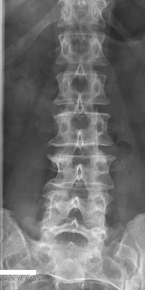
Block vertebrae on:
[Wikipedia]
[Google]
[Amazon]
Congenital vertebral anomalies are a collection of malformations of the
 ''Lumbarization'' is an anomaly in the
  ''Sacralization of the fifth lumbar vertebra'' (or ''sacralization'') is a
File:Blockwirbel degenerativ.jpg, Block vertebrae of the cervical spine (vertebrae 4 and 5). Probably based on degenerative or inflammatory changes.
File:Block- und Halbwirbel.jpg, Several congenital block vertebrae in the transition from the thoracic to the lumbar spine and hemivertebrae.
File:Partieller Blockwirbel.jpg, Congenital block vertebra in the lumbar spine (partial vertebrae 3 and 4). The rear portion of the disc still exists.
File:Blockwirbel CT VR frontal.jpg, Congenital block vertebra of the lumbar spine. CT volume rendering.
File:Blockwirbel CT VR seitlich.jpg, Congenital block vertebra of the lumbar spine. CT volume rendering.
File:Schmetterlingswirbel.jpg, Butterfly vertebra (red). Normal vertebra for comparison (blue).
File:Volume rendering of CT of butterfly vertebrae.jpg, Volume rendering of a CT scan of the lumbar vertebral column, showing butterfly vertebrae at several levels, most typically in L1.
spine
Spine or spinal may refer to:
Science Biology
* Vertebral column, also known as the backbone
* Dendritic spine, a small membranous protrusion from a neuron's dendrite
* Thorns, spines, and prickles, needle-like structures in plants
* Spine (zoolo ...
. Most, around 85%, are not clinically significant, but they can cause compression of the spinal cord by deforming the vertebral canal or causing instability. This condition occurs in the womb. Congenital vertebral anomalies include alterations of the shape and number of vertebra
The spinal column, a defining synapomorphy shared by nearly all vertebrates,Hagfish are believed to have secondarily lost their spinal column is a moderately flexible series of vertebrae (singular vertebra), each constituting a characteristic ...
e.
Lumbarization and sacralization
 ''Lumbarization'' is an anomaly in the
 spine
Spine or spinal may refer to:
Science Biology
* Vertebral column, also known as the backbone
* Dendritic spine, a small membranous protrusion from a neuron's dendrite
* Thorns, spines, and prickles, needle-like structures in plants
* Spine (zoolo ...
. It is defined by the nonfusion of the first and second segments of the sacrum. The lumbar spine subsequently appears to have six vertebrae
The spinal column, a defining synapomorphy shared by nearly all vertebrates,Hagfish are believed to have secondarily lost their spinal column is a moderately flexible series of vertebrae (singular vertebra), each constituting a characteristic i ...
or segments, not five. This sixth lumbar vertebra
The lumbar vertebrae are, in human anatomy, the five vertebrae between the rib cage and the pelvis. They are the largest segments of the vertebral column and are characterized by the absence of the foramen transversarium within the transverse ...
is known as a transitional vertebra. Conversely the sacrum appears to have only four segments instead of its designated five segments. Lumbosacral transitional vertebrae consist of the process of the last lumbar vertebra fusing with the first sacral segment
The spinal cord is a long, thin, tubular structure made up of nervous tissue, which extends from the medulla oblongata in the brainstem to the lumbar region of the vertebral column (backbone). The backbone encloses the central canal of the spina ...
. While only around 10 percent of adults have a spinal abnormality due to genetics, a sixth lumbar vertebra is one of the more common abnormalities. Dorland's Medical Dictionary
 ''Sacralization of the fifth lumbar vertebra'' (or ''sacralization'') is a
 congenital anomaly
A birth defect, also known as a congenital disorder, is an abnormal condition that is present at birth regardless of its cause. Birth defects may result in disabilities that may be physical, intellectual, or developmental. The disabilities can r ...
, in which the transverse process
The spinal column, a defining synapomorphy shared by nearly all vertebrates, Hagfish are believed to have secondarily lost their spinal column is a moderately flexible series of vertebrae (singular vertebra), each constituting a characteristic ...
of the last lumbar vertebra
The lumbar vertebrae are, in human anatomy, the five vertebrae between the rib cage and the pelvis. They are the largest segments of the vertebral column and are characterized by the absence of the foramen transversarium within the transverse ...
(L5) fuses to the sacrum on one side or both, or to ilium, or both. These anomalies are observed at about 3.5 percent of people, and it is usually bilateral but can be unilateral or incomplete ( ipsilateral or contralateral
Standard anatomical terms of location are used to unambiguously describe the anatomy of animals, including humans. The terms, typically derived from Latin or Greek roots, describe something in its standard anatomical position. This position prov ...
rudimentary facets) as well. Although sacralization may be a cause of low back pain, it is asymptomatic in many cases (especially bilateral type). Low back pain in these cases most likely occurs due to biomechanics
Biomechanics is the study of the structure, function and motion of the mechanical aspects of biological systems, at any level from whole organisms to organs, cells and cell organelles, using the methods of mechanics. Biomechanics is a branch of ...
. In sacralization, the L5-S1 intervertebral disc may be thin and narrow. This abnormality is found by X-ray
An X-ray, or, much less commonly, X-radiation, is a penetrating form of high-energy electromagnetic radiation. Most X-rays have a wavelength ranging from 10 picometers to 10 nanometers, corresponding to frequencies in the range 30 ...
.
Sacralization of L6 means L6 attaches to S1 via a rudimentary joint. This L6-S1 joint creates additional motion, increasing the potential for motion-related stress and lower back pain/conditions. This condition can usually be treated without surgery, injecting steroid medication at the pseudoarticulation instead. Additionally, if L6 fuses to another vertebra this is increasingly likely to cause lower back pain. The presence of a sixth vertebra in the space where five vertebrae normally reside also decreases the flexibility of the spine and increases the likelihood of injury.
Hemivertebrae
Hemivertebrae are wedge-shaped vertebrae and therefore can cause an angle in the spine (such askyphosis
Kyphosis is an abnormally excessive convex curvature of the spine as it occurs in the thoracic and sacral regions. Abnormal inward concave ''lordotic'' curving of the cervical and lumbar regions of the spine is called lordosis. It can result ...
, scoliosis, and lordosis
Lordosis is historically defined as an ''abnormal'' inward curvature of the lumbar spine. However, the terms ''lordosis'' and ''lordotic'' are also used to refer to the normal inward curvature of the lumbar and cervical regions of the human spi ...
).
Among the congenital vertebral anomalies, hemivertebrae are the most likely to cause neurologic
Neurology (from el, νεῦρον (neûron), "string, nerve" and the suffix -logia, "study of") is the branch of medicine dealing with the diagnosis and treatment of all categories of conditions and disease involving the brain, the spinal c ...
problems. The most common location is the mid thoracic vertebrae, especially the eighth (T8). Neurologic signs result from severe angulation of the spine, narrowing of the spinal canal, instability of the spine, and luxation or fracture of the vertebrae. Signs include rear limb weakness or paralysis, urinary or fecal incontinence, and spinal pain. Most cases of hemivertebrae have no or mild symptoms, so treatment is usually conservative. Severe cases may respond to surgical spinal cord decompression and vertebral stabilization.
Associations
Recognised associations are many and include:
Aicardi syndrome
Aicardi syndrome is a rare genetic malformation syndrome characterized by the partial or complete absence of a key structure in the brain called the corpus callosum, the presence of retinal lacunes, and epileptic seizures in the form of infan ...
,
cleidocranial dysostosis
Cleidocranial dysostosis (CCD), also called cleidocranial dysplasia, is a birth defect that mostly affects the bones and teeth. The collarbones are typically either poorly developed or absent, which allows the shoulders to be brought close togeth ...
,
gastroschisis
Gastroschisis is a birth defect in which the baby's intestines extend outside of the abdomen through a hole next to the belly button. The size of the hole is variable, and other organs including the stomach and liver may also occur outside the ...
3,
Gorlin syndrome Gorlin may refer to:
People
* Dan Gorlin, computer game programmer, designer and founder of Dan Gorlin Productions
* Eitan Gorlin, filmmaker, author and actor
* Mikhail Gorlin, Russian emigre poet
* Richard Gorlin, American cardiologist, co-devel ...
,
fetal pyelectasis 3,
Jarcho-Levin syndrome
Spondylocostal dysostosis, also known as Jarcho-Levin syndrome (JLS), is a rare, heritable axial skeleton growth disorder. It is characterized by widespread and sometimes severe malformations of the vertebral column and ribs, shortened thorax, a ...
,
OEIS complex
Cloacal exstrophy (EC) is a severe birth defect wherein much of the abdominal organs (the bladder and intestines) are exposed. It often causes the splitting of the bladder, genitalia, and the anus. It is sometimes called OEIS complex.
Diagnostic ...
,
VACTERL association
The VACTERL association (also VATER association, and less accurately VACTERL syndrome) refers to a recognized group of birth defects which tend to co-occur (see below). This pattern is a recognized association, as opposed to a syndrome, because t ...
.
The probable cause of hemivertebrae is a lack of blood supply causing part of the vertebrae not to form.
Hemivertebrae in dog
The dog (''Canis familiaris'' or ''Canis lupus familiaris'') is a domesticated descendant of the wolf. Also called the domestic dog, it is derived from the extinct Pleistocene wolf, and the modern wolf is the dog's nearest living relative. ...
s are most common in the tail, resulting in a screw shape.
Block vertebrae
Block vertebrae occur when there is improper segmentation of the vertebrae, leading to parts of or the entire vertebrae being fused. The adjacent vertebrae fuse through their intervertebral discs and also through other intervertebral joints so that it can lead to blocking or stretching of the exiting nerve roots from that segment. It may lead to certain neurological problems depending on the severity of the block. It can increase stress on the inferior and the superior intervertebral joints. It can lead to an abnormal angle in the spine, there are certain syndromes associated with block vertebrae; for example,Klippel–Feil syndrome
Klippel–Feil syndrome (KFS), also known as cervical vertebral fusion syndrome, is a rare congenital condition characterized by the abnormal fusion of any two of the seven bones in the neck (cervical vertebrae). It results in a limited ability ...
. The sacrum is a normal block vertebra.
Fossil record
Evidence for block vertebrae found in the fossil record is studied bypaleopathologists
Paleopathology, also spelled palaeopathology, is the study of ancient diseases and injuries in organisms through the examination of fossils, mummified tissue, skeletal remains, and analysis of coprolites. Specific sources in the study of ancient ...
, specialists in ancient disease and injury. A block vertebra has been documented in ''T. rex''. This suggests that the basic development pattern of vertebrae goes at least as far back as the most recent common ancestor of archosaurs
Archosauria () is a clade of diapsids, with birds and crocodilians as the only living representatives. Archosaurs are broadly classified as reptiles, in the cladistic sense of the term which includes birds. Extinct archosaurs include non-avian ...
and mammals. The tyrannosaur's block vertebra was probably caused by a "failure of the resegmentation of the sclerotome
The somites (outdated term: primitive segments) are a set of bilaterally paired blocks of paraxial mesoderm that form in the embryonic stage of somitogenesis, along the head-to-tail axis in segmented animals. In vertebrates, somites subdivide in ...
s".Molnar, R. E., 2001, Theropod paleopathology: a literature survey: In: Mesozoic Vertebrate Life, edited by Tanke, D. H., and Carpenter, K., Indiana University Press, p. 337-363.
Butterfly vertebrae
Butterfly vertebrae have a sagittal cleft through the body of the vertebrae and a funnel shape at the ends. This gives the appearance of abutterfly
Butterflies are insects in the macrolepidopteran clade Rhopalocera from the order Lepidoptera, which also includes moths. Adult butterflies have large, often brightly coloured wings, and conspicuous, fluttering flight. The group comprises ...
on an x-ray
An X-ray, or, much less commonly, X-radiation, is a penetrating form of high-energy electromagnetic radiation. Most X-rays have a wavelength ranging from 10 picometers to 10 nanometers, corresponding to frequencies in the range 30 ...
. It is caused by persistence of the notochord (which usually only remains as the center of the intervertebral disc) during vertebrae formation. There are usually no symptoms. There are also coronal clefts mainly in skeletal dysplasias such as chondrodysplasia punctata. In dogs, butterfly vertebrae occur most often in Bulldogs, Pugs, and Boston Terriers.
Transitional vertebrae
Transitional vertebrae have the characteristics of two types of vertebra. The condition usually involves the vertebral arch ortransverse processes
The spinal column, a defining synapomorphy shared by nearly all vertebrates,Hagfish are believed to have secondarily lost their spinal column is a moderately flexible series of vertebrae (singular vertebra), each constituting a characteristic i ...
. It occurs at the cervicothoracic, thoracolumbar, or lumbosacral junction. For instance, the transverse process of the last cervical vertebra
In tetrapods, cervical vertebrae (singular: vertebra) are the vertebrae of the neck, immediately below the skull. Truncal vertebrae (divided into thoracic and lumbar vertebrae in mammals) lie caudal (toward the tail) of cervical vertebrae. In sau ...
may resemble a rib. A transitional vertebra at the lumbosacral junction can cause arthritis
Arthritis is a term often used to mean any disorder that affects joints. Symptoms generally include joint pain and stiffness. Other symptoms may include redness, warmth, swelling, and decreased range of motion of the affected joints. In some ...
, disk changes, or thecal sac compression. Back pain associated with lumbosacral transitional vertebrae (LSTV) is known as Bertolotti's syndrome. One study found that male German Shepherd Dogs with a lumbosacral transitional vertebra are at greater risk for cauda equina syndrome
Cauda equina syndrome (CES) is a condition that occurs when the bundle of nerves below the end of the spinal cord known as the cauda equina is damaged. Signs and symptoms include low back pain, pain that radiates down the leg, numbness around th ...
, which can cause rear limb weakness and incontinence.
The significance of transitional vertebrae has been questioned by one study finding similar prevalence in the general population as those with low back pain, but more recent study found a large difference.
Spina bifida
Spina bifida is characterized by a midline cleft in the vertebral arch. It usually causes no symptoms in dogs. It is seen most commonly in Bulldogs and Manx cats. In Manx it accompanies a condition known as sacrocaudal dysgenesis that gives these cats their characteristic tailless or stumpy tail appearance. It is inherited in Manx as anautosomal dominant
In genetics, dominance is the phenomenon of one variant (allele) of a gene on a chromosome masking or overriding the effect of a different variant of the same gene on the other copy of the chromosome. The first variant is termed dominant and ...
trait.
Associations
Vertebral anomalies is associated with an increased incidence of some other specific anomalies as well, together being called theVACTERL association
The VACTERL association (also VATER association, and less accurately VACTERL syndrome) refers to a recognized group of birth defects which tend to co-occur (see below). This pattern is a recognized association, as opposed to a syndrome, because t ...
:
* V - ''Vertebral anomalies''
* A - Anal atresia
An imperforate anus or anorectal malformations (ARMs) are birth defects in which the rectum is malformed. ARMs are a spectrum of different congenital anomalies which vary from fairly minor lesions to complex anomalies. The cause of ARMs is unknow ...
* C - Cardiovascular anomalies
Cardiology () is a branch of medicine that deals with disorders of the heart and the cardiovascular system. The field includes medical diagnosis and treatment of congenital heart defects, coronary artery disease, heart failure, valvular heart d ...
* T - Tracheoesophageal fistula
A tracheoesophageal fistula (TEF, or TOF; see spelling differences) is an abnormal connection ( fistula) between the esophagus and the trachea. TEF is a common congenital abnormality, but when occurring late in life is usually the sequela of surg ...
* E - Esophageal atresia Esophageal can refer to:
* The esophagus
* Esophageal arteries
* Esophageal glands
* Esophageal cancer
Esophageal cancer is cancer arising from the esophagus—the food pipe that runs between the throat and the stomach. Symptoms often includ ...
* R - Renal (Kidney) and/or radial anomalies
* L - Limb defects
References
External links
{{Congenital malformations and deformations of musculoskeletal system Dog musculoskeletal disorders Congenital disorders of musculoskeletal system