|
Vesicular Glands
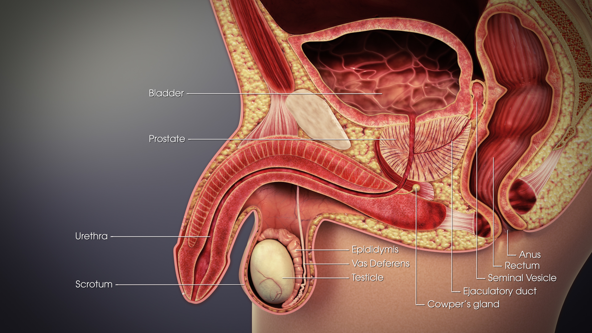
The seminal vesicles (also called vesicular glands, or seminal glands) are a pair of two convoluted tubular glands that lie behind the urinary bladder of some male mammals. They secrete fluid that partly composes the semen. The vesicles are 5–10 cm in size, 3–5 cm in diameter, and are located between the bladder and the rectum. They have multiple outpouchings which contain secretory glands, which join together with the vas deferens at the ejaculatory duct. They receive blood from the vesiculodeferential artery, and drain into the vesiculodeferential veins. The glands are lined with column-shaped and cuboidal cells. The vesicles are present in many groups of mammals, but not marsupials, monotremes or carnivores. Inflammation of the seminal vesicles is called seminal vesiculitis, most often is due to bacterial infection as a result of a sexually transmitted disease or following a surgical procedure. Seminal vesiculitis can cause pain in the lower abdomen, scro ... [...More Info...] [...Related Items...] OR: [Wikipedia] [Google] [Baidu] |
Abdomen
The abdomen (colloquially called the belly, tummy, midriff, tucky or stomach) is the part of the body between the thorax (chest) and pelvis, in humans and in other vertebrates. The abdomen is the front part of the abdominal segment of the torso. The area occupied by the abdomen is called the abdominal cavity. In arthropods it is the posterior tagma of the body; it follows the thorax or cephalothorax. In humans, the abdomen stretches from the thorax at the thoracic diaphragm to the pelvis at the pelvic brim. The pelvic brim stretches from the lumbosacral joint (the intervertebral disc between L5 and S1) to the pubic symphysis and is the edge of the pelvic inlet. The space above this inlet and under the thoracic diaphragm is termed the abdominal cavity. The boundary of the abdominal cavity is the abdominal wall in the front and the peritoneal surface at the rear. In vertebrates, the abdomen is a large body cavity enclosed by the abdominal muscles, at front and to ... [...More Info...] [...Related Items...] OR: [Wikipedia] [Google] [Baidu] |
Vesiculodeferential Vein
The seminal vesicles (also called vesicular glands, or seminal glands) are a pair of two convoluted tubular glands that lie behind the urinary bladder of some male mammals. They secrete fluid that partly composes the semen. The vesicles are 5–10 cm in size, 3–5 cm in diameter, and are located between the bladder and the rectum. They have multiple outpouchings which contain secretory glands, which join together with the vas deferens at the ejaculatory duct. They receive blood from the vesiculodeferential artery, and drain into the vesiculodeferential veins. The glands are lined with column-shaped and cuboidal cells. The vesicles are present in many groups of mammals, but not marsupials, monotremes or carnivores. Inflammation of the seminal vesicles is called seminal vesiculitis, most often is due to bacterial infection as a result of a sexually transmitted disease or following a surgical procedure. Seminal vesiculitis can cause pain in the lower abdomen, scrotu ... [...More Info...] [...Related Items...] OR: [Wikipedia] [Google] [Baidu] |
Internal Iliac Arteries
The internal iliac artery (formerly known as the hypogastric artery) is the main artery of the pelvis. Structure The internal iliac artery supplies the walls and viscera of the pelvis, the buttock, the reproductive organs, and the medial compartment of the thigh. The vesicular branches of the internal iliac arteries supply the bladder. It is a short, thick vessel, smaller than the external iliac artery, and about 3 to 4 cm in length. Course The internal iliac artery arises at the bifurcation of the common iliac artery, opposite the lumbosacral articulation, and, passing downward to the upper margin of the greater sciatic foramen, divides into two large trunks, an anterior and a posterior. It is posterior to the ureter, anterior to the internal iliac vein, anterior to the lumbosacral trunk, and anterior to the piriformis muscle. Near its origin, it is medial to the external iliac vein, which lies between it and the psoas major muscle. It is above the obturator nerve. ... [...More Info...] [...Related Items...] OR: [Wikipedia] [Google] [Baidu] |
Umbilical Arteries
The umbilical artery is a paired artery (with one for each half of the body) that is found in the abdominal and pelvic regions. In the fetus, it extends into the umbilical cord. Structure Development The umbilical arteries supply deoxygenated blood from the fetus to the placenta. Although this blood is typically referred to as deoxygenated, this blood is fetal systemic arterial blood and will have the same amount of oxygen and nutrients as blood distributed to the other fetal tissues. There are usually two umbilical arteries present together with one umbilical vein in the umbilical cord. The umbilical arteries surround the urinary bladder and then carry all the deoxygenated blood out of the fetus through the umbilical cord. Inside the placenta, the umbilical arteries connect with each other at a distance of approximately 5 mm from the cord insertion in what is called the ''Hyrtl anastomosis''. Subsequently, they branch into chorionic arteries or ''intraplacental fetal art ... [...More Info...] [...Related Items...] OR: [Wikipedia] [Google] [Baidu] |
Inferior Vesical Artery
The inferior vesical artery (or inferior vesicle artery) is an artery of the pelvis which arises from the internal iliac artery and supplies parts of the urinary bladder as well as other structures of the urinary system and structures of the male reproductive system. Some sources consider this vessel to be present only in males, and cite the vaginal artery as the homologous structure in females; others consider it to be present in both sexes, with the vessel taking the form of a small branch of a vaginal artery in females. Structure Origin The inferior vesical artery is a branch of the anterior division of the internal iliac artery. It frequently has a common origin with the middle rectal artery. Course The inferior vesical artery passes medially across the pelvic floor. Distribution The inferior vesical artery is distributed to the trigone and inferior portion of the urinary bladder, the ureter, prostate, vas deferens, and seminal vesicles.vas deferens The vas defe ... [...More Info...] [...Related Items...] OR: [Wikipedia] [Google] [Baidu] |
Urethra
The urethra (from Greek οὐρήθρα – ''ourḗthrā'') is a tube that connects the urinary bladder to the urinary meatus for the removal of urine from the body of both females and males. In human females and other primates, the urethra connects to the urinary meatus above the vagina, whereas in marsupials, the female's urethra empties into the urogenital sinus. Females use their urethra only for urinating, but males use their urethra for both urination and ejaculation. The external urethral sphincter is a striated muscle that allows voluntary control over urination. The internal sphincter, formed by the involuntary smooth muscles lining the bladder neck and urethra, receives its nerve supply by the sympathetic division of the autonomic nervous system. The internal sphincter is present both in males and females. Structure The urethra is a fibrous and muscular tube which connects the urinary bladder to the external urethral meatus. Its length differs between ... [...More Info...] [...Related Items...] OR: [Wikipedia] [Google] [Baidu] |
Seminal Colliculus
The seminal colliculus (Latin ''colliculus seminalis''), or verumontanum, of the prostatic urethra is a landmark distal to the entrance of the ejaculatory ducts (on both sides, corresponding vas deferens and seminal vesicle feed into corresponding ejaculatory duct). ''Verumontanum'' is translated from Latin to mean 'mountain ridge', a reference to the distinctive median elevation of urothelium that characterizes the landmark on magnified views. Embryologically, it is derived from the uterovaginal primordium. The landmark is important in classification of several urethral developmental disorders. The margins of seminal colliculus are the following: * the orifices of the prostatic utricle * the slit-like openings of the ejaculatory ducts. * the openings of the prostatic ducts Posterior urethral valves The verumontanum is an important anatomic landmark for pathology in a congenital anomaly known as posterior urethral valves, in which there is a developmental obstruction of th ... [...More Info...] [...Related Items...] OR: [Wikipedia] [Google] [Baidu] |
Vasa Deferentia
The vas deferens or ductus deferens is part of the male reproductive system of many vertebrates. The ducts transport sperm from the epididymis to the ejaculatory ducts in anticipation of ejaculation. The vas deferens is a partially coiled tube which exits the abdominal cavity through the inguinal canal. Etymology ''Vas deferens'' is Latin, meaning "carrying-away vessel"; the plural version is ''vasa deferentia''. ''Ductus deferens'' is also Latin, meaning "carrying-away duct"; the plural version is ''ducti deferentes''. Structure There are two vasa deferentia, connecting the left and right epididymis with the seminal vesicles to form the ejaculatory duct in order to move sperm. The (human) vas deferens measures 30–35 cm in length, and 2–3 mm in diameter. The vas deferens is continuous proximally with the tail of the epididymis. The vas deferens exhibits a tortuous, convoluted initial/proximal section (which measures 2–3 cm in length). Distally, it forms ... [...More Info...] [...Related Items...] OR: [Wikipedia] [Google] [Baidu] |
Galen
Aelius Galenus or Claudius Galenus ( el, Κλαύδιος Γαληνός; September 129 – c. AD 216), often Anglicized as Galen () or Galen of Pergamon, was a Greek physician, surgeon and philosopher in the Roman Empire. Considered to be one of the most accomplished of all medical researchers of antiquity, Galen influenced the development of various scientific disciplines, including anatomy, physiology, pathology, pharmacology, and neurology, as well as philosophy and logic. The son of Aelius Nicon, a wealthy Greek architect with scholarly interests, Galen received a comprehensive education that prepared him for a successful career as a physician and philosopher. Born in the ancient city of Pergamon (present-day Bergama, Turkey), Galen traveled extensively, exposing himself to a wide variety of medical theories and discoveries before settling in Rome, where he served prominent members of Roman society and eventually was given the position of personal physician to sever ... [...More Info...] [...Related Items...] OR: [Wikipedia] [Google] [Baidu] |
Haematospermia
Hematospermia (also known as haematospermia, hemospermia, or haemospermia) is the presence of blood in ejaculation. It is most often a benign symptom. Among men age 40 or older, hematospermia is a slight predictor of cancer, typically prostate cancer. No specific cause is found in up to 70% of cases. Cause Though haematospermia may cause considerable distress to patients, it is often a benign and self-limiting condition caused by infections, particularly in younger patients. An isolated episode is usually considered benign and not likely to be associated with malignancy. Recurrent haematospermia may indicate a more serious underlying pathology particularly in patients over 40 years of age. Infection and inflammation Infection or inflammation is considered the most common cause of the condition. Implicated pathogens include; Gram-negative bacteria (often ''E. coli''), gonococci, ''T. pallidum'', '' C. trachomatis'', ''N. gonorrhoeae,'' echinococcus (rarely), HSV type 1 or 2, an ... [...More Info...] [...Related Items...] OR: [Wikipedia] [Google] [Baidu] |
Ejaculation
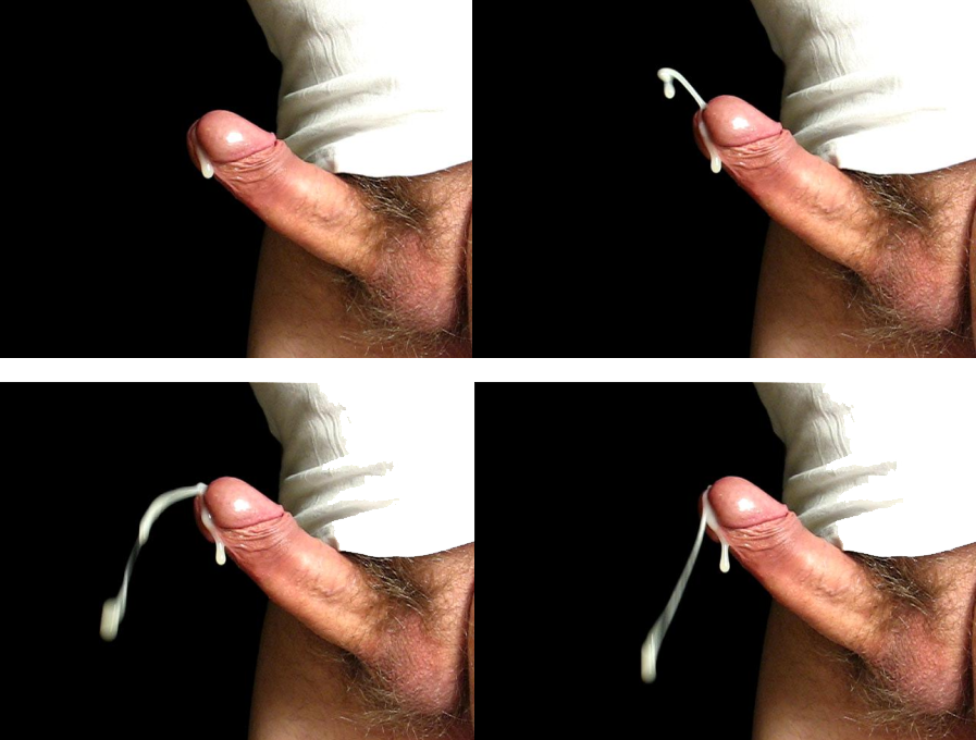
Ejaculation is the discharge of semen (the ''ejaculate''; normally containing sperm) from the male reproductory tract as a result of an orgasm. It is the final stage and natural objective of male sexual stimulation, and an essential component of natural conception. In rare cases, ejaculation occurs because of prostatic disease. Ejaculation may also occur spontaneously during sleep (a nocturnal emission or "wet dream"). '' Anejaculation'' is the condition of being unable to ejaculate. Ejaculation is usually very pleasurable for men; '' dysejaculation'' is an ejaculation that is painful or uncomfortable. Retrograde ejaculation is the condition where semen travels backwards into the bladder rather than out the urethra. Phases Stimulation A usual precursor to ejaculation is the sexual arousal of the male, leading to the erection of the penis, though not every arousal nor erection leads to ejaculation. Penile sexual stimulation during masturbation or vaginal, anal ... [...More Info...] [...Related Items...] OR: [Wikipedia] [Google] [Baidu] |
Peritoneum
The peritoneum is the serous membrane forming the lining of the abdominal cavity or coelom in amniotes and some invertebrates, such as annelids. It covers most of the intra-abdominal (or coelomic) organs, and is composed of a layer of mesothelium supported by a thin layer of connective tissue. This peritoneal lining of the cavity supports many of the abdominal organs and serves as a conduit for their blood vessels, lymphatic vessels, and nerves. The abdominal cavity (the space bounded by the vertebrae, abdominal muscles, diaphragm, and pelvic floor) is different from the intraperitoneal space (located within the abdominal cavity but wrapped in peritoneum). The structures within the intraperitoneal space are called "intraperitoneal" (e.g., the stomach and intestines), the structures in the abdominal cavity that are located behind the intraperitoneal space are called " retroperitoneal" (e.g., the kidneys), and those structures below the intraperitoneal space are calle ... [...More Info...] [...Related Items...] OR: [Wikipedia] [Google] [Baidu] |


_-_Veloso_Salgado.png)
