|
Venae Comitantes
Vena comitans (Latin for accompanying vein, also known as a satellite vein) refers to a vein that is usually paired, with both veins lying on the sides of an artery. Because they are generally found in pairs, they are often referred to by their plural form: venae comitantes. Venae comitantes are usually found with certain smaller arteries, especially those in the extremities. Larger arteries, on the other hand, generally do not have venae comitantes. They usually have a single, similarly sized vein which is not as intimately associated with the artery. Function As the vein is found in close proximity to an artery the pulsations of the artery aid venous return. Claude Bernard suggested the interchange of heat between the arteries and adjacent veins might moderate cooling of the arterial blood, for which there is experimental evidence. Examples Examples of arteries and their venae comitantes: * Radial artery and radial veins * Ulnar artery and ulnar veins * Brachial artery a ... [...More Info...] [...Related Items...] OR: [Wikipedia] [Google] [Baidu] |
Deep Vein
A deep vein is a vein that is deep in the body. This contrasts with superficial veins that are close to the body's surface. Deep veins are almost always beside an artery with the same name (e.g. the femoral vein is beside the femoral artery). Collectively, they carry the vast majority of the blood. Occlusion of a deep vein can be life-threatening and is most often caused by thrombosis. Occlusion of a deep vein by thrombosis is called ''deep vein thrombosis''. Because of their location deep within the body, operation on these veins can be difficult. List *Internal jugular vein Upper limb * Brachial vein * Axillary vein *Subclavian vein Lower limb *Common femoral vein *Femoral vein * Profunda femoris vein * Popliteal vein * Peroneal vein * Anterior tibial vein *Posterior tibial vein The posterior tibial veins are veins of the leg in humans. They drain the posterior compartment of the leg and the plantar surface of the foot to the popliteal vein. Structure The poste ... [...More Info...] [...Related Items...] OR: [Wikipedia] [Google] [Baidu] |
Anterior Tibial Artery
The anterior tibial artery is an artery of the leg. It carries blood to the anterior compartment of the leg and dorsal surface of the foot, from the popliteal artery. Structure Course The anterior tibial artery is a branch of the popliteal artery. It originates at the distal end of the popliteus muscle posterior to the tibia. The artery typically passes anterior to the popliteus muscle prior to passing between the tibia and fibula through an oval opening at the superior aspect of the interosseus membrane. The artery then descends between the tibialis anterior and extensor digitorum longus muscles. It is accompanied by the anterior tibial vein, and the deep peroneal nerve, along its course. It crosses the anterior aspect of the ankle joint, at which point it becomes the dorsalis pedis artery. Branches The branches of the anterior tibial artery are: * posterior tibial recurrent artery * anterior tibial recurrent artery * muscular branches * anterior medial malleolar ar ... [...More Info...] [...Related Items...] OR: [Wikipedia] [Google] [Baidu] |
Subclavian Artery
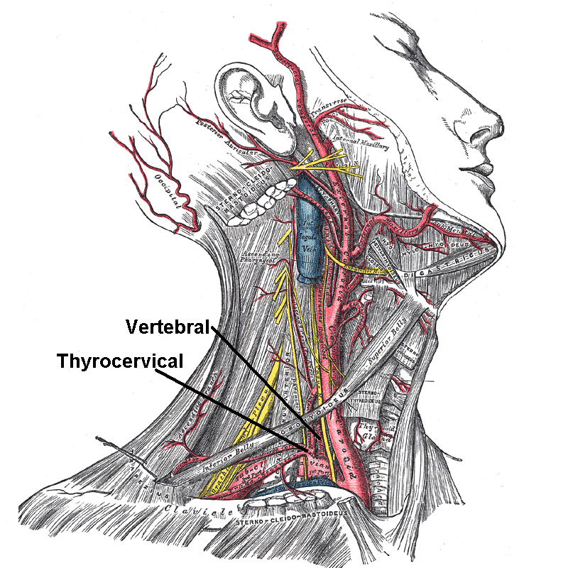
In human anatomy, the subclavian arteries are paired major arteries of the upper thorax, below the clavicle. They receive blood from the aortic arch. The left subclavian artery supplies blood to the left arm and the right subclavian artery supplies blood to the right arm, with some branches supplying the head and thorax. On the left side of the body, the subclavian comes directly off the aortic arch, while on the right side it arises from the relatively short brachiocephalic artery when it bifurcates into the subclavian and the right common carotid artery. The usual branches of the subclavian on both sides of the body are the vertebral artery, the internal thoracic artery, the thyrocervical trunk, the costocervical trunk and the dorsal scapular artery, which may branch off the transverse cervical artery, which is a branch of the thyrocervical trunk. The subclavian becomes the axillary artery at the lateral border of the first rib. Structure From its origin, the subclavian art ... [...More Info...] [...Related Items...] OR: [Wikipedia] [Google] [Baidu] |
Axillary Vein
In human anatomy, the axillary vein is a large blood vessel that conveys blood from the lateral aspect of the thorax, axilla (armpit) and upper limb toward the heart. There is one axillary vein on each side of the body. Structure Its origin is at the lower margin of the teres major muscle and a continuation of the brachial vein. This large vein is formed by the brachial vein and the basilic vein. At its terminal part, it is also joined by the cephalic vein. Other tributaries include the subscapular vein, circumflex humeral vein, lateral thoracic vein and thoraco-acromial vein. It terminates at the lateral margin of the first rib, at which it becomes the subclavian vein. It is accompanied along its course by a similarly named artery, the axillary artery In human anatomy, the axillary artery is a large blood vessel that conveys oxygenated blood to the lateral aspect of the thorax, the axilla (armpit) and the upper limb. Its origin is at the lateral margin of the fi ... [...More Info...] [...Related Items...] OR: [Wikipedia] [Google] [Baidu] |
Axillary Artery
In human anatomy, the axillary artery is a large blood vessel that conveys oxygenated blood to the lateral aspect of the thorax, the axilla (armpit) and the upper limb. Its origin is at the lateral margin of the first rib, before which it is called the subclavian artery. After passing the lower margin of teres major muscle, teres major it becomes the brachial artery. Structure The axillary artery is often referred to as having three parts, with these divisions based on its location relative to the pectoralis minor muscle, which is superficial to the artery. * First part – the part of the artery superior to the pectoralis minor * Second part – the part of the artery posterior to the pectoralis minor * Third part – the part of the artery inferior to the pectoralis minor. Relations The axillary artery is accompanied by the axillary vein, which lies medial to the artery, along its length. In the axilla, the axillary artery is surrounded by the brachial plexus. The second ... [...More Info...] [...Related Items...] OR: [Wikipedia] [Google] [Baidu] |
Fibular Veins
In anatomy, the fibular veins (also known as peroneal veins) are accompanying veins (venae comitantes) of the fibular artery. Structure The fibular veins are deep veins that help carry blood from the lateral compartment of the leg. They drain into the posterior tibial veins, which in turn drain into the popliteal vein. The fibular veins accompany the fibular artery. See also * Fibular artery * Common fibular nerve * Venae comitantes Additional images File:Gray440_color.png, Cross-section through middle of leg. File:Ultrasonography of thrombosis of the fibular veins, coronal plane, annotated.jpg, Coronal plane The dorsal plane (also known as the coronal plane or frontal plane, especially in human anatomy) is an anatomical plane that divides the body into Anatomical terms of location#Dorsal and ventral, dorsal and ventral sections. It is perpendicular t ... (seen from medial side of lower leg) ultrasonography of deep vein thrombosis of the fibular veins, seen as hypere ... [...More Info...] [...Related Items...] OR: [Wikipedia] [Google] [Baidu] |
Fibular Artery
In anatomy, the fibular artery, also known as the peroneal artery, supplies blood to the lateral compartment of the leg. It arises from the tibial-fibular trunk. Structure The fibular artery arises from the bifurcation of tibial-fibular trunk into the fibular and posterior tibial arteries in the upper part of the leg proper, just below the knee. It runs towards the foot in the deep posterior compartment of the leg, just medial to the fibula. It supplies a perforating branch to both the lateral and anterior compartments of the leg; it also provides a nutrient artery to the fibula. Some sources claim that the fibular artery arises directly from the posterior tibial artery, but vascular and plastic surgeons note the clinical significance of the tibial-fibular trunk. The fibular artery is accompanied by small veins ( venae comitantes) known as fibular veins. Branches Communication branch to posterior tibial artery. Perforating branch to anterior lateral malleolar artery. A ... [...More Info...] [...Related Items...] OR: [Wikipedia] [Google] [Baidu] |
Posterior Tibial Veins
The posterior tibial veins are veins of the leg in humans. They drain the posterior compartment of the leg and the plantar surface of the foot to the popliteal vein. Structure The posterior tibial veins receive blood from the medial and lateral plantar veins. They drain the posterior compartment of the leg and the plantar surface of the foot to the popliteal vein, which it forms when it joins with the anterior tibial vein. The posterior tibial vein is accompanied by an homonym artery, the posterior tibial artery, along its course. It lies posterior to the medial malleolus in the ankle. They receive the most important perforator vein Perforator veins are so called because they perforate the deep fascia of muscles, to connect the superficial veins to the deep veins where they drain. Perforator veins play an essential role in maintaining normal blood draining. They have venous ...s: the Cockett perforators, superior, medial and inferior. Additional images File:Gray440_col ... [...More Info...] [...Related Items...] OR: [Wikipedia] [Google] [Baidu] |
Posterior Tibial Artery
The posterior tibial artery of the lower limb is an artery that carries blood to the posterior compartment of the leg and plantar surface of the foot. It branches from the popliteal artery via the tibial-fibular trunk. Structure The posterior tibial artery arises from the popliteal artery in the popliteal fossa. It is accompanied by a deep vein, the posterior tibial vein, along its course. It passes just posterior to the medial malleolus of the tibia, but anterior to the Achilles tendon. It passes into the foot deep to the flexor retinaculum of the foot. It runs through the tarsal tunnel. Branches The posterior tibial artery gives rise to: * medial plantar artery. * lateral plantar artery. * fibular artery, which is said to rise from the bifurcation of the tibial-fibular trunk and the posterior tibial artery. * calcaneal branch to the medial aspect of the calcaneus. Function The posterior tibial artery supplies oxygenated blood to the posterior compartment of th ... [...More Info...] [...Related Items...] OR: [Wikipedia] [Google] [Baidu] |
Anterior Tibial Veins
The anterior tibial vein is a vein in the lower leg. In human anatomy, there are two anterior tibial veins. They originate and receive blood from the dorsal venous arch, on the back of the foot and empties into the popliteal vein. The anterior tibial veins drain the ankle joint, knee joint, tibiofibular joint, and the anterior portion of the lower leg. The two anterior tibial veins ascend in the interosseous membrane between the tibia and fibula and unite with the posterior tibial veins to form the popliteal vein. Like most deep veins in legs, anterior tibial veins are accompanied by the homonym artery, the anterior tibial artery The anterior tibial artery is an artery of the leg. It carries blood to the anterior compartment of the leg and dorsal surface of the foot, from the popliteal artery. Structure Course The anterior tibial artery is a branch of the popliteal ..., along its course. References Veins of the lower limb {{Circulatory-stub ... [...More Info...] [...Related Items...] OR: [Wikipedia] [Google] [Baidu] |
Brachial Veins
In human anatomy, the brachial veins are venae comitantes of the brachial artery in the arm proper. Because they are deep to muscle, they are considered deep veins. Their course is that of the brachial artery (in reverse): they begin where radial veins and ulnar veins join (corresponding to the bifurcation of the brachial artery). They end at the inferior border of the teres major muscle. At this point, the brachial veins join the basilic vein to form the axillary vein. The brachial veins also have small tributaries that drain the muscles of the upper arm, such as biceps brachii muscle and triceps brachii muscle. Additional images File:Slide8UUU.JPG, Brachial vein File:Gray576.png, The veins of the right axilla The axilla (: axillae or axillas; also known as the armpit, underarm or oxter) is the area on the human body directly under the shoulder joint. It includes the axillary space, an anatomical space within the shoulder girdle between the arm a ..., view ... [...More Info...] [...Related Items...] OR: [Wikipedia] [Google] [Baidu] |
Elsevier
Elsevier ( ) is a Dutch academic publishing company specializing in scientific, technical, and medical content. Its products include journals such as ''The Lancet'', ''Cell (journal), Cell'', the ScienceDirect collection of electronic journals, ''Trends (journals), Trends'', the ''Current Opinion (Elsevier), Current Opinion'' series, the online citation database Scopus, the SciVal tool for measuring research performance, the ClinicalKey search engine for clinicians, and the ClinicalPath evidence-based cancer care service. Elsevier's products and services include digital tools for Data management platform, data management, instruction, research analytics, and assessment. Elsevier is part of the RELX Group, known until 2015 as Reed Elsevier, a publicly traded company. According to RELX reports, in 2022 Elsevier published more than 600,000 articles annually in over 2,800 journals. As of 2018, its archives contained over 17 million documents and 40,000 Ebook, e-books, with over one b ... [...More Info...] [...Related Items...] OR: [Wikipedia] [Google] [Baidu] |

