|
Umbilical Line
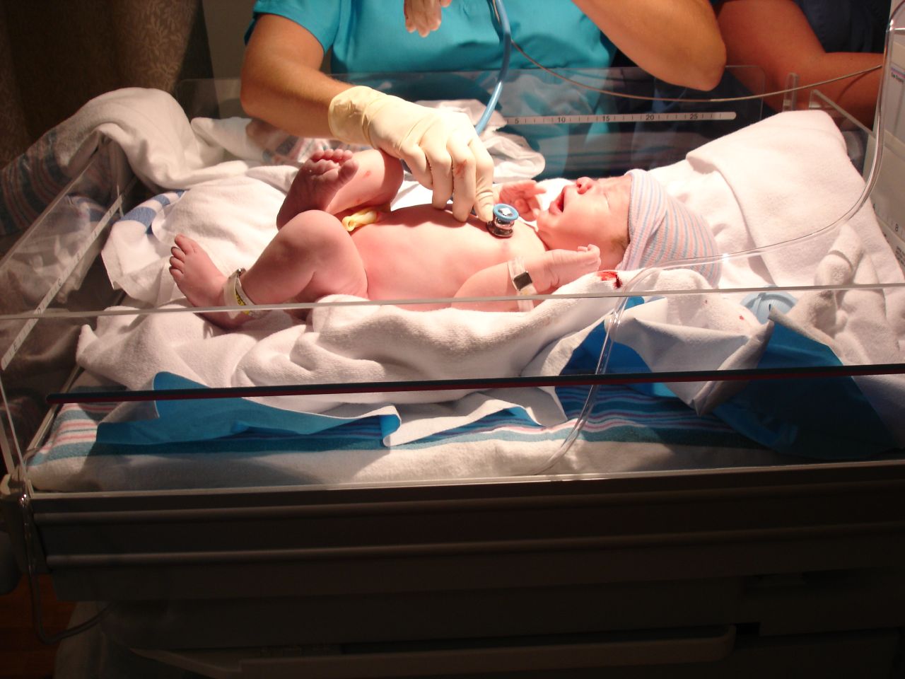
An umbilical line is a catheter that is inserted into one of the two arteries or the vein of the umbilical cord. Generally the UAC/UVC (Umbilical Artery Catheter/Umbilical Vein Catheter) is used in Neonatal Intensive Care Units (NICU) as it provides quick access to the central circulation of premature infants. UAC/UVC lines can be placed at the time of birth and allow medical staff to quickly infuse fluids, inotropic drugs, and blood if required. It is sometimes used in term or near-term newborns in whom the umbilical cord stump is still connected to the circulatory system. Medications, fluids, and blood can be given through this catheter and it allows monitoring of blood gasses and withdrawing of blood samples. ''Transumbilical catheter intervention'' is also a method of gaining access to the heart, for example to surgically correct a patent ductus arteriosus. Complications The complications of umbilical lines are similar to those of central venous catheters mainly infections ... [...More Info...] [...Related Items...] OR: [Wikipedia] [Google] [Baidu] |
Neonatology
Neonatology is a subspecialty of pediatrics that consists of the medical care of newborn infants, especially the ill or premature newborn. It is a hospital-based specialty and is usually practised in neonatal intensive care units (NICUs). The principal patients of neonatologists are newborn infants who are ill or require special medical care due to prematurity, low birth weight, intrauterine growth restriction, congenital malformations ( birth defects), sepsis, pulmonary hypoplasia, or birth asphyxia. Historical developments Though high infant mortality rates were recognized by the medical community at least as early as the 1860s, advances in modern neonatal intensive care have led to a significant decline in infant mortality in the modern era. This has been achieved through a combination of technological advances, enhanced understanding of newborn physiology, improved sanitation practices, and development of specialized units for neonatal intensive care. Around the mid ... [...More Info...] [...Related Items...] OR: [Wikipedia] [Google] [Baidu] |
Staphylococcus Epidermidis
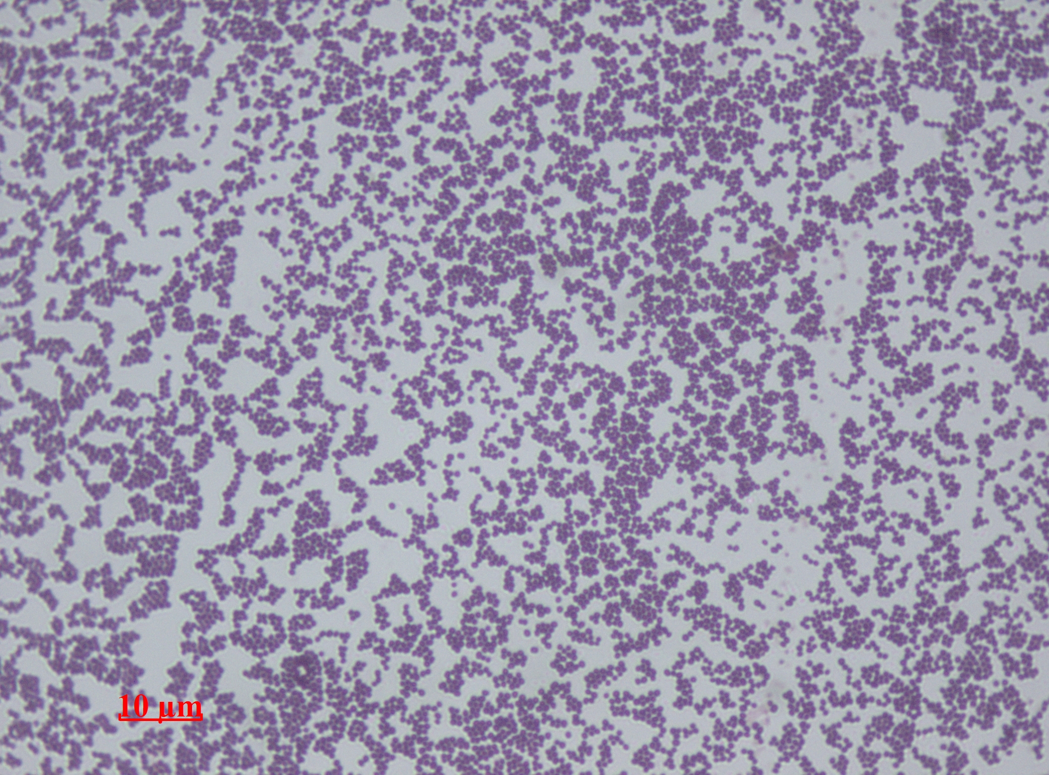
''Staphylococcus epidermidis'' is a Gram-positive bacterium, and one of over 40 species belonging to the genus ''Staphylococcus''. It is part of the human flora, normal human microbiota, typically the skin flora, skin microbiota, and less commonly the mucosal microbiota and also found in marine sponges. It is a facultative anaerobic bacteria. Although ''S. epidermidis'' is not usually Pathogenic bacteria, pathogenic, patients with compromised immune systems are at risk of developing infection. These infections are generally Hospital-acquired infection, hospital-acquired. ''S. epidermidis'' is a particular concern for people with catheters or other surgical implants because it is known to form biofilms that grow on these devices. Being part of the normal skin microbiota, ''S. epidermidis'' is a frequent contaminant of specimens sent to the diagnostic laboratory. Some strains of ''S. epidermidis'' are highly salt tolerant and commonly found in marine environments. S.I. Paul et al ... [...More Info...] [...Related Items...] OR: [Wikipedia] [Google] [Baidu] |
Thoracic Diaphragm
The thoracic diaphragm, or simply the diaphragm (; ), is a sheet of internal Skeletal striated muscle, skeletal muscle in humans and other mammals that extends across the bottom of the thoracic cavity. The diaphragm is the most important Muscles of respiration, muscle of respiration, and separates the thoracic cavity, containing the heart and lungs, from the abdominal cavity: as the diaphragm contracts, the volume of the thoracic cavity increases, creating a negative pressure there, which draws air into the lungs. Its high oxygen consumption is noted by the many mitochondria and capillaries present; more than in any other skeletal muscle. The term ''diaphragm'' in anatomy, created by Gerard of Cremona, can refer to other flat structures such as the urogenital diaphragm or Pelvic floor, pelvic diaphragm, but "the diaphragm" generally refers to the thoracic diaphragm. In humans, the diaphragm is slightly asymmetric—its right half is higher up (superior) to the left half, since th ... [...More Info...] [...Related Items...] OR: [Wikipedia] [Google] [Baidu] |
Right Atrium
The atrium (; : atria) is one of the two upper chambers in the heart that receives blood from the circulatory system. The blood in the atria is pumped into the heart ventricles through the atrioventricular mitral and tricuspid heart valves. There are two atria in the human heart – the left atrium receives blood from the pulmonary circulation, and the right atrium receives blood from the venae cavae of the systemic circulation. During the cardiac cycle, the atria receive blood while relaxed in diastole, then contract in systole to move blood to the ventricles. Each atrium is roughly cube-shaped except for an ear-shaped projection called an atrial appendage, previously known as an auricle. All animals with a closed circulatory system have at least one atrium. The atrium was formerly called the 'auricle'. That term is still used to describe this chamber in some other animals, such as the ''Mollusca''. Auricles in this modern terminology are distinguished by having thicker ... [...More Info...] [...Related Items...] OR: [Wikipedia] [Google] [Baidu] |
Inferior Vena Cava
The inferior vena cava is a large vein that carries the deoxygenated blood from the lower and middle body into the right atrium of the heart. It is formed by the joining of the right and the left common iliac veins, usually at the level of the fifth Lumbar vertebrae, lumbar vertebra. The inferior vena cava is the lower ("anatomical terms of location#Superior and inferior, inferior") of the two venae cavae, the two large veins that carry deoxygenated blood from the body to the right atrium of the heart: the inferior vena cava carries blood from the lower half of the body whilst the superior vena cava carries blood from the upper half of the body. Together, the venae cavae (in addition to the coronary sinus, which carries blood from the muscle of the heart itself) form the venous counterparts of the aorta. It is a large retroperitoneal vein that lies Posterior (anatomy), posterior to the abdominal cavity and runs along the right side of the vertebral column. It enters the right a ... [...More Info...] [...Related Items...] OR: [Wikipedia] [Google] [Baidu] |
Ductus Venosus
In the fetus, the ''ductus venosus'' (''"DV"''; Arantius' duct after Julius Caesar Aranzi) shunts a portion of umbilical vein blood flow directly to the inferior vena cava. Thus, it allows oxygenated blood from the placenta to bypass the liver. Compared to the 50% shunting of umbilical blood through the ''ductus venosus'' found in animal experiments, the degree of shunting in the human fetus under physiological conditions is considerably less, 30% at 20 weeks, which decreases to 18% at 32 weeks, suggesting a higher priority of the fetal liver than previously realized. In conjunction with the other fetal shunts, the ''foramen ovale'' and ''ductus arteriosus'', it plays a critical role in preferentially shunting oxygenated blood to the fetal brain. It is a part of fetal circulation. Anatomic course The pathway of fetal umbilical venous flow is umbilical vein \rightarrow left portal vein \rightarrow ''ductus venosus'' \rightarrow ''inferior vena cava'' \rightarrow eventually r ... [...More Info...] [...Related Items...] OR: [Wikipedia] [Google] [Baidu] |
Portal Vein
The portal vein or hepatic portal vein (HPV) is a blood vessel that carries blood from the gastrointestinal tract, gallbladder, pancreas and spleen to the liver. This blood contains nutrients and toxins extracted from digested contents. Approximately 75% of total liver blood flow is through the portal vein, with the remainder coming from the hepatic artery proper. The blood leaves the liver to the heart in the hepatic veins. The portal vein is not a true vein, because it conducts blood to capillary beds in the liver and not directly to the heart. It is a major component of the hepatic portal system, one of three portal venous systems in the human body; the others being the hypophyseal portal system, hypophyseal and Renal portal system, renal portal systems. The portal vein is usually formed by the confluence of the superior mesenteric vein, superior mesenteric, splenic veins, inferior mesenteric vein, inferior mesenteric, left gastric vein, left, right gastric veins and the pancr ... [...More Info...] [...Related Items...] OR: [Wikipedia] [Google] [Baidu] |
Navel
The navel (clinically known as the umbilicus; : umbilici or umbilicuses; also known as the belly button or tummy button) is a protruding, flat, or hollowed area on the abdomen at the attachment site of the umbilical cord. Structure The umbilicus is used to visually separate the abdomen into quadrants. The umbilicus is a prominent Scar#Umbilical, scar on the abdomen, with its position being relatively consistent among humans. The skin around the waist at the level of the umbilicus is supplied by the tenth thoracic spinal nerve (T10 dermatome (anatomy), dermatome). The umbilicus itself typically lies at a vertical level corresponding to the junction between the L3 and L4 vertebrae, with a normal variation among people between the L3 and L5 vertebrae. Parts of the adult navel include the "umbilical cord remnant" or "umbilical tip", which is the often protruding scar left by the detachment of the umbilical cord. This is located in the center of the navel, sometimes described ... [...More Info...] [...Related Items...] OR: [Wikipedia] [Google] [Baidu] |
Umbilical Vein
The umbilical vein is a vein present during fetal development that carries oxygenated blood from the placenta into the growing fetus. The umbilical vein provides convenient access to the central circulation of a neonate for restoration of blood volume and for administration of glucose and drugs. The blood pressure inside the umbilical vein is approximately 20 mmHg.Wang, Y. Vascular biology of the placenta. in Colloquium Series on Integrated Systems Physiology: from Molecule to Function. 2010. Morgan & Claypool Life Sciences. Fetal circulation The unpaired umbilical vein carries oxygen and nutrient rich blood derived from fetal-maternal blood exchange at the chorionic villi. More than two-thirds of fetal hepatic circulation is via the main portal vein, while the remainder is shunted from the left portal vein via the ductus venosus to the inferior vena cava, eventually being delivered to the fetal right atrium. Closure Closure of the umbilical vein usually occurs after the umbilic ... [...More Info...] [...Related Items...] OR: [Wikipedia] [Google] [Baidu] |
Vasospasm
Vasospasm refers to a condition in which an arterial spasm leads to vasoconstriction. This can lead to tissue ischemia (insufficient blood flow) and tissue death (necrosis). Along with physical resistance, vasospasm is a main cause of ischemia. Like physical resistance, vasospasms can occur due to atherosclerosis. Vasospasm is the major cause of Prinzmetal's angina. Cerebral vasospasm may arise in the context of subarachnoid hemorrhage as symptomatic vasospasm (or delayed cerebral ischemia), where it is a major contributor to post-operative stroke and mortality. Vasospasm typically appears 4 to 10 days after subarachnoid hemorrhage, however the relationship between radiological arterial spasm (seen on angiography) and clinical neurological deterioration is nuanced and uncertain. Pathophysiology Normally endothelial cells release prostacyclin and nitric oxide (NO) which induce relaxation of the smooth muscle cells, and reduce aggregation of platelets. Aggregating platelets ... [...More Info...] [...Related Items...] OR: [Wikipedia] [Google] [Baidu] |
Lumbar Vertebra
The lumbar vertebrae are located between the thoracic vertebrae and pelvis. They form the lower part of the back in humans, and the tail end of the back in quadrupeds. In humans, there are five lumbar vertebrae. The term is used to describe the anatomy of humans and quadrupeds, such as horses, pigs, or cattle. These bones are found in particular cuts of meat, including tenderloin or sirloin steak. Human anatomy In human anatomy, the five vertebrae are between the rib cage and the pelvis. They are the largest segments of the vertebral column and are characterized by the absence of the foramen transversarium within the transverse process (since it is only found in the cervical region) and by the absence of facets on the sides of the body (as found only in the thoracic region). They are designated L1 to L5, starting at the top. The lumbar vertebrae help support the weight of the body, and permit movement. General characteristics The adjacent figure depicts the general charac ... [...More Info...] [...Related Items...] OR: [Wikipedia] [Google] [Baidu] |
Thoracic Vertebra
In vertebrates, thoracic vertebrae compose the middle segment of the vertebral column, between the cervical vertebrae and the lumbar vertebrae. In humans, there are twelve thoracic vertebra (anatomy), vertebrae of intermediate size between the cervical and lumbar vertebrae; they increase in size going towards the lumbar vertebrae. They are distinguished by the presence of Zygapophysial joint, facets on the sides of the bodies for Articulation (anatomy), articulation with the head of rib, heads of the ribs, as well as facets on the transverse processes of all, except the eleventh and twelfth, for articulation with the tubercle (rib), tubercles of the ribs. By convention, the human thoracic vertebrae are numbered T1–T12, with the first one (T1) located closest to the skull and the others going down the spine toward the lumbar region. General characteristics These are the general characteristics of the second through eighth thoracic vertebrae. The first and ninth through twelfth v ... [...More Info...] [...Related Items...] OR: [Wikipedia] [Google] [Baidu] |


