|
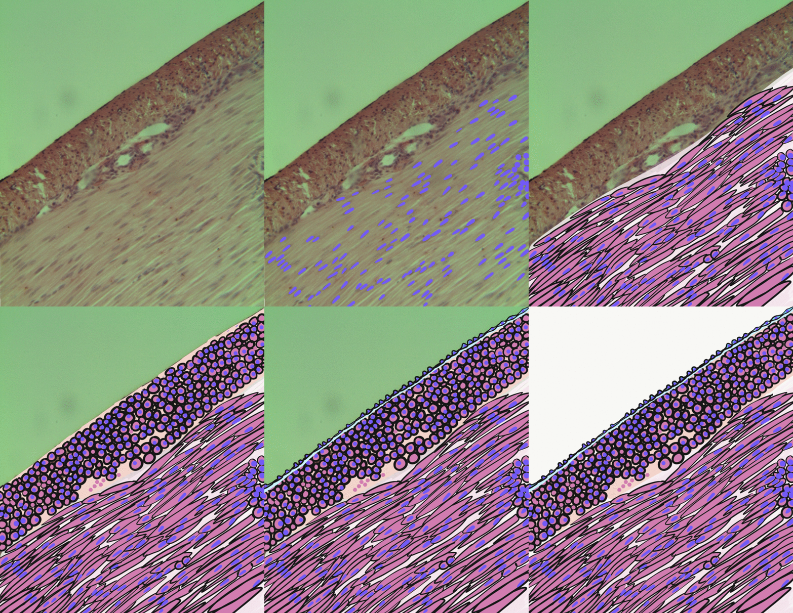
Tunica Intima
The tunica intima ( New Latin "inner coat"), or intima for short, is the innermost tunica (layer) of an artery or vein. It is made up of one layer of endothelial cells and is supported by an internal elastic lamina. The endothelial cells are in direct contact with the blood flow. The three layers of a blood vessel are an inner layer (the tunica intima), a middle layer (the tunica media), and an outer layer (the tunica externa). In dissection, the inner coat (tunica intima) can be separated from the middle (tunica media) by a little maceration, or it may be stripped off in small pieces; but, because of its friability, it cannot be separated as a complete membrane. It is a fine, transparent, colorless structure which is highly elastic, and, after death, is commonly corrugated into longitudinal wrinkles. Structure The structure of the tunica intima depends on the blood vessel type. Elastic arteries – A single layer of Endothelial and a supporting layer of elastin-rich ... [...More Info...] [...Related Items...] OR: [Wikipedia] [Google] [Baidu] |
Artery
An artery (plural arteries) () is a blood vessel in humans and most animals that takes blood away from the heart to one or more parts of the body (tissues, lungs, brain etc.). Most arteries carry oxygenated blood; the two exceptions are the pulmonary and the umbilical arteries, which carry deoxygenated blood to the organs that oxygenate it (lungs and placenta, respectively). The effective arterial blood volume is that extracellular fluid which fills the arterial system. The arteries are part of the circulatory system, that is responsible for the delivery of oxygen and nutrients to all cells, as well as the removal of carbon dioxide and waste products, the maintenance of optimum blood pH, and the circulation of proteins and cells of the immune system. Arteries contrast with veins, which carry blood back towards the heart. Structure The anatomy of arteries can be separated into gross anatomy, at the macroscopic level, and microanatomy, which must be studied with a mic ... [...More Info...] [...Related Items...] OR: [Wikipedia] [Google] [Baidu] |
Endothelium
The endothelium is a single layer of squamous endothelial cells that line the interior surface of blood vessels and lymphatic vessels. The endothelium forms an interface between circulating blood or lymph in the lumen and the rest of the vessel wall. Endothelial cells form the barrier between vessels and tissue and control the flow of substances and fluid into and out of a tissue. Endothelial cells in direct contact with blood are called vascular endothelial cells whereas those in direct contact with lymph are known as lymphatic endothelial cells. Vascular endothelial cells line the entire circulatory system, from the heart to the smallest capillaries. These cells have unique functions that include fluid filtration, such as in the glomerulus of the kidney, blood vessel tone, hemostasis, neutrophil recruitment, and hormone trafficking. Endothelium of the interior surfaces of the heart chambers is called endocardium. An impaired function can lead to serious health iss ... [...More Info...] [...Related Items...] OR: [Wikipedia] [Google] [Baidu] |
Fenestra
A fenestra (fenestration; plural fenestrae or fenestrations) is any small opening or pore, commonly used as a term in the biological sciences. It is the Latin word for "window", and is used in various fields to describe a pore in an anatomical structure. Biological morphology In morphology, fenestrae are found in cancellous bones, particularly in the skull. In anatomy, the round window and oval window are also known as the ''fenestra rotunda'' and the ''fenestra ovalis''. In microanatomy, fenestrae are found in endothelium of fenestrated capillaries, enabling the rapid exchange of molecules between the blood and surrounding tissue. The elastic layer of the tunica intima is a fenestrated membrane. In surgery, a fenestration is a new opening made in a part of the body to enable drainage or access. Plant biology and mycology In plant biology, the perforations in a perforate leaf are also described as fenestrae, and the leaf is called a fenestrate leaf. The leaf window i ... [...More Info...] [...Related Items...] OR: [Wikipedia] [Google] [Baidu] |
Connective Tissue
Connective tissue is one of the four primary types of animal tissue, along with epithelial tissue, muscle tissue, and nervous tissue. It develops from the mesenchyme derived from the mesoderm the middle embryonic germ layer. Connective tissue is found in between other tissues everywhere in the body, including the nervous system. The three meninges, membranes that envelop the brain and spinal cord are composed of connective tissue. Most types of connective tissue consists of three main components: elastic and collagen fibers, ground substance, and cells. Blood, and lymph are classed as specialized fluid connective tissues that do not contain fiber. All are immersed in the body water. The cells of connective tissue include fibroblasts, adipocytes, macrophages, mast cells and leucocytes. The term "connective tissue" (in German, ''Bindegewebe'') was introduced in 1830 by Johannes Peter Müller. The tissue was already recognized as a distinct class in the 18th century. ... [...More Info...] [...Related Items...] OR: [Wikipedia] [Google] [Baidu] |
Stellate Cells
Stellate cells are neurons in the central nervous system, named for their star-like shape formed by dendritic processes radiating from the cell body. Many stellate cells are GABAergic and are located in the molecular layer of the cerebellum. Stellate cells are derived from dividing progenitor cells in the white matter of postnatal cerebellum. Dendritic trees can vary between neurons. There are two types of dendritic trees in the cerebral cortex, which include pyramidal cells, which are pyramid shaped and stellate cells which are star shaped. Dendrites can also aid neuron classification. Dendrites with spines are classified as spiny, those without spines are classified as aspinous. Stellate cells can be spiny or aspinous, while pyramidal cells are always spiny. Most common stellate cells are the inhibitory interneurons found within the upper half of the molecular layer in the cerebellum. Cerebellar stellate cells synapse onto the dendritic trees of Purkinje cells and send inhib ... [...More Info...] [...Related Items...] OR: [Wikipedia] [Google] [Baidu] |
Silver Nitrate
Silver nitrate is an inorganic compound with chemical formula . It is a versatile precursor to many other silver compounds, such as those used in photography. It is far less sensitive to light than the halides. It was once called ''lunar caustic'' because silver was called ''luna'' by ancient alchemists who associated silver with the moon. In solid silver nitrate, the silver ions are three- coordinated in a trigonal planar arrangement. Synthesis and structure Albertus Magnus, in the 13th century, documented the ability of nitric acid to separate gold and silver by dissolving the silver. Indeed silver nitrate can be prepared by dissolving silver in nitric acid followed by evaporation of the solution. The stoichiometry of the reaction depends upon the concentration of nitric acid used. :3 Ag + 4 HNO3 (cold and diluted) → 3 AgNO3 + 2 H2O + NO :Ag + 2 HNO3 (hot and concentrated) → AgNO3 + H2O + NO2 The structure of silver nitrate has been examined by X-ray crystallography ... [...More Info...] [...Related Items...] OR: [Wikipedia] [Google] [Baidu] |
Vein
Veins are blood vessels in humans and most other animals that carry blood towards the heart. Most veins carry deoxygenated blood from the tissues back to the heart; exceptions are the pulmonary and umbilical veins, both of which carry oxygenated blood to the heart. In contrast to veins, arteries carry blood away from the heart. Veins are less muscular than arteries and are often closer to the skin. There are valves (called ''pocket valves'') in most veins to prevent backflow. Structure Veins are present throughout the body as tubes that carry blood back to the heart. Veins are classified in a number of ways, including superficial vs. deep, pulmonary vs. systemic, and large vs. small. *Superficial veins are those closer to the surface of the body, and have no corresponding arteries. *Deep veins are deeper in the body and have corresponding arteries. * Perforator veins drain from the superficial to the deep veins. These are usually referred to in the lower limbs and feet. *Comm ... [...More Info...] [...Related Items...] OR: [Wikipedia] [Google] [Baidu] |
Arteriole
An arteriole is a small-diameter blood vessel in the microcirculation that extends and branches out from an artery and leads to capillaries. Arterioles have muscular walls (usually only one to two layers of smooth muscle cells) and are the primary site of vascular resistance. The greatest change in blood pressure and velocity of blood flow occurs at the transition of arterioles to capillaries.This function is extremely important because it prevents the thin, one-layer capillaries from exploding upon pressure. The arterioles achieve this decrease in pressure, as they are the site with the highest resistance (a large contributor to total peripheral resistance) which translates to a large decrease in the pressure. Structure Microanatomy In a healthy vascular system the endothelium lines all blood-contacting surfaces, including arteries, arterioles, veins, venules, capillaries, and heart chambers. This healthy condition is promoted by the ample production of nitric oxide by the en ... [...More Info...] [...Related Items...] OR: [Wikipedia] [Google] [Baidu] |
Distributing Artery
A muscular artery (or distributing artery) is a medium-sized artery that draws blood from an elastic artery and branches into "resistance vessels" including small arteries and arterioles. Their walls contain larger number of smooth muscles, allowing them to contract and expand depending on peripheral blood demand. This contrasts to the mechanism of elastic arteries, which use their elastic properties to store the energy generated by the heart's contraction for a brief moment (elastic recoil). Under the microscope, muscular arteries can be identified by their clearly defined internal elastic lamina. In constricted vessels, the elastic lamina of muscular arteries appears thick and kinky. The elastic lamina is best visualized using Verhoeff's stain, but can be easily detected in specimens stained using other techniques as a well-defined negative staining region. Examples of muscular arteries include the radial artery, femoral artery and the splenic artery In human anatomy, the s ... [...More Info...] [...Related Items...] OR: [Wikipedia] [Google] [Baidu] |
Smooth Muscle
Smooth muscle is an involuntary non- striated muscle, so-called because it has no sarcomeres and therefore no striations (''bands'' or ''stripes''). It is divided into two subgroups, single-unit and multiunit smooth muscle. Within single-unit muscle, the whole bundle or sheet of smooth muscle cells contracts as a syncytium. Smooth muscle is found in the walls of hollow organs, including the stomach, intestines, bladder and uterus; in the walls of passageways, such as blood, and lymph vessels, and in the tracts of the respiratory, urinary, and reproductive systems. In the eyes, the ciliary muscles, a type of smooth muscle, dilate and contract the iris and alter the shape of the lens. In the skin, smooth muscle cells such as those of the arrector pili cause hair to stand erect in response to cold temperature or fear. Structure Gross anatomy Smooth muscle is grouped into two types: single-unit smooth muscle, also known as visceral smooth muscle, and multiunit ... [...More Info...] [...Related Items...] OR: [Wikipedia] [Google] [Baidu] |
Fibroblast
A fibroblast is a type of biological cell that synthesizes the extracellular matrix and collagen, produces the structural framework ( stroma) for animal tissues, and plays a critical role in wound healing. Fibroblasts are the most common cells of connective tissue in animals. Structure Fibroblasts have a branched cytoplasm surrounding an elliptical, speckled nucleus having two or more nucleoli. Active fibroblasts can be recognized by their abundant rough endoplasmic reticulum. Inactive fibroblasts (called fibrocytes) are smaller, spindle-shaped, and have a reduced amount of rough endoplasmic reticulum. Although disjointed and scattered when they have to cover a large space, fibroblasts, when crowded, often locally align in parallel clusters. Unlike the epithelial cells lining the body structures, fibroblasts do not form flat monolayers and are not restricted by a polarizing attachment to a basal lamina on one side, although they may contribute to basal lamina components ... [...More Info...] [...Related Items...] OR: [Wikipedia] [Google] [Baidu] |
Collagen
Collagen () is the main structural protein in the extracellular matrix found in the body's various connective tissues. As the main component of connective tissue, it is the most abundant protein in mammals, making up from 25% to 35% of the whole-body protein content. Collagen consists of amino acids bound together to form a triple helix of elongated fibril known as a collagen helix. It is mostly found in connective tissue such as cartilage, bones, tendons, ligaments, and skin. Depending upon the degree of mineralization, collagen tissues may be rigid (bone) or compliant (tendon) or have a gradient from rigid to compliant (cartilage). Collagen is also abundant in corneas, blood vessels, the gut, intervertebral discs, and the dentin in teeth. In muscle tissue, it serves as a major component of the endomysium. Collagen constitutes one to two percent of muscle tissue and accounts for 6% of the weight of the skeletal muscle tissue. The fibroblast is the most common ... [...More Info...] [...Related Items...] OR: [Wikipedia] [Google] [Baidu] |


.jpg)