|
Triradiate Cartilages
The triradiate cartilage (in Latin cartilago ypsiloformis) is the 'Y'-shaped epiphyseal plate between the ilium, ischium and pubis to form the acetabulum of the os coxae. Human development In children, the triradiate cartilage closes at an approximate bone age of 12 years for girls and 14 years for boys. Clinical use Evaluating the position of the triradiate cartilage on an AP radiograph of the pelvis with both Perkin's line and Hilgenreiner's line Hilgenreiner's line is a horizontal line drawn on an AP radiograph of the pelvis running between the inferior aspects of both triradiate cartilages of the acetabulums. It is named for Heinrich Hilgenreiner. Clinical Use Used in conjunction with ... can help establish a diagnosis of developmental dysplasia of the hip. References See also {{Pelvis Pelvis ... [...More Info...] [...Related Items...] OR: [Wikipedia] [Google] [Baidu] |
Epiphyseal Plate
The epiphyseal plate (or epiphysial plate, physis, or growth plate) is a hyaline cartilage plate in the metaphysis at each end of a long bone. It is the part of a long bone where new bone growth takes place; that is, the whole bone is alive, with maintenance remodeling throughout its existing bone tissue, but the growth plate is the place where the long bone grows longer (adds length). The plate is only found in children and adolescents; in adults, who have stopped growing, the plate is replaced by an epiphyseal line. This replacement is known as epiphyseal closure or growth plate fusion. Complete fusion can occur as early as 12 for girls (with the most common being 14-15 years for girls) and as early as 14 for boys (with the most common being 15–17 years for boys). Structure Development Endochondral ossification is responsible for the initial bone development from cartilage in utero and infants and the longitudinal growth of long bones in the epiphyseal plate. The plate's ... [...More Info...] [...Related Items...] OR: [Wikipedia] [Google] [Baidu] |
Ilium (bone)
The ilium () (plural ilia) is the uppermost and largest part of the hip bone, and appears in most vertebrates including mammals and birds, but not bony fish. All reptiles have an ilium except snakes, although some snake species have a tiny bone which is considered to be an ilium. The ilium of the human is divisible into two parts, the body and the wing; the separation is indicated on the top surface by a curved line, the arcuate line, and on the external surface by the margin of the acetabulum. The name comes from the Latin ('' ile'', ''ilis''), meaning "groin" or "flank". Structure The ilium consists of the body and wing. Together with the ischium and pubis, to which the ilium is connected, these form the pelvic bone, with only a faint line indicating the place of union. The body ( la, corpus) forms less than two-fifths of the acetabulum; and also forms part of the acetabular fossa. The internal surface of the body is part of the wall of the lesser pelvis and give ... [...More Info...] [...Related Items...] OR: [Wikipedia] [Google] [Baidu] |
Ischium
The ischium () forms the lower and back region of the hip bone (''os coxae''). Situated below the ilium and behind the pubis, it is one of three regions whose fusion creates the coxal bone. The superior portion of this region forms approximately one-third of the . Structure The i ...[...More Info...] [...Related Items...] OR: [Wikipedia] [Google] [Baidu] |
Pubis (bone)
In vertebrates, the pubic region ( la, pubis) is the most forward-facing (ventral and anterior) of the three main regions making up the coxal bone. The left and right pubic regions are each made up of three sections, a superior ramus, inferior ramus, and a body. Structure The pubic region is made up of a ''body'', ''superior ramus'', and ''inferior ramus'' (). The left and right coxal bones join at the pubic symphysis. It is covered by a layer of fat, which is covered by the mons pubis. The pubis is the lower limit of the suprapubic region. In the female, the pubic region is anterior to the urethral sponge. Body The body forms the wide, strong, middle and flat part of the pubic region. The bodies of the left and right pubic regions join at the pubic symphysis. The rough upper edge is the pubic crest, ending laterally in the pubic tubercle. This tubercle, found roughly 3 cm from the pubic symphysis, is a distinctive feature on the lower part of the abdominal wall; important ... [...More Info...] [...Related Items...] OR: [Wikipedia] [Google] [Baidu] |
Acetabulum
The acetabulum (), also called the cotyloid cavity, is a concave surface of the pelvis. The head of the femur meets with the pelvis at the acetabulum, forming the hip joint. Structure There are three bones of the ''os coxae'' (hip bone) that come together to form the ''acetabulum''. Contributing a little more than two-fifths of the structure is the ischium, which provides lower and side boundaries to the acetabulum. The ilium forms the upper boundary, providing a little less than two-fifths of the structure of the acetabulum. The rest is formed by the pubis, near the midline. It is bounded by a prominent uneven rim, which is thick and strong above, and serves for the attachment of the acetabular labrum, which reduces its opening, and deepens the surface for formation of the hip joint. At the lower part of the ''acetabulum'' is the acetabular notch, which is continuous with a circular depression, the acetabular fossa, at the bottom of the cavity of the ''acetabulum''. The res ... [...More Info...] [...Related Items...] OR: [Wikipedia] [Google] [Baidu] |
Os Coxae
The hip bone (os coxae, innominate bone, pelvic bone or coxal bone) is a large flat bone, constricted in the center and expanded above and below. In some vertebrates (including humans before puberty) it is composed of three parts: the ilium, ischium, and the pubis. The two hip bones join at the pubic symphysis and together with the sacrum and coccyx (the pelvic part of the spine) comprise the skeletal component of the pelvis – the pelvic girdle which surrounds the pelvic cavity. They are connected to the sacrum, which is part of the axial skeleton, at the sacroiliac joint. Each hip bone is connected to the corresponding femur (thigh bone) (forming the primary connection between the bones of the lower limb and the axial skeleton) through the large ball and socket joint of the hip. Structure The hip bone is formed by three parts: the ilium, ischium, and pubis. At birth, these three components are separated by hyaline cartilage. They join each other in a Y-shaped portion of c ... [...More Info...] [...Related Items...] OR: [Wikipedia] [Google] [Baidu] |
Bone Age
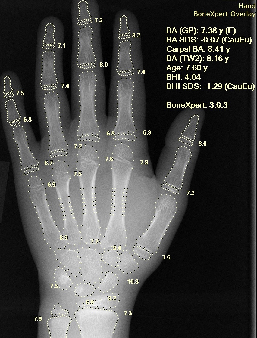
Bone age is the degree of a person's skeletal development. In children, bone age serves as a measure of physiological maturity and aids in the diagnosis of growth abnormalities, endocrine disorders, and other medical conditions. As a person grows from fetal life through childhood, puberty, and finishes growth as a young adult, the bones of the skeleton change in size and shape. These changes can be seen by x-ray and other imaging techniques. A comparison between the appearance of a patient's bones to a standard set of bone images known to be representative of the average bone shape and size for a given age can be used to assign a "bone age" to the patient. Bone age is distinct from an individual's biological or chronological age, which is the amount of time that has elapsed since birth. Discrepancies between bone age and biological age can be seen in people with stunted growth, where bone age may be less than biological age. Similarly, a bone age that is older than a person's chronol ... [...More Info...] [...Related Items...] OR: [Wikipedia] [Google] [Baidu] |
Anteroposterior
Standard anatomical terms of location are used to unambiguously describe the anatomy of animals, including humans. The terms, typically derived from Latin or Greek roots, describe something in its standard anatomical position. This position provides a definition of what is at the front ("anterior"), behind ("posterior") and so on. As part of defining and describing terms, the body is described through the use of anatomical planes and anatomical axes. The meaning of terms that are used can change depending on whether an organism is bipedal or quadrupedal. Additionally, for some animals such as invertebrates, some terms may not have any meaning at all; for example, an animal that is radially symmetrical will have no anterior surface, but can still have a description that a part is close to the middle ("proximal") or further from the middle ("distal"). International organisations have determined vocabularies that are often used as standard vocabularies for subdisciplines of anatom ... [...More Info...] [...Related Items...] OR: [Wikipedia] [Google] [Baidu] |
Medical Radiography
Radiography is an imaging technique using X-rays, gamma rays, or similar ionizing radiation and non-ionizing radiation to view the internal form of an object. Applications of radiography include medical radiography ("diagnostic" and "therapeutic") and industrial radiography. Similar techniques are used in airport security (where "body scanners" generally use backscatter X-ray). To create an image in conventional radiography, a beam of X-rays is produced by an X-ray generator and is projected toward the object. A certain amount of the X-rays or other radiation is absorbed by the object, dependent on the object's density and structural composition. The X-rays that pass through the object are captured behind the object by a detector (either photographic film or a digital detector). The generation of flat two dimensional images by this technique is called projectional radiography. In computed tomography (CT scanning) an X-ray source and its associated detectors rotate around ... [...More Info...] [...Related Items...] OR: [Wikipedia] [Google] [Baidu] |
Human Pelvis
The pelvis (plural pelves or pelvises) is the lower part of the trunk, between the abdomen and the thighs (sometimes also called pelvic region), together with its embedded skeleton (sometimes also called bony pelvis, or pelvic skeleton). The pelvic region of the trunk includes the bony pelvis, the pelvic cavity (the space enclosed by the bony pelvis), the pelvic floor, below the pelvic cavity, and the perineum, below the pelvic floor. The pelvic skeleton is formed in the area of the back, by the sacrum and the coccyx and anteriorly and to the left and right sides, by a pair of hip bones. The two hip bones connect the spine with the lower limbs. They are attached to the sacrum posteriorly, connected to each other anteriorly, and joined with the two femurs at the hip joints. The gap enclosed by the bony pelvis, called the pelvic cavity, is the section of the body underneath the abdomen and mainly consists of the reproductive organs (sex organs) and the rectum, while the pel ... [...More Info...] [...Related Items...] OR: [Wikipedia] [Google] [Baidu] |
Perkin's Line
Perkin's line is a line drawn on an AP radiograph of the pelvis perpendicular to Hilgenreiner's line at the lateral aspects of the triradiate cartilage of the acetabulum. Clinical use Used in conjunction with Hilgenreiner's line, Perkin's line is useful in the diagnosis of developmental dysplasia of the hip; the upper femoral epiphysis The epiphysis () is the rounded end of a long bone, at its joint with adjacent bone(s). Between the epiphysis and diaphysis (the long midsection of the long bone) lies the metaphysis, including the epiphyseal plate (growth plate). At the jo ... should be in the inferomedial quadrant on a normal radiograph. Lateral displacement relative to Perkin's line is indicative of DDH. References External links Wheeless Online Musculoskeletal radiographic signs {{Orthopedics-stub ... [...More Info...] [...Related Items...] OR: [Wikipedia] [Google] [Baidu] |
Hilgenreiner's Line
Hilgenreiner's line is a horizontal line drawn on an AP radiograph of the pelvis running between the inferior aspects of both triradiate cartilages of the acetabulums. It is named for Heinrich Hilgenreiner. Clinical Use Used in conjunction with Perkin's line Perkin's line is a line drawn on an AP radiograph of the pelvis perpendicular to Hilgenreiner's line at the lateral aspects of the triradiate cartilage of the acetabulum. Clinical use Used in conjunction with Hilgenreiner's line, Perkin's l ... or the acetabular angle, Hilgenreiner's line is useful in the diagnosis of developmental dysplasia of the hip. References {{Orthopedics-stub Musculoskeletal radiographic signs ... [...More Info...] [...Related Items...] OR: [Wikipedia] [Google] [Baidu] |


