|
Tissue Calcification
Calcification is the accumulation of calcium salts in a body tissue. It normally occurs in the formation of bone, but calcium can be deposited abnormally in soft tissue,Miller, J. D. Cardiovascular calcification: Orbicular origins. ''Nature Materials'' 12, 476-478 (2013). causing it to harden. Calcifications may be classified on whether there is mineral balance or not, and the location of the calcification. Calcification may also refer to the processes of normal mineral deposition in biological systems, such as the formation of stromatolites or mollusc shells (see Biomineralization). Signs and symptoms Calcification can manifest itself in many ways in the body depending on the location. In the pulpal structure of a tooth, calcification often presents asymptomatically, and is diagnosed as an incidental finding during radiographic interpretation. Individual teeth with calcified pulp will typically respond negatively to vitality testing; teeth with calcified pulp often lack sen ... [...More Info...] [...Related Items...] OR: [Wikipedia] [Google] [Baidu] |
Cardiovascular Calcification - Sergio Bertazzo
The blood circulatory system is a system of organs that includes the heart, blood vessels, and blood which is circulated throughout the entire body of a human or other vertebrate. It includes the cardiovascular system, or vascular system, that consists of the heart and blood vessels (from Greek ''kardia'' meaning ''heart'', and from Latin ''vascula'' meaning ''vessels''). The circulatory system has two divisions, a systemic circulation or circuit, and a pulmonary circulation or circuit. Some sources use the terms ''cardiovascular system'' and ''vascular system'' interchangeably with the ''circulatory system''. The network of blood vessels are the great vessels of the heart including large elastic arteries, and large veins; other arteries, smaller arterioles, capillaries that join with venules (small veins), and other veins. The circulatory system is closed in vertebrates, which means that the blood never leaves the network of blood vessels. Some invertebrates such as arthr ... [...More Info...] [...Related Items...] OR: [Wikipedia] [Google] [Baidu] |
Metastatic Calcification
Metastatic calcification is deposition of calcium salts in otherwise normal tissue, because of elevated serum levels of calcium, which can occur because of deranged metabolism as well as increased absorption or decreased excretion of calcium and related minerals, as seen in hyperparathyroidism. In contrast, dystrophic calcification is caused by abnormalities or degeneration of tissues resulting in mineral deposition, though blood levels of calcium remain normal. These differences in pathology also mean that metastatic calcification is often found in many tissues throughout a person or animal, whereas dystrophic calcification is localized. Metastatic calcification can occur widely throughout the body but principally affects the interstitial tissues of the vasculature, kidneys, lungs, and gastric mucosa. For the latter three, acid secretions or rapid changes in pH levels contribute to the formation of salts. Causes Hypercalcemia, elevated blood calcium, has numerous causes, inclu ... [...More Info...] [...Related Items...] OR: [Wikipedia] [Google] [Baidu] |
Breast Disease
Breast diseases make up a number of conditions. The most common symptoms are a breast mass, breast pain, and nipple discharge. A majority of breast diseases are noncancerous. Tumor A breast tumor is an abnormal mass of tissue in the breast as a result of neoplasia. A breast neoplasm may be benign, as in fibroadenoma, or it may be malignant, in which case it is termed breast cancer. Either case commonly presents as a breast lump. Approximately 7% of breast lumps are fibroadenomas and 10% are breast cancer, the rest being other benign conditions or no disease.Page 739 in: 8th edition. Phyllodes tumor is a fibroepithelial tumor which can be benign, borderline or malignant. Breast cancer Breast cancer is cancer of the breast tissues, most commonly arising from the milk ducts. Worldwide, breast cancer is the leading type of cancer in women, accounting for 25% of all cases. It is most common in women over age 50. Signs of breast cancer may include a lump in the breast, a chan ... [...More Info...] [...Related Items...] OR: [Wikipedia] [Google] [Baidu] |
Pulp Stone
Pulp stones (also denticles or endoliths) are nodular, calcified masses appearing in either or both the coronal and root portion of the pulp organ in teeth. Pulp stones are not painful unless they impinge on nerves. They are classified: :A) On the basis of structure ::1) True pulp stones: formed of dentin by odontoblasts ::2) False pulp stones: formed by mineralization of degenerating pulp cells, often in a concentric pattern :B) On the basis of location ::1) Free: entirely surrounded by pulp tissue ::2) Adherent: partly fused with dentin ::3) Embedded: entirely surrounded by dentin Introduction Pulp stones are discrete calcifications found in the pulp chamber of the tooth which may undergo changes to become diffuse pulp calcifications such as dystrophic calcification. They are usually noticed by radiographic examination and appeared as round or ovoid radiopaque lesions. Clinically, a tooth with a pulp stone has normal appearance like any other tooth. The number of pulp stone ... [...More Info...] [...Related Items...] OR: [Wikipedia] [Google] [Baidu] |
Tonsil Stones
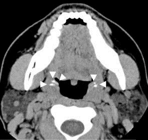
Tonsil stones, also known as tonsilloliths, are mineralizations of debris within the crevices of the tonsils. When not mineralized, the presence of debris is known as chronic caseous tonsillitis (CCT). Symptoms may include bad breath. Generally there is no pain, though there may be the feeling of something present. Risk factors may include recurrent throat infections. Tonsil stones contain a biofilm composed of a number of different bacteria. While they most commonly occur in the palatine tonsils, they may also occur in the lingual tonsils. Tonsil stones have been recorded weighing from 0.3g to 42g. They are often discovered during medical imaging for other reasons. If tonsil stones do not bother the patient, no treatment is needed. Otherwise gargling salt water and manual removal may be tried. Chlorhexidine or cetylpyridinium chloride may also be tried. Surgical treatment may include partial or complete tonsil removal. Up to 10% of people have tonsil stones. Biological sex ... [...More Info...] [...Related Items...] OR: [Wikipedia] [Google] [Baidu] |
Heterotopic Bone
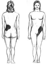
Heterotopic ossification (HO) is the process by which bone tissue forms outside of the skeleton in muscles and soft tissue. Symptoms In traumatic heterotopic ossification (traumatic myositis ossificans), the patient may complain of a warm, tender, firm swelling in a muscle and decreased range of motion in the joint served by the muscle involved. There is often a history of a blow or other trauma to the area a few weeks to a few months earlier. Patients with traumatic neurological injuries, severe neurologic disorders or severe burns who develop heterotopic ossification experience limitation of motion in the areas affected. Causes Heterotopic ossification of varying severity can be caused by surgery or trauma to the hips and legs. About every third patient who has total hip arthroplasty (joint replacement) or a severe fracture of the long bones of the lower leg will develop heterotopic ossification, but is uncommonly symptomatic. Between 50% and 90% of patients who develope ... [...More Info...] [...Related Items...] OR: [Wikipedia] [Google] [Baidu] |
Gall Stones
A gallstone is a stone formed within the gallbladder from precipitated bile components. The term cholelithiasis may refer to the presence of gallstones or to any disease caused by gallstones, and choledocholithiasis refers to the presence of migrated gallstones within bile ducts. Most people with gallstones (about 80%) are asymptomatic. However, when a gallstone obstructs the bile duct and causes acute cholestasis, a reflexive smooth muscle spasm often occurs, resulting in an intense cramp-like visceral pain in the right upper part of the abdomen known as a biliary colic (or "gallbladder attack"). This happens in 1–4% of those with gallstones each year. Complications from gallstones may include inflammation of the gallbladder (cholecystitis), inflammation of the pancreas (pancreatitis), obstructive jaundice, and infection in bile ducts ( cholangitis). Symptoms of these complications may include pain that lasts longer than five hours, fever, yellowish skin, vomiting, dark ur ... [...More Info...] [...Related Items...] OR: [Wikipedia] [Google] [Baidu] |
Kidney Stones
Kidney stone disease, also known as nephrolithiasis or urolithiasis, is a crystallopathy where a calculus (medicine), solid piece of material (kidney stone) develops in the urinary tract. Kidney stones typically form in the kidney and leave the body in the urine stream. A small stone may pass without causing symptoms. If a stone grows to more than , it can cause blockage of the ureter, resulting in renal colic, sharp and severe pain in the lower back or abdomen. A stone may also result in Hematuria, blood in the urine, vomiting, or Dysuria, painful urination. About half of people who have had a kidney stone will have another within ten years. Most stones form by a combination of genetics and environmental factors. Risk factors include hypercalciuria, high urine calcium levels, obesity, certain foods, some medications, calcium supplements, hyperparathyroidism, gout and not drinking enough fluids. Stones form in the kidney when minerals in urine are at high concentration. The Med ... [...More Info...] [...Related Items...] OR: [Wikipedia] [Google] [Baidu] |
Bone Spurs
An exostosis, also known as bone spur, is the formation of new bone on the surface of a bone. Exostoses can cause chronic pain ranging from mild to debilitatingly severe, depending on the shape, size, and location of the lesion. It is most commonly found in places like the ribs, where small bone growths form, but sometimes larger growths can grow on places like the ankles, knees, shoulders, elbows and hips. Very rarely are they on the skull. Exostoses are sometimes shaped like spurs, such as calcaneal spurs. Osteomyelitis, a bone infection, may leave the adjacent bone with exostosis formation. Charcot foot, the neuropathic breakdown of the feet seen primarily in diabetics, can also leave bone spurs that may then become symptomatic. They normally form on the bones of joints, and can grow upwards. For example, if an extra bone formed on the ankle, it might grow up to the shin. When used in the phrases "cartilaginous exostosis" or "osteocartilaginous exostosis", the term is consid ... [...More Info...] [...Related Items...] OR: [Wikipedia] [Google] [Baidu] |
Lateral Ventricles
The lateral ventricles are the two largest ventricles of the brain and contain cerebrospinal fluid (CSF). Each cerebral hemisphere contains a lateral ventricle, known as the left or right ventricle, respectively. Each lateral ventricle resembles a C-shaped cavity that begins at an inferior horn in the temporal lobe, travels through a body in the parietal lobe and frontal lobe, and ultimately terminates at the interventricular foramina where each lateral ventricle connects to the single, central third ventricle. Along the path, a posterior horn extends backward into the occipital lobe, and an anterior horn extends farther into the frontal lobe. Structure Each lateral ventricle takes the form of an elongated curve, with an additional anterior-facing continuation emerging inferiorly from a point near the posterior end of the curve; the junction is known as the ''trigone of the lateral ventricle''. The centre of the superior curve is referred to as the ''body'', while the three ... [...More Info...] [...Related Items...] OR: [Wikipedia] [Google] [Baidu] |
Choroid Plexus
The choroid plexus, or plica choroidea, is a plexus of cells that arises from the tela choroidea in each of the ventricles of the brain. Regions of the choroid plexus produce and secrete most of the cerebrospinal fluid (CSF) of the central nervous system. The choroid plexus consists of modified ependymal cells surrounding a core of capillaries and loose connective tissue. Multiple cilia on the ependymal cells move to circulate the cerebrospinal fluid. Structure Location There is a choroid plexus in each of the four ventricles. In the lateral ventricles it is found in the body, and continued in an enlarged amount in the atrium. There is no choroid plexus in the anterior horn. In the third ventricle there is a small amount in the roof that is continuous with that in the body, via the interventricular foramina, the channels that connect the lateral ventricles with the third ventricle. A choroid plexus is in part of the roof of the fourth ventricle. Microanatomy T ... [...More Info...] [...Related Items...] OR: [Wikipedia] [Google] [Baidu] |
Primary Familial Brain Calcification
Primary Indiana familial brain calcification Initial Posting: April 18, 2004; Last Update: August 24, 2017. (PFBC), also known as familial idiopathic basal ganglia calcification (FIBGC) and Fahr's disease, is a rare, genetically dominant, inherited neurological disorder characterized by abnormal deposits of calcium in areas of the brain that control movement. Through the use of CT scans, calcifications are seen primarily in the basal ganglia and in other areas such as the cerebral cortex. Signs and symptoms Symptoms of this disease include deterioration of motor functions and speech, seizures, and other involuntary movement. Other symptoms are headaches, dementia, and vision impairment. Characteristics of Parkinson's Disease are also similar to PFBC. The disease usually manifests itself in the third to fifth decade of life but may appear in childhood or later in life.Sobrido MJ, Hopfer S, Geschwind DH (2007)Familial idiopathic basal ganglia calcification" In: Pagon RA, Bird T ... [...More Info...] [...Related Items...] OR: [Wikipedia] [Google] [Baidu] |

_(14734336166).jpg)


