|
Transverse Atlantal Ligament
In anatomy, the transverse ligament of the atlas is a broad, tough ligament which arches across the ring of the atlas (first cervical vertebra) posterior to the dens to keep the dens (odontoid process) in contact with the atlas. It forms the transverse component of the cruciform ligament of atlas. Structure The length of the ligament is variable; its mean length is 2 cm. The ligament broadens and thickensGray's anatomy, 1918 medially. The anterior medial aspect of the ligament is lined by a thin layer of articular cartilage. The neck of the odontoid process is constricted where it is embraced posteriorly by the transverse ligament so it retains the dens in position even after all other ligaments have been sectioned. Attachments The ligament attaches on either side onto a small yet prominent tubercle upon the medial aspect of either lateral mass of atlas. Cruciate ligament A strong median band (''crus superius'') extends superiorly from the superior margin of the ligamen ... [...More Info...] [...Related Items...] OR: [Wikipedia] [Google] [Baidu] |
Joint
A joint or articulation (or articular surface) is the connection made between bones, ossicles, or other hard structures in the body which link an animal's skeletal system into a functional whole.Saladin, Ken. Anatomy & Physiology. 7th ed. McGraw-Hill Connect. Webp.274/ref> They are constructed to allow for different degrees and types of movement. Some joints, such as the knee, elbow, and shoulder, are self-lubricating, almost frictionless, and are able to withstand compression and maintain heavy loads while still executing smooth and precise movements. Other joints such as suture (joint), sutures between the bones of the skull permit very little movement (only during birth) in order to protect the brain and the sense organs. The connection between a tooth and the jawbone is also called a joint, and is described as a fibrous joint known as a gomphosis. Joints are classified both structurally and functionally. Joints play a vital role in the human body, contributing to movement, sta ... [...More Info...] [...Related Items...] OR: [Wikipedia] [Google] [Baidu] |
Axis (anatomy)
In anatomy, the axis (from Latin ''axis'', "axle") is the second cervical vertebra (C2) of the spine, immediately inferior to the atlas, upon which the head rests. The spinal cord passes through the axis. The defining feature of the axis is its strong bony protrusion known as the dens, which rises from the superior aspect of the bone. Structure The body is deeper in front or in the back and is prolonged downward anteriorly to overlap the upper and front part of the third vertebra. It presents a median longitudinal ridge in front, separating two lateral depressions for the attachment of the longus colli muscles. Dens The dens, also called the odontoid process, or the peg, is the most pronounced projecting feature of the axis. The dens exhibits a slight constriction where it joins the main body of the vertebra. The condition where the dens is separated from the body of the axis is called ''os odontoideum'' and may cause nerve and circulation compression syndrome. On its an ... [...More Info...] [...Related Items...] OR: [Wikipedia] [Google] [Baidu] |
Atlanto-axial Joint
The atlanto-axial joint is a joint in the upper part of the neck between the atlas bone and the axis bone, which are the first and second cervical vertebrae. It is a pivot joint, that can start from C2 To C7. Structure The atlanto-axial joint is a joint between the atlas bone and the axis bone, which are the first and second cervical vertebrae. It is a pivot joint that provides 40 to 70% of axial rotation of the head. There is a pivot articulation between the odontoid process of the axis and the ring formed by the anterior arch and the transverse ligament of the atlas. Lateral and median joints There are three atlanto-axial joints: one median and two lateral: * The median atlanto-axial joint is sometimes considered a triple joint: ** one between the posterior surface of the anterior arch of atlas and the front of the odontoid process ** one between the anterior surface of the ligament and the back of the odontoid process * The lateral atlantoaxial joint involves the ... [...More Info...] [...Related Items...] OR: [Wikipedia] [Google] [Baidu] |
Cervical Myelopathy
Myelopathy describes any neurologic deficit related to the spinal cord. When due to Physical trauma, trauma, myelopathy is known as (acute) spinal cord injury. When inflammatory, it is known as myelitis. Disease that is vascular in nature is known as vascular myelopathy. The most common form of myelopathy in humans, cervical spondylotic myelopathy, cervical spondylotic myelopathy (CSM) also called ''degenerative cervical myelopathy'', results from narrowing of the spinal canal (spinal stenosis) ultimately causing compression of the spinal cord. In Asian populations, spinal cord compression often occurs due to a different, inflammatory process affecting the posterior longitudinal ligament. Presentation Clinical signs and symptoms depend on which spinal cord level (cervical, thoracic, or lumbar) is affected and the extent (anterior, posterior, or lateral) of the pathology, and may include: * Upper motor neuron syndrome, Upper motor neuron signs—weakness, spasticity, clumsiness, ... [...More Info...] [...Related Items...] OR: [Wikipedia] [Google] [Baidu] |
Ehlers–Danlos Syndrome
Ehlers–Danlos syndromes (EDS) is a group of 14 genetic connective-tissue disorders. Symptoms often include loose joints, joint pain, stretchy velvety skin, and abnormal scar formation. These may be noticed at birth or in early childhood. Complications may include aortic dissection, joint dislocations, scoliosis, chronic pain, or early osteoarthritis. The existing classification was last updated in 2017, when a number of rarer forms of EDS were added. EDS occurs due to mutations in one or more particular genes—there are 19 genes that can contribute to the condition. The specific gene affected determines the type of EDS, though the genetic causes of hypermobile Ehlers–Danlos syndrome are still unknown. Some cases result from a new variation occurring during early development, while others are inherited in an autosomal dominant or recessive manner. Typically, these variations result in defects in the structure or processing of the protein collagen or tenascin. Diagnos ... [...More Info...] [...Related Items...] OR: [Wikipedia] [Google] [Baidu] |
Atlantoaxial Instability
In anatomy, the transverse ligament of the atlas is a broad, tough ligament which arches across the ring of the atlas (first cervical vertebra) posterior to the dens to keep the dens (odontoid process) in contact with the atlas. It forms the transverse component of the cruciform ligament of atlas. Structure The length of the ligament is variable; its mean length is 2 cm. The ligament broadens and thickensGray's anatomy, 1918 medially. The anterior medial aspect of the ligament is lined by a thin layer of articular cartilage. The neck of the odontoid process is constricted where it is embraced posteriorly by the transverse ligament so it retains the dens in position even after all other ligaments have been sectioned. Attachments The ligament attaches on either side onto a small yet prominent tubercle upon the medial aspect of either lateral mass of atlas. Cruciate ligament A strong median band (''crus superius'') extends superiorly from the superior margin of the ligamen ... [...More Info...] [...Related Items...] OR: [Wikipedia] [Google] [Baidu] |
Accessory Nerve
The accessory nerve, also known as the eleventh cranial nerve, cranial nerve XI, or simply CN XI, is a cranial nerve that supplies the sternocleidomastoid and trapezius muscles. It is classified as the eleventh of twelve pairs of cranial nerves because part of it was formerly believed to originate in the brain. The sternocleidomastoid muscle tilts and rotates the head, whereas the trapezius muscle, connecting to the scapula, acts to shrug the shoulder. Traditional descriptions of the accessory nerve divide it into a spinal part and a cranial part. The cranial component rapidly joins the vagus nerve, and there is ongoing debate about whether the cranial part should be considered part of the accessory nerve proper. Consequently, the term "accessory nerve" usually refers only to nerve supplying the sternocleidomastoid and trapezius muscles, also called the spinal accessory nerve. Strength testing of these muscles can be measured during a neurological examination to assess func ... [...More Info...] [...Related Items...] OR: [Wikipedia] [Google] [Baidu] |
Vertebral Foramen
In a typical vertebra, the vertebral foramen is the foramen (opening) of a vertebra bounded ventrally/anteriorly by the body of the vertebra, and the dorsally/posteriorly by the vertebral arch. In the articulated spine, the successive vertebral foramina of the stacked vertebrae (together with adjacent structures) collectively form the spinal canal (vertebral canal) which lodges the spinal cord and its meninges In anatomy, the meninges (; meninx ; ) are the three membranes that envelop the brain and spinal cord. In mammals, the meninges are the dura mater, the arachnoid mater, and the pia mater. Cerebrospinal fluid is located in the subarachnoid spac ... as well as spinal nerve roots and blood vessels. See also * Atlas (anatomy)#Vertebral foramen References External links * - "Superior and lateral views of typical vertebrae"Vertebral foramen- BlueLink Anatomy - University of Michigan Medical School * - "Typical Lumbar Vertebra, Superior View; Lumbar Vertebral Column, O ... [...More Info...] [...Related Items...] OR: [Wikipedia] [Google] [Baidu] |
Cruciate Ligament Of Atlas
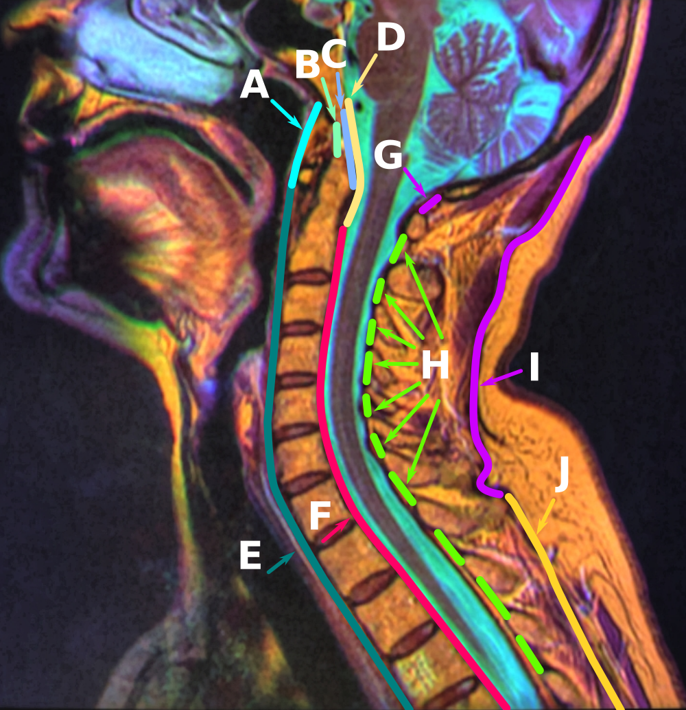
The cruciate ligament of the atlas (cruciform ligament) is a cross-shaped (thus the name) ligament in the neck forming part of the atlanto-axial joint. It consists of the transverse ligament of atlas, a superior longitudinal band, and an inferior longitudinal band. The cruciate ligament of the atlas prevents abnormal movement of the atlanto-axial joint. It may be torn, such as by fractures of the atlas bone. Structure The cruciate ligament of the atlas consists of the transverse ligament of the atlas, a superior longitudinal band, and an inferior longitudinal band. The superior longitudinal band connects the transverse ligament to the anterior side of the foramen magnum (near the basilar part) in the occipital bone of the skull. The inferior longitudinal band connects the transverse ligament to the body of the axis bone (C2). Variation The inferior longitudinal band may be absent in some people; the rest of the ligament is invariably present. Gerber's ligament In about h ... [...More Info...] [...Related Items...] OR: [Wikipedia] [Google] [Baidu] |
Tectorial Membrane Of Atlanto-axial Joint
The tectorial membrane of atlanto-axial joint (occipitoaxial ligaments) is a tough membrane/broad, strong band representing the superior-ward prolongation of the posterior longitudinal ligament (the two being continuous). It attaches inferiorly onto (the posterior aspect of) the body of axis. It broadens superiorly. Superiorly, the membrane extends deep to the median atlanto-axial joint and its associated ligaments, then through the foramen magnum into the cranial cavity where it ends by attaching onto the basilar part of occipital bone superior to the foramen magnum. Anatomy The membrane broadens superiorly. Structure The membrane consists of two laminae - superficial and deep. The superficial lamina broadens superiorly before attaching onto the superior/internal surface of the basilar part of occipital bone superior to the foramen magnum, here blending with the cranial dura mater. The deep lamina consists of a strong medial band which extends superiorly to the foramen m ... [...More Info...] [...Related Items...] OR: [Wikipedia] [Google] [Baidu] |
Odontoid Process
In anatomy, the axis (from Latin ''axis'', "axle") is the second cervical vertebra (C2) of the spine, immediately inferior to the atlas, upon which the head rests. The spinal cord passes through the axis. The defining feature of the axis is its strong bony protrusion known as the dens, which rises from the superior aspect of the bone. Structure The body is deeper in front or in the back and is prolonged downward anteriorly to overlap the upper and front part of the third vertebra. It presents a median longitudinal ridge in front, separating two lateral depressions for the attachment of the longus colli muscles. Dens The dens, also called the odontoid process, or the peg, is the most pronounced projecting feature of the axis. The dens exhibits a slight constriction where it joins the main body of the vertebra. The condition where the dens is separated from the body of the axis is called ''os odontoideum'' and may cause nerve and circulation compression syndrome. On its ante ... [...More Info...] [...Related Items...] OR: [Wikipedia] [Google] [Baidu] |


