|
Transcytosis
Transcytosis (also known as cytopempsis) is a type of transcellular transport in which various macromolecules are transported across the interior of a cell. Macromolecules are captured in vesicles on one side of the cell, drawn across the cell, and ejected on the other side. Examples of macromolecules transported include IgA, transferrin, and insulin. While transcytosis is most commonly observed in epithelial cells, the process is also present elsewhere. Blood capillaries are a well-known site for transcytosis, though it occurs in other cells, including neurons, osteoclasts and M cells of the intestine. Regulation The regulation of transcytosis varies greatly due to the many different tissues in which this process is observed. Various tissue-specific mechanisms of transcytosis have been identified. Brefeldin A, a commonly used inhibitor of ER-to-Golgi apparatus transport, has been shown to inhibit transcytosis in dog kidney cells, which provided the first clues as to the nat ... [...More Info...] [...Related Items...] OR: [Wikipedia] [Google] [Baidu] |
Transcellular Transport
Transcellular transport involves the transportation of solutes by a cell ''through'' a cell. Transcellular transport can occur in three different ways active transport, passive transport, and transcytosis. Active Transport Main article: Active transport Active transport is the process of moving molecules from an area of low concentrations to an area of high concentration. There are two types of active transport, primary active transport and secondary active transport. Primary active transport uses adenosine triphosphate (ATP) to move specific molecules and solutes against its concentration gradient. Examples of molecules that follow this process are potassium K+, sodium Na+, and calcium Ca2+. A place in the human body where this occurs is in the intestines with the uptake of glucose. Secondary active transport is when one solute moves down the electrochemical gradient to produce enough energy to force the transport of another solute from low concentration to high concentrat ... [...More Info...] [...Related Items...] OR: [Wikipedia] [Google] [Baidu] |
Osteoclast
An osteoclast () is a type of bone cell that breaks down bone tissue. This function is critical in the maintenance, repair, and remodeling of bones of the vertebral skeleton. The osteoclast disassembles and digests the composite of hydrated protein and mineral at a molecular level by secreting acid and a collagenase, a process known as '' bone resorption''. This process also helps regulate the level of blood calcium. Osteoclasts are found on those surfaces of bone that are undergoing resorption. On such surfaces, the osteoclasts are seen to be located in shallow depressions called ''resorption bays (Howship's lacunae)''. The resorption bays are created by the erosive action of osteoclasts on the underlying bone. The border of the lower part of an osteoclast exhibits finger-like processes due to the presence of deep infoldings of the cell membrane; this border is called ''ruffled border''. The ruffled border lies in contact with the bone surface within a resorption bay. The p ... [...More Info...] [...Related Items...] OR: [Wikipedia] [Google] [Baidu] |
Microfold Cell
Microfold cells (or M cells) are found in the gut-associated lymphoid tissue (GALT) of the Peyer's patches in the small intestine, and in the mucosa-associated lymphoid tissue (MALT) of other parts of the gastrointestinal tract. These cells are known to initiate mucosal immunity responses on the apical membrane of the M cells and allow for transport of microbes and particles across the epithelial cell layer from the gut lumen to the lamina propria where interactions with immune cells can take place. Unlike their neighbor cells, M cells have the unique ability to take up antigen from the lumen of the small intestine via endocytosis, phagocytosis, or transcytosis. Antigens are delivered to antigen-presenting cells, such as dendritic cells, and B lymphocytes. M cells express the protease cathepsin E, similar to other antigen-presenting cells. This process takes place in a unique pocket-like structure on their basolateral side. Antigens are recognized via expression of cell surface ... [...More Info...] [...Related Items...] OR: [Wikipedia] [Google] [Baidu] |
RAB17
Ras-related protein Rab-17 is a protein that in humans is encoded by the ''RAB17'' gene. In melanocytic cells RAB17 gene expression may be regulated by MITF Microphthalmia-associated transcription factor also known as class E basic helix-loop-helix protein 32 or bHLHe32 is a protein that in humans is encoded by the ''MITF'' gene. MITF is a basic helix-loop-helix leucine zipper transcription factor .... References Further reading * * * * * * * * * * * * * * {{gene-2-stub ... [...More Info...] [...Related Items...] OR: [Wikipedia] [Google] [Baidu] |
Immunoglobulin A
Immunoglobulin A (Ig A, also referred to as sIgA in its secretory form) is an antibody that plays a role in the immune function of mucous membranes. The amount of IgA produced in association with mucosal membranes is greater than all other types of antibody combined. In absolute terms, between three and five grams are secreted into the intestinal lumen each day. This represents up to 15% of total immunoglobulins produced throughout the body. IgA has two subclasses (IgA1 and IgA2) and can be produced as a monomeric as well as a dimeric form. The IgA dimeric form is the most prevalent and is also called ''secretory IgA'' (sIgA). sIgA is the main immunoglobulin found in mucous secretions, including tears, saliva, sweat, colostrum and secretions from the genitourinary tract, gastrointestinal tract, prostate and respiratory epithelium. It is also found in small amounts in blood. The secretory component of sIgA protects the immunoglobulin from being degraded by proteolytic ... [...More Info...] [...Related Items...] OR: [Wikipedia] [Google] [Baidu] |
Signal Transduction
Signal transduction is the process by which a chemical or physical signal is transmitted through a cell as a series of molecular events, most commonly protein phosphorylation catalyzed by protein kinases, which ultimately results in a cellular response. Proteins responsible for detecting stimuli are generally termed receptors, although in some cases the term sensor is used. The changes elicited by ligand binding (or signal sensing) in a receptor give rise to a biochemical cascade, which is a chain of biochemical events known as a signaling pathway. When signaling pathways interact with one another they form networks, which allow cellular responses to be coordinated, often by combinatorial signaling events. At the molecular level, such responses include changes in the transcription or translation of genes, and post-translational and conformational changes in proteins, as well as changes in their location. These molecular events are the basic mechanisms controlling cell gro ... [...More Info...] [...Related Items...] OR: [Wikipedia] [Google] [Baidu] |
Thyroid Epithelial Cell
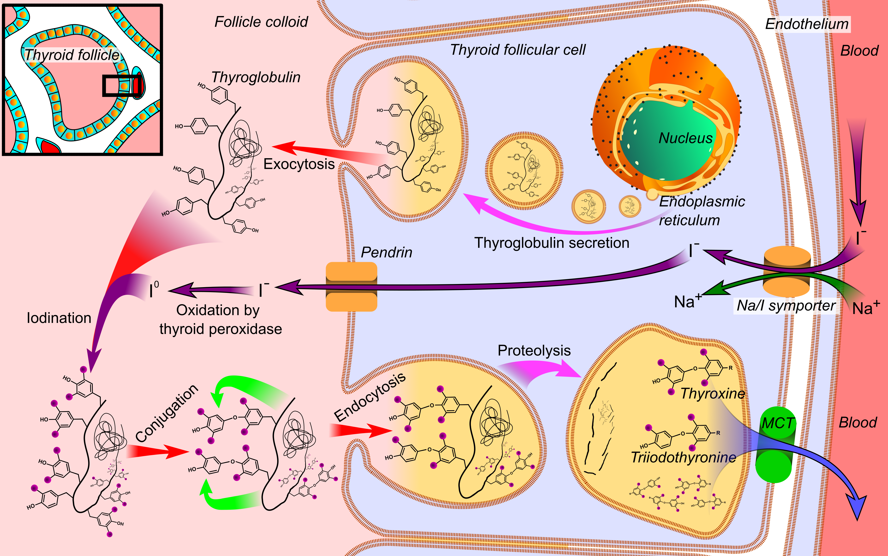
Thyroid follicular cells (also called thyroid epithelial cells or thyrocytes) are the major cell type in the thyroid gland, and are responsible for the production and secretion of the thyroid hormones thyroxine (T4) and triiodothyronine (T3). They form the single layer of cuboidal epithelium that makes up the outer structure of the almost spherical thyroid follicle. Structure Location Thyroid follicular cells form a simple cuboidal epithelium and are arranged in spherical thyroid follicles surrounding a fluid filled space known as the colloid. The interior space formed by the follicular cells is known as the follicular lumen. The basolateral membrane of follicular cells contains thyrotropin receptors which bind to thyroid-stimulating hormone (TSH) found circulating in the blood. Calcitonin-producing parafollicular cells are also found along the basement membrane of the thyroid follicle, interspersed between follicular cells; and in spaces between the spherical follicles. Par ... [...More Info...] [...Related Items...] OR: [Wikipedia] [Google] [Baidu] |
Thyroid
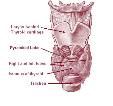
The thyroid, or thyroid gland, is an endocrine gland in vertebrates. In humans it is in the neck and consists of two connected lobes. The lower two thirds of the lobes are connected by a thin band of tissue called the thyroid isthmus. The thyroid is located at the front of the neck, below the Adam's apple. Microscopically, the functional unit of the thyroid gland is the spherical thyroid follicle, lined with follicular cells (thyrocytes), and occasional parafollicular cells that surround a lumen containing colloid. The thyroid gland secretes three hormones: the two thyroid hormones triiodothyronine (T3) and thyroxine (T4)and a peptide hormone, calcitonin. The thyroid hormones influence the metabolic rate and protein synthesis, and in children, growth and development. Calcitonin plays a role in calcium homeostasis. Secretion of the two thyroid hormones is regulated by thyroid-stimulating hormone (TSH), which is secreted from the anterior pituitary gland. TSH is reg ... [...More Info...] [...Related Items...] OR: [Wikipedia] [Google] [Baidu] |
Pregnancy (mammals)
In mammals, pregnancy is the period of reproduction during which a female carries one or more live offspring from implantation in the uterus through gestation. It begins when a fertilized zygote implants in the female's uterus, and ends once it leaves the uterus. Fertilization and implantation During copulation, the male inseminates the female. The spermatozoon fertilizes an ovum or various ova in the uterus or fallopian tubes, and this results in one or multiple zygotes. Sometimes, a zygote can be created by humans outside of the animal's body in the artificial process of in-vitro fertilization. After fertilization, the newly formed zygote then begins to divide through mitosis, forming an embryo, which implants in the female's endometrium. At this time, the embryo usually consists of 50 cells. Development After implantation A blastocele is a small cavity on the center of the embryo, and the developing embryonary cells will grow around it. Then, a flat layer cell forms on ... [...More Info...] [...Related Items...] OR: [Wikipedia] [Google] [Baidu] |
Mammary Gland
A mammary gland is an exocrine gland in humans and other mammals that produces milk to feed young offspring. Mammals get their name from the Latin word ''mamma'', "breast". The mammary glands are arranged in organs such as the breasts in primates (for example, humans and chimpanzees), the udder in ruminants (for example, cows, goats, sheep, and deer), and the dugs of other animals (for example, dogs and cats). Lactorrhea, the occasional production of milk by the glands, can occur in any mammal, but in most mammals, lactation, the production of enough milk for nursing, occurs only in phenotypic females who have gestated in recent months or years. It is directed by hormonal guidance from sex steroids. In a few mammalian species, male lactation can occur. With humans, male lactation can occur only under specific circumstances. Mammals are divided into 3 groups: prototherians, metatherians, and eutherians. In the case of prototherians, both males and females have functional ... [...More Info...] [...Related Items...] OR: [Wikipedia] [Google] [Baidu] |
Prolactin
Prolactin (PRL), also known as lactotropin, is a protein best known for its role in enabling mammals to produce milk. It is influential in over 300 separate processes in various vertebrates, including humans. Prolactin is secreted from the pituitary gland in response to eating, mating, estrogen treatment, ovulation and nursing. It is secreted heavily in pulses in between these events. Prolactin plays an essential role in metabolism, regulation of the immune system and pancreatic development. Discovered in non-human animals around 1930 by Oscar Riddle and confirmed in humans in 1970 by Henry Friesen, prolactin is a peptide hormone, encoded by the ''PRL'' gene. In mammals, prolactin is associated with milk production; in fish it is thought to be related to the control of water and salt balance. Prolactin also acts in a cytokine-like manner and as an important regulator of the immune system. It has important cell cycle-related functions as a growth-, differentiating- and a ... [...More Info...] [...Related Items...] OR: [Wikipedia] [Google] [Baidu] |
Estradiol
Estradiol (E2), also spelled oestradiol, is an estrogen steroid hormone and the major female sex hormone. It is involved in the regulation of the estrous and menstrual female reproductive cycles. Estradiol is responsible for the development of female secondary sexual characteristics such as the breasts, widening of the hips and a female-associated pattern of fat distribution. It is also important in the development and maintenance of female reproductive tissues such as the mammary glands, uterus and vagina during puberty, adulthood and pregnancy. It also has important effects in many other tissues including bone, fat, skin, liver, and the brain. Though estradiol levels in males are much lower than in females, estradiol has important roles in males as well. Apart from humans and other mammals, estradiol is also found in most vertebrates and crustaceans, insects, fish, and other animal species. Estradiol is produced especially within the follicles of the ovaries, but ... [...More Info...] [...Related Items...] OR: [Wikipedia] [Google] [Baidu] |


