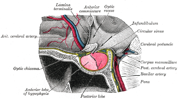
|
Thalamic
The thalamus (: thalami; from Greek θάλαμος, "chamber") is a large mass of gray matter on the lateral wall of the third ventricle forming the dorsal part of the diencephalon (a division of the forebrain). Nerve fibers project out of the thalamus to the cerebral cortex in all directions, known as the thalamocortical radiations, allowing hub-like exchanges of information. It has several functions, such as the relaying of sensory and motor signals to the cerebral cortex and the regulation of consciousness, sleep, and alertness. Anatomically, the thalami are paramedian symmetrical structures (left and right), within the vertebrate brain, situated between the cerebral cortex and the midbrain. It forms during embryonic development as the main product of the diencephalon, as first recognized by the Swiss embryologist and anatomist Wilhelm His Sr. in 1893. Anatomy The thalami are paired structures of gray matter about four centimetres long and ovoid in appearance, located in ... [...More Info...] [...Related Items...] OR: [Wikipedia] [Google] [Baidu] [Amazon] |
Thalamocortical Radiations
In neuroanatomy, thalamocortical radiations, also known as thalamocortical fibers, are the efferent fibers that project from the thalamus to distinct areas of the cerebral cortex. They form fiber bundles that emerge from the lateral surface of the thalamus. Structure Thalamocortical fibers (TC fibers) have been referred to as one of the two constituents of the isothalamus, the other being microneurons. Thalamocortical fibers have a bush or tree-like appearance as they extend into the internal capsule and project to the layers of the cortex. The thalamus supplies all parts of the neocortex with afferents. The main thalamocortical fibers extend from different nuclei of the thalamus and project to the visual cortex, somatosensory (and associated sensori-motor) cortex, and the auditory cortex in the brain. Thalamocortical radiations also innervate gustatory and olfactory pathways, as well as pre-frontal motor areas. Visual input from the optic tract is processed by the lateral ge ... [...More Info...] [...Related Items...] OR: [Wikipedia] [Google] [Baidu] [Amazon] |
Thalamic Reticular Nucleus
The thalamic reticular nucleus is part of the ventral thalamus that forms a capsule around the thalamus laterally. However, recent evidence from mice and fish question this statement and define it as a dorsal thalamic structure. It is separated from the thalamus by the external medullary lamina. Reticular nucleus cells are all GABAergic, and have discoid dendritic arbors in the plane of the nucleus. Thalamic Reticular Nucleus is variously abbreviated TRN, RTN, NRT, and RT. The TRN is found in all mammals. Input and output The thalamic reticular nucleus receives input from the cerebral cortex and dorsal thalamic nuclei. Most input comes from collaterals of fibers passing through the thalamic reticular nucleus. The outputs from the primary thalamic reticular nucleus project to dorsal thalamic nuclei, but never to the cerebral cortex. This is the only thalamic nucleus that does not project to the cerebral cortex. Instead it modulates the information from other nuclei in the thala ... [...More Info...] [...Related Items...] OR: [Wikipedia] [Google] [Baidu] [Amazon] |
Interthalamic Adhesion
The interthalamic adhesion (also known as the massa intermedia, intermediate mass or middle commissure) is a flattened band of tissue that connects both parts of the thalamus at their medial surfaces. The medial surfaces form the upper part of the lateral wall to the third ventricle. In humans, it is only about one centimeter long – though in females, it is about 50% larger on average. Sometimes, it is in two parts – and 20% of the time, it is absent. In other mammals, it is larger. In 1889, a Portuguese anatomist by the name of Macedo examined 215 brains, showing that male humans are approximately twice as likely to lack an interthalamic adhesion as are female humans. He also reported its absence, still reported today in about 20% of humans. Its absence is seen to be of no consequence. The interthalamic adhesion contains nerve cells and nerve fibers; a few of the latter may cross the middle line, but most of them pass toward the middle line and then curve laterally on the ... [...More Info...] [...Related Items...] OR: [Wikipedia] [Google] [Baidu] [Amazon] |
Third Ventricle
The third ventricle is one of the four connected cerebral ventricles of the ventricular system within the mammalian brain. It is a slit-like cavity formed in the diencephalon between the two thalami, in the midline between the right and left lateral ventricles, and is filled with cerebrospinal fluid (CSF). Running through the third ventricle is the interthalamic adhesion, which contains thalamic neurons and fibers that may connect the two thalami. Structure The third ventricle is a narrow, laterally flattened, vaguely rectangular region, filled with cerebrospinal fluid, and lined by ependyma. It is connected at the superior anterior corner to the lateral ventricles, by the interventricular foramina, and becomes the cerebral aqueduct (''aqueduct of Sylvius'') at the posterior caudal corner. Since the interventricular foramina are on the lateral edge, the corner of the third ventricle itself forms a bulb, known as the ''anterior recess'' (it is also known as the ''bulb ... [...More Info...] [...Related Items...] OR: [Wikipedia] [Google] [Baidu] [Amazon] |
Brain
The brain is an organ (biology), organ that serves as the center of the nervous system in all vertebrate and most invertebrate animals. It consists of nervous tissue and is typically located in the head (cephalization), usually near organs for special senses such as visual perception, vision, hearing, and olfaction. Being the most specialized organ, it is responsible for receiving information from the sensory nervous system, processing that information (thought, cognition, and intelligence) and the coordination of motor control (muscle activity and endocrine system). While invertebrate brains arise from paired segmental ganglia (each of which is only responsible for the respective segmentation (biology), body segment) of the ventral nerve cord, vertebrate brains develop axially from the midline dorsal nerve cord as a brain vesicle, vesicular enlargement at the rostral (anatomical term), rostral end of the neural tube, with centralized control over all body segments. All vertebr ... [...More Info...] [...Related Items...] OR: [Wikipedia] [Google] [Baidu] [Amazon] |
List Of Thalamic Nuclei
This traditional list does not accord strictly with human thalamic anatomy. Nucleus (neuroanatomy), Nuclear groups of the thalamus include: *anterior nuclei of thalamus, anterior nuclear group (anteroventral, anterodorsal, anteromedial) *medial nuclear group (medial dorsal nucleus, dorsomedial) **medial dorsal nucleus#Parts of nucleus, parvocellular part ( parvicellular part) **medial dorsal nucleus#Parts of nucleus, magnocellular part *midline nuclear group or paramedian **paratenial nucleus **paraventricular thalamus **reuniens nucleus ( medioventral nucleus) **rhomboidal nucleus **interanteromedial **intermediodorsal *intralaminar nuclear group **anterior (rostral) group ***intralaminar thalamic nuclei#Structure, paracentral nucleus ***central lateral nucleus ***central medial nucleus (''not'' called "centromedial") **posterior (caudal) intralaminar group ***centromedian nucleus ***parafascicular nucleus *lateral nuclear group is replaced by **posterior region ***Pulvinar nucle ... [...More Info...] [...Related Items...] OR: [Wikipedia] [Google] [Baidu] [Amazon] |
Neothalamus
The paleothalamus is an obsolete term for the portion of the thalamus that is believed to be phylogenetically (or evolutionarily) older than other parts of the thalamus. Specifically, the midline and medial nuclei of the thalamus, as well as the intralaminar nucleus, are considered to belong to the paleothalamus The term "paleothalamus" is opposed to the paired term neothalamus (also considered to be obsolete), which designates the phylogenetically (or evolutionarily) newer or younger parts of the thalamus, specifically, lateral nuclei of the thalamus, the pulvinar and the geniculate bodies. Initially, the paleothalamus was distinguished from other parts of the thalamus (i.e. from the neothalamus) on the basis that paleothalamic nuclei were believed to lack reciprocal connections with neocortex The neocortex, also called the neopallium, isocortex, or the six-layered cortex, is a set of layers of the mammalian cerebral cortex involved in higher-order brain functions such ... [...More Info...] [...Related Items...] OR: [Wikipedia] [Google] [Baidu] [Amazon] |
Cerebral Cortex
The cerebral cortex, also known as the cerebral mantle, is the outer layer of neural tissue of the cerebrum of the brain in humans and other mammals. It is the largest site of Neuron, neural integration in the central nervous system, and plays a key role in attention, perception, awareness, thought, memory, language, and consciousness. The six-layered neocortex makes up approximately 90% of the Cortex (anatomy), cortex, with the allocortex making up the remainder. The cortex is divided into left and right parts by the longitudinal fissure, which separates the two cerebral hemispheres that are joined beneath the cortex by the corpus callosum and other commissural fibers. In most mammals, apart from small mammals that have small brains, the cerebral cortex is folded, providing a greater surface area in the confined volume of the neurocranium, cranium. Apart from minimising brain and cranial volume, gyrification, cortical folding is crucial for the Neural circuit, brain circuitry ... [...More Info...] [...Related Items...] OR: [Wikipedia] [Google] [Baidu] [Amazon] |
Lateral Nuclear Group
The lateral nuclear group is a collection of nuclei on the lateral side of the thalamus. This nucleus group is one of the three regions of the thalamus which result from trisection by the Y-shaped internal medullary lamina. The name "lateral nuclear group" is also given to a subset of the lateral group of nuclei which result from trisection by the internal medullary lamina. The lateral nuclear group consists of the following: * lateral dorsal nucleus * lateral posterior nucleus * pulvinar nuclei The lateral region of the thalamus which results from trisection by the internal medullary lamina also includes the ventral nuclear group and the lateral Lateral is a geometric term of location which may also refer to: Biology and healthcare * Lateral (anatomy), a term of location meaning "towards the side" * Lateral cricoarytenoid muscle, an intrinsic muscle of the larynx * Lateral release ( ... and medial geniculate nuclei. References Thalamic nuclei {{Neuroana ... [...More Info...] [...Related Items...] OR: [Wikipedia] [Google] [Baidu] [Amazon] |
Pulvinar Nuclei
The pulvinar nuclei or nuclei of the pulvinar (nuclei pulvinares) are the nuclei ( cell bodies of neurons) located in the thalamus (a part of the vertebrate brain). As a group they make up the collection called the pulvinar of the thalamus (pulvinar thalami), usually just called the pulvinar. The pulvinar is usually grouped as one of the ''lateral thalamic nuclei'' in rodents and carnivores, and stands as an independent complex in primates. Pulvinar acts as an association nucleus that, along with medial dorsal nucleus, connected with parietal, occipital, and temporal lobes, but the function is largely unknown. No distinctive syndrome or obvious sensory deficit can be linked to either one. Structure By convention, the pulvinar is divided into four nuclei: Their connectomic details are as follows: * The ''lateral'' and ''inferior'' pulvinar nuclei have widespread connections with early visual cortical areas. * The dorsal part of the ''lateral'' pulvinar nucleus predominant ... [...More Info...] [...Related Items...] OR: [Wikipedia] [Google] [Baidu] [Amazon] |
Posterior Cerebral Artery
The posterior cerebral artery (PCA) is one of a pair of cerebral arteries that supply oxygenated blood to the occipital lobe, as well as the medial and inferior aspects of the temporal lobe of the human brain. The two arteries originate from the distal end of the basilar artery, where it bifurcates into the left and right posterior cerebral arteries. These anastomose with the middle cerebral artery, middle cerebral arteries and internal carotid artery, internal carotid arteries via the posterior communicating arteries. Structure The posterior cerebral artery is subdivided into 4 segments: P1: pre-communicating segment * Originated at the termination of the basilar artery * May give rise to the artery of Percheron if present P2: post-communicating segment * From the PCOM around the midbrain * Terminates as it enters the quadrigeminal ganglion * Gives rise to the choroidal branches (medial and lateral posterior choroidal arteries) P3: quadrigeminal segment * Courses poster ... [...More Info...] [...Related Items...] OR: [Wikipedia] [Google] [Baidu] [Amazon] |
Midbrain
The midbrain or mesencephalon is the uppermost portion of the brainstem connecting the diencephalon and cerebrum with the pons. It consists of the cerebral peduncles, tegmentum, and tectum. It is functionally associated with vision, hearing, motor control, sleep and wakefulness, arousal (alertness), and temperature regulation.Breedlove, Watson, & Rosenzweig. Biological Psychology, 6th Edition, 2010, pp. 45-46 The name ''mesencephalon'' comes from the Greek ''mesos'', "middle", and ''enkephalos'', "brain". Structure The midbrain is the shortest segment of the brainstem, measuring less than 2cm in length. It is situated mostly in the posterior cranial fossa, with its superior part extending above the tentorial notch. The principal regions of the midbrain are the tectum, the cerebral aqueduct, tegmentum, and the cerebral peduncles. Rostral and caudal, Rostrally the midbrain adjoins the diencephalon (thalamus, hypothalamus, etc.), while Rostral and caudal, cau ... [...More Info...] [...Related Items...] OR: [Wikipedia] [Google] [Baidu] [Amazon] |


