|
Submucosa
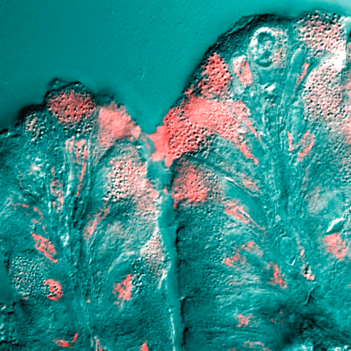
The submucosa (or tela submucosa) is a thin layer of tissue in various organs of the gastrointestinal, respiratory, and genitourinary tracts. It is the layer of dense irregular connective tissue that supports the mucosa (mucous membrane) and joins it to the muscular layer, the bulk of overlying smooth muscle (fibers running circularly within layer of longitudinal muscle). The submucosa ('' sub-'' + ''mucosa'') is to a mucous membrane what the subserosa ('' sub-'' + ''serosa'') is to a serous membrane. Structure Blood vessels, lymphatic vessels, and nerves (all supplying the mucosa) will run through here. In the intestinal wall, tiny parasympathetic ganglia are scattered around forming the submucous plexus (or "Meissner's plexus") where preganglionic parasympathetic neurons synapse with postganglionic nerve fibers that supply the muscularis mucosae. Histologically, the wall of the alimentary canal shows four distinct layers (from the lumen moving out): mucosa, submucosa, ... [...More Info...] [...Related Items...] OR: [Wikipedia] [Google] [Baidu] |
Submucosal Gland
Submucosal glands can refer to various racemose exocrine glands of the mucous type. These glands secrete mucus to facilitate the movement of particles along the body's various tubes, such as the throat and intestines. The mucosa is the lining of the tubes, like a kind of ''skin''. ''Submucosal'' means that the actual gland resides in the connecting tissue below the mucosa. The submucosa is the tissue that connects the mucosa to the muscle outside the tube. The glands themselves are quite complex. The mucus factory is at the bottom, in the submucosa, it is composed of many little sacs (acini) where the mucus originates. Each sac (acinus) has one end that can open and close (dilate) to allow the mucus out. The acini empty into little tubes (tubules) that lead to a reservoir (collecting duct) that has a portal through the ''skin'' (mucosa) that can open and close allowing the mucus into the main tube. The submucosal glands are a companion to goblet cells which also produce mucus, an ... [...More Info...] [...Related Items...] OR: [Wikipedia] [Google] [Baidu] |
Submucous Plexus
The submucosal plexus (Meissner's plexus, plexus of the submucosa, plexus submucosus) lies in the submucosa of the intestinal wall. The nerves of this plexus are derived from the myenteric plexus which itself is derived from the plexuses of parasympathetic nerves around the superior mesenteric artery. Branches from the myenteric plexus perforate the circular muscle fibers to form the submucosal plexus. Ganglia from the plexus extend into the muscularis mucosae and also extend into the mucous membrane. They contain Dogiel cells. The nerve bundles of the submucous plexus are finer than those of the myenteric plexus. Its function is to innervate cells in the epithelial layer and the smooth muscle of the muscularis mucosae. 14% of submucosal plexus neurons are sensory neurons - Dogiel type II, also known as enteric primary afferent neurons or intrinsic primary afferent neurons. History Meissners' plexus was described by German professor Georg Meissner George Meissner (19 Novembe ... [...More Info...] [...Related Items...] OR: [Wikipedia] [Google] [Baidu] |
Meissner's Plexus
The submucosal plexus (Meissner's plexus, plexus of the submucosa, plexus submucosus) lies in the submucosa of the intestinal wall. The nerves of this plexus are derived from the myenteric plexus which itself is derived from the plexuses of parasympathetic nerves around the superior mesenteric artery. Branches from the myenteric plexus perforate the circular muscle fibers to form the submucosal plexus. Ganglia from the plexus extend into the muscularis mucosae and also extend into the mucous membrane. They contain Dogiel cells. The nerve bundles of the submucous plexus are finer than those of the myenteric plexus. Its function is to innervate cells in the epithelial layer and the smooth muscle of the muscularis mucosae. 14% of submucosal plexus neurons are sensory neurons - Dogiel type II, also known as enteric primary afferent neurons or intrinsic primary afferent neurons. History Meissners' plexus was described by German professor Georg Meissner George Meissner (19 Novemb ... [...More Info...] [...Related Items...] OR: [Wikipedia] [Google] [Baidu] |
Mucus
Mucus ( ) is a slippery aqueous secretion produced by, and covering, mucous membranes. It is typically produced from cells found in mucous glands, although it may also originate from mixed glands, which contain both serous and mucous cells. It is a viscous colloid containing inorganic salts, antimicrobial enzymes (such as lysozymes), immunoglobulins (especially IgA), and glycoproteins such as lactoferrin and mucins, which are produced by goblet cells in the mucous membranes and submucosal glands. Mucus serves to protect epithelial cells in the linings of the respiratory, digestive, and urogenital systems, and structures in the visual and auditory systems from pathogenic fungi, bacteria and viruses. Most of the mucus in the body is produced in the gastrointestinal tract. Amphibians, fish, snails, slugs, and some other invertebrates also produce external mucus from their epidermis as protection against pathogens, and to help in movement and is also produced ... [...More Info...] [...Related Items...] OR: [Wikipedia] [Google] [Baidu] |
Gastrointestinal Tract
The gastrointestinal tract (GI tract, digestive tract, alimentary canal) is the tract or passageway of the digestive system that leads from the mouth to the anus. The GI tract contains all the major organs of the digestive system, in humans and other animals, including the esophagus, stomach, and intestines. Food taken in through the mouth is digested to extract nutrients and absorb energy, and the waste expelled at the anus as feces. ''Gastrointestinal'' is an adjective meaning of or pertaining to the stomach and intestines. Most animals have a "through-gut" or complete digestive tract. Exceptions are more primitive ones: sponges have small pores ( ostia) throughout their body for digestion and a larger dorsal pore ( osculum) for excretion, comb jellies have both a ventral mouth and dorsal anal pores, while cnidarians and acoels have a single pore for both digestion and excretion. The human gastrointestinal tract consists of the esophagus, stomach, and intestines, and i ... [...More Info...] [...Related Items...] OR: [Wikipedia] [Google] [Baidu] |
Esophagus
The esophagus (American English) or oesophagus (British English; both ), non-technically known also as the food pipe or gullet, is an organ in vertebrates through which food passes, aided by peristaltic contractions, from the pharynx to the stomach. The esophagus is a fibromuscular tube, about long in adults, that travels behind the trachea and heart, passes through the diaphragm, and empties into the uppermost region of the stomach. During swallowing, the epiglottis tilts backwards to prevent food from going down the larynx and lungs. The word ''oesophagus'' is from Ancient Greek οἰσοφάγος (oisophágos), from οἴσω (oísō), future form of φέρω (phérō, “I carry”) + ἔφαγον (éphagon, “I ate”). The wall of the esophagus from the lumen outwards consists of mucosa, submucosa (connective tissue), layers of muscle fibers between layers of fibrous tissue, and an outer layer of connective tissue. The mucosa is a stratified squamous epi ... [...More Info...] [...Related Items...] OR: [Wikipedia] [Google] [Baidu] |
Dense Irregular Connective Tissue
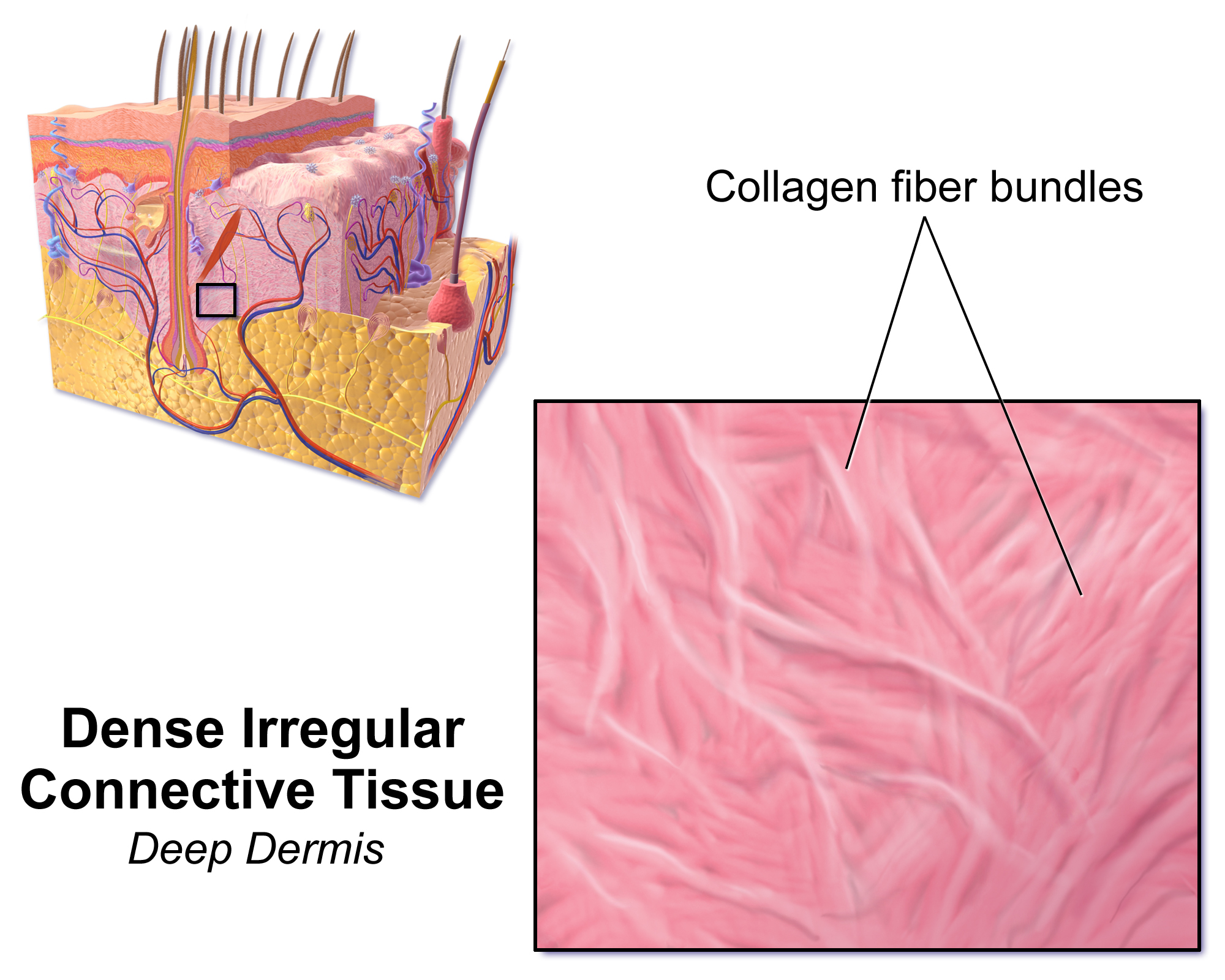
Dense irregular connective tissue has fibers that are not arranged in parallel bundles as in dense regular connective tissue. Dense irregular connective tissue consists of mostly collagen fibers. It has less ground substance than loose connective tissue. Fibroblasts are the predominant cell type, scattered sparsely across the tissue. Function This type of connective tissue is found mostly in the reticular layer (or deep layer) of the dermis. It is also in the sclera and in the deeper skin layers. Due to high portions of collagenous fibers, dense irregular connective tissue provides strength, making the skin resistant to tearing by stretching forces from different directions. Dense irregular connective tissue also makes up submucosa of the digestive tract, lymph nodes, and some types of fascia. Other examples include periosteum and perichondrium The perichondrium (from Greek el, περί, peri, around, label=none and el, χόνδρος, chondros, cartilage, label=none ... [...More Info...] [...Related Items...] OR: [Wikipedia] [Google] [Baidu] |
Muscularis Mucosae
The lamina muscularis mucosae (or muscularis mucosae) is a thin layer ( lamina) of muscle of the gastrointestinal tract, located outside the lamina propria, and separating it from the submucosa. It is present in a continuous fashion from the esophagus to the upper rectum (the exact nomenclature of the rectum's muscle layers is still being debated). A discontinuous muscularis mucosae–like muscle layer is present in the urinary tract, from the renal pelvis to the bladder; as it is discontinuous, it should not be regarded as a true muscularis mucosae. In the gastrointestinal tract, the term ''mucosa'' or ''mucous membrane'' refers to the combination of epithelium, lamina propria, and (where it occurs) muscularis mucosae.H.G. Burkitt et al., ''Wheater's Functional Histology, 3rd ed.'' The etymology suggests this, since the Latin names translate to "the mucosa's own special layer" (''lamina propria mucosae'') and "muscular layer of the mucosa" (''lamina muscularis mucosae''). ... [...More Info...] [...Related Items...] OR: [Wikipedia] [Google] [Baidu] |
Subserosa
The subserosa or tela subserosa, is a thin layer of tissue in the walls of various organs. It is a layer of connective tissue (usually of the areolar type) between the muscular layer (muscularis externa) and the serosa ( serous membrane). The subserosa has clinical importance particularly in cancer staging (for example, in staging stomach cancer or uterine cancer). The subserosa ('' sub-'' + ''serosa'') is to a serous membrane what the submucosa ('' sub-'' + ''mucosa'') is to a mucous membrane A mucous membrane or mucosa is a membrane that lines various cavities in the body of an organism and covers the surface of internal organs. It consists of one or more layers of epithelial cells overlying a layer of loose connective tissue. It is .... References External links * - "Female Reproductive System: oviduct; infundibulum" Histology at uio.noDiagram at uniklinik-saarland.de Membrane biology {{Digestive-stub ... [...More Info...] [...Related Items...] OR: [Wikipedia] [Google] [Baidu] |
Muscular Layer
The muscular layer (muscular coat, muscular fibers, muscularis propria, muscularis externa) is a region of muscle in many organs in the vertebrate body, adjacent to the submucosa. It is responsible for gut movement such as peristalsis. The Latin, tunica muscularis, may also be used. Structure It usually has two layers of smooth muscle: * inner and "circular" * outer and "longitudinal" However, there are some exceptions to this pattern. * In the stomach there are three layers to the muscular layer. Stomach contains an additional oblique muscle layer just interior to circular muscle layer. * In the upper esophagus, part of the externa is ''skeletal muscle'', rather than smooth muscle. * In the vas deferens of the spermatic cord, there are three layers: inner longitudinal, middle circular, and outer longitudinal. * In the ureter the smooth muscle orientation is opposite that of the GI tract. There is an inner longitudinal and an outer circular layer. The inner layer of the muscul ... [...More Info...] [...Related Items...] OR: [Wikipedia] [Google] [Baidu] |
Muscular Coat
The muscular layer (muscular coat, muscular fibers, muscularis propria, muscularis externa) is a region of muscle in many organs in the vertebrate body, adjacent to the submucosa. It is responsible for gut movement such as peristalsis. The Latin, tunica muscularis, may also be used. Structure It usually has two layers of smooth muscle: * inner and "circular" * outer and "longitudinal" However, there are some exceptions to this pattern. * In the stomach there are three layers to the muscular layer. Stomach contains an additional oblique muscle layer just interior to circular muscle layer. * In the upper esophagus, part of the externa is '' skeletal muscle'', rather than smooth muscle. * In the vas deferens of the spermatic cord, there are three layers: inner longitudinal, middle circular, and outer longitudinal. * In the ureter the smooth muscle orientation is opposite that of the GI tract. There is an inner longitudinal and an outer circular layer. The inner layer of the m ... [...More Info...] [...Related Items...] OR: [Wikipedia] [Google] [Baidu] |


