|
Straight Sinus
The straight sinus, also known as tentorial sinus or the , is an area within the skull beneath the brain. It receives blood from the inferior sagittal sinus and the great cerebral vein, and drains into the confluence of sinuses. Structure The straight sinus is situated within the dura mater, where the falx cerebri meets the midline of tentorium cerebelli. It forms from the confluence of the inferior sagittal sinus and the great cerebral vein. It may also drain blood from the superior cerebellar veins and veins from the falx cerebri. In cross-section, it is triangular, contains a few transverse bands across its interior, and increases in size as it proceeds backward. It is usually around 5 cm long. Variation The straight sinus is usually an unpaired structure. However, there may be two straight sinuses, which may be one on top of the other or parallel. Function The straight sinus allows blood to drain from the inferior center of the head outwards posteriorly. It receives ... [...More Info...] [...Related Items...] OR: [Wikipedia] [Google] [Baidu] |
Dura Mater
In neuroanatomy, dura mater is a thick membrane made of dense irregular connective tissue that surrounds the brain and spinal cord. It is the outermost of the three layers of membrane called the meninges that protect the central nervous system. The other two meningeal layers are the arachnoid mater and the pia mater. It envelops the arachnoid mater, which is responsible for keeping in the cerebrospinal fluid. It is derived primarily from the neural crest cell population, with postnatal contributions of the paraxial mesoderm. Structure The dura mater has several functions and layers. The dura mater is a membrane that envelops the arachnoid mater. It surrounds and supports the dural sinuses (also called dural venous sinuses, cerebral sinuses, or cranial sinuses) and carries blood from the brain toward the heart. Cranial dura mater has two layers called '' lamellae'', a superficial layer (also called the periosteal layer), which serves as the skull's inner periosteum, called t ... [...More Info...] [...Related Items...] OR: [Wikipedia] [Google] [Baidu] |
Human Skull
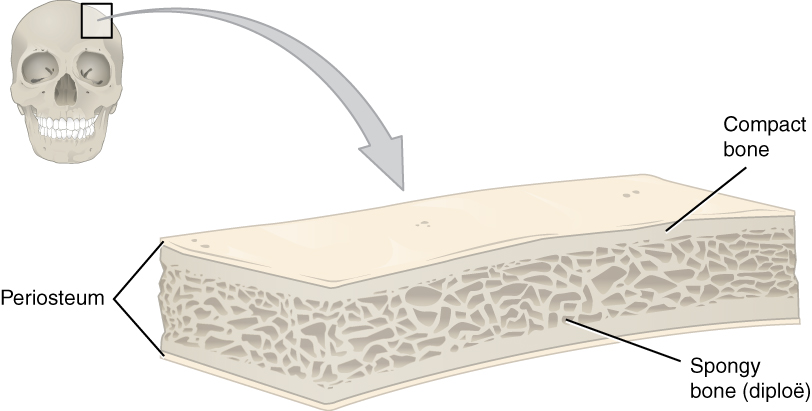
The skull is a bone protective cavity for the brain. The skull is composed of four types of bone i.e., cranial bones, facial bones, ear ossicles and hyoid bone. However two parts are more prominent: the cranium and the mandible. In humans, these two parts are the neurocranium and the viscerocranium ( facial skeleton) that includes the mandible as its largest bone. The skull forms the anterior-most portion of the skeleton and is a product of cephalisation—housing the brain, and several sensory structures such as the eyes, ears, nose, and mouth. In humans these sensory structures are part of the facial skeleton. Functions of the skull include protection of the brain, fixing the distance between the eyes to allow stereoscopic vision, and fixing the position of the ears to enable sound localisation of the direction and distance of sounds. In some animals, such as horned ungulates (mammals with hooves), the skull also has a defensive function by providing the mount (on the ... [...More Info...] [...Related Items...] OR: [Wikipedia] [Google] [Baidu] |
Brain
A brain is an organ that serves as the center of the nervous system in all vertebrate and most invertebrate animals. It is located in the head, usually close to the sensory organs for senses such as vision. It is the most complex organ in a vertebrate's body. In a human, the cerebral cortex contains approximately 14–16 billion neurons, and the estimated number of neurons in the cerebellum is 55–70 billion. Each neuron is connected by synapses to several thousand other neurons. These neurons typically communicate with one another by means of long fibers called axons, which carry trains of signal pulses called action potentials to distant parts of the brain or body targeting specific recipient cells. Physiologically, brains exert centralized control over a body's other organs. They act on the rest of the body both by generating patterns of muscle activity and by driving the secretion of chemicals called hormones. This centralized control allows rapid and coordinated responses ... [...More Info...] [...Related Items...] OR: [Wikipedia] [Google] [Baidu] |
Inferior Sagittal Sinus
The inferior sagittal sinus (also known as inferior longitudinal sinus), within the human head, is an area beneath the brain which allows blood to drain outwards posteriorly from the center of the head. It drains (from the center of the brain) to the straight sinus (at the back of the head), which connects to the transverse sinuses. ''See diagram (at right)'': labeled in the brain as "" (for Latin: ''sinus sagittalis inferior''). The inferior sagittal sinus courses along the inferior border of the falx cerebri, superior to the corpus callosum. It receives blood from the deep and medial aspects of the cerebral hemispheres and drains into the straight sinus. Additional images File:Gray568.png, Sagittal section of the skull, showing the sinuses of the dura. File:Human brain dura mater (reflections) description.JPG, Human brain dura mater (reflections) See also * Dural venous sinuses * Occipital sinus The occipital sinus is the smallest of the dural venous sinuses. It is us ... [...More Info...] [...Related Items...] OR: [Wikipedia] [Google] [Baidu] |
Great Cerebral Vein
The great cerebral vein is one of the large blood vessels in the skull draining the cerebrum of the brain. It is also known as the "vein of Galen", named for its discoverer, the Greek physician Galen. However, it is not the only vein with this eponym. Structure The great cerebral vein is considered one of the deep cerebral veins. Other deep cerebral veins are the internal cerebral veins, formed by the union of the superior thalamostriate vein and the superior choroid vein at the interventricular foramina. The internal cerebral veins can be seen on the superior surfaces of the caudate nuclei and thalami just under the corpus callosum. The veins at the anterior poles of the thalami merge posterior to the pineal gland to form the great cerebral vein. Most of the blood in the deep cerebral veins collects into the great cerebral vein. This comes from the inferior side of the posterior end of the corpus callosum and empties ie similarities, there are also differences between thes ... [...More Info...] [...Related Items...] OR: [Wikipedia] [Google] [Baidu] |
Confluence Of Sinuses
The confluence of sinuses (Latin: confluens sinuum), torcular Herophili, or torcula is the connecting point of the superior sagittal sinus, straight sinus, and occipital sinus. It is below the internal occipital protuberance of the skull. It drains venous blood from the brain into the transverse sinuses. It may be affected by arteriovenous fistulas, a thrombus, major trauma, or surgical damage, and may be imaged with many radiology techniques. Structure The confluence of sinuses is found deep to the internal occipital protuberance of the occipital bone of the skull. This puts it inferior to the occipital lobes of the brain, and posterosuperior to the cerebellum. It connects the ends of the superior sagittal sinus, the straight sinus, and the occipital sinus. Blood from it can drain into the left and right transverse sinuses. It is lined with endothelium, with some smooth muscle. Variation The confluence of sinuses shows significant variation. Most commonly, there i ... [...More Info...] [...Related Items...] OR: [Wikipedia] [Google] [Baidu] |
Skull
The skull is a bone protective cavity for the brain. The skull is composed of four types of bone i.e., cranial bones, facial bones, ear ossicles and hyoid bone. However two parts are more prominent: the cranium and the mandible. In humans, these two parts are the neurocranium and the viscerocranium ( facial skeleton) that includes the mandible as its largest bone. The skull forms the anterior-most portion of the skeleton and is a product of cephalisation—housing the brain, and several sensory structures such as the eyes, ears, nose, and mouth. In humans these sensory structures are part of the facial skeleton. Functions of the skull include protection of the brain, fixing the distance between the eyes to allow stereoscopic vision, and fixing the position of the ears to enable sound localisation of the direction and distance of sounds. In some animals, such as horned ungulates (mammals with hooves), the skull also has a defensive function by providing the mount (on the ... [...More Info...] [...Related Items...] OR: [Wikipedia] [Google] [Baidu] |
Dura Mater
In neuroanatomy, dura mater is a thick membrane made of dense irregular connective tissue that surrounds the brain and spinal cord. It is the outermost of the three layers of membrane called the meninges that protect the central nervous system. The other two meningeal layers are the arachnoid mater and the pia mater. It envelops the arachnoid mater, which is responsible for keeping in the cerebrospinal fluid. It is derived primarily from the neural crest cell population, with postnatal contributions of the paraxial mesoderm. Structure The dura mater has several functions and layers. The dura mater is a membrane that envelops the arachnoid mater. It surrounds and supports the dural sinuses (also called dural venous sinuses, cerebral sinuses, or cranial sinuses) and carries blood from the brain toward the heart. Cranial dura mater has two layers called '' lamellae'', a superficial layer (also called the periosteal layer), which serves as the skull's inner periosteum, called t ... [...More Info...] [...Related Items...] OR: [Wikipedia] [Google] [Baidu] |
Falx Cerebri
The falx cerebri (also known as the cerebral falx) is a large, crescent-shaped fold of dura mater that descends vertically into the longitudinal fissure between the cerebral hemispheres of the human brain,Saladin K. "Anatomy & Physiology: The Unity of Form and Function. New York: McGraw Hill, 2014. Print. pp 512, 770-773 separating the two hemispheres and supporting dural sinuses that provide venous and CSF drainage to the brain. The falx cerebri is often subject to age-related calcification, and a site of falcine meningiomas. The falx cerebri is named for its sickle-like form. Anatomy The falx cerebri is a strong, crescent-shaped sheet lying in the sagittal plane. It is a dural formation (one of four dural partitions of the brain along with the falx cerebelli, tentorium cerebelli, and diaphragma sellae); it is formed through invagination of the dura mater into the longitudinal fissure between the cerebral hemispheres. Anteriorly, the falx cerebri is narrower, thinner, an ... [...More Info...] [...Related Items...] OR: [Wikipedia] [Google] [Baidu] |
Tentorium Cerebelli
The cerebellar tentorium or tentorium cerebelli (Latin for "tent of the cerebellum") is an extension of the dura mater that separates the cerebellum from the inferior portion of the occipital lobes. Structure The cerebellar tentorium is an arched lamina, elevated in the middle, and inclining downward toward the circumference. It covers the top of the cerebellum, and supports the occipital lobes of the brain. Its anterior border is free and concave, and bounds a large oval opening, the tentorial incisure, through which pass the cerebral peduncles. It is attached, behind, by its convex border, to the transverse ridges upon the inner surface of the occipital bone, and there encloses the transverse sinuses; in front, to the superior angle of the petrous part of the temporal bone on either side, enclosing the superior petrosal sinuses. At the apex of the petrous part of the temporal bone the free and attached borders meet, and, crossing one another, are continued forward to ... [...More Info...] [...Related Items...] OR: [Wikipedia] [Google] [Baidu] |
Journal Of Neurosurgery
The ''Journal of Neurosurgery'' is a monthly peer-reviewed medical journal covering all aspects of neurosurgery. It is published by the American Association of Neurological Surgeons and the editor-in-chief is James Rutka. It was established in 1944, with Louise Eisenhardt as founding editor. Originally published bimonthly, it switched to a monthly schedule in 1962. All content is freely available online after 12 months, until it is 10 years old. Editors-in-chief The following persons have been editors-in-chief of the journal: * James Rutka James Rutka (born January 14, 1956) is a Canadian neurosurgeon from Toronto, Canada. Rutka served as RS McLaughlin Professor and Chair of the Department of Surgery in the Faculty of Medicine at the University of Toronto from 2011 – 2022. He ... (2013–present) * John A. Jane (1992–2013) * Thoralf Sundt, Jr. (1989–1992) * William Collins Jr. (1985–1989) * Henry Schwartz (1975–1985) * Henry Heyl (1965–1975) * Louise Eisenhardt ... [...More Info...] [...Related Items...] OR: [Wikipedia] [Google] [Baidu] |
Superior Cerebellar Veins
The cerebellar veins are veins which drain the cerebellum. They consist of the superior cerebellar veins and the inferior cerebellar veins (dorsal cerebellar veins). The superior cerebellar veins drain to the straight sinus and the internal cerebral veins. The inferior cerebellar veins drain to the transverse sinus, the superior petrosal sinus, and the occipital sinus. Structure The superior cerebellar veins pass partly forward and medialward, across the superior cerebellar vermis. They end in the straight sinus, and the internal cerebral veins, partly lateralward to the transverse and superior petrosal sinuses. The inferior cerebellar veins are larger. They end in the transverse sinus, the superior petrosal sinus, and the occipital sinus. Clinical significance The cerebellar veins may be affected by infarction or thrombosis Thrombosis (from Ancient Greek "clotting") is the formation of a blood clot inside a blood vessel, obstructing the flow of blood through the ci ... [...More Info...] [...Related Items...] OR: [Wikipedia] [Google] [Baidu] |


