|
Solenoglypha
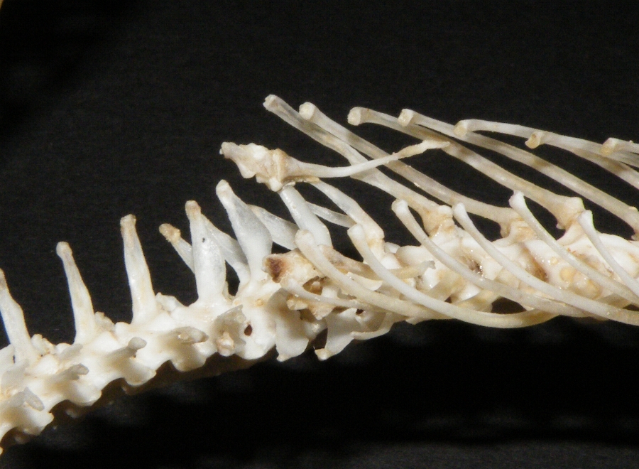
A snake skeleton consists primarily of the skull, vertebrae, and ribs, with only vestigial remnants of the limbs. Skull The skull of a snake is a very complex structure, with numerous joints to allow the snake to swallow prey far larger than its head. The typical snake skull has a solidly ossified braincase, with the separate frontal bones and the united parietal bones extending downward to the basisphenoid, which is large and extends forward into a rostrum extending to the ethmoidal region. The nose is less ossified, and the paired nasal bones are often attached only at their base. The occipital condyle is either trilobate and formed by the basioccipital and the exoccipitals, or a simple knob formed by the basioccipital; the supraoccipital is excluded from the foramen magnum. The basioccipital may bear a curved ventral process or hypapophysis in the vipers. The prefrontal bone is situated, on each side, between the frontal bone and the maxilla, and may or may not b ... [...More Info...] [...Related Items...] OR: [Wikipedia] [Google] [Baidu] |
Snake Skeleton
A snake skeleton consists primarily of the skull, vertebrae, and ribs, with only vestigial remnants of the limbs. Skull The skull of a snake is a very complex structure, with numerous joints to allow the snake to swallow prey far larger than its head. The typical snake skull has a solidly ossified braincase, with the separate frontal bones and the united parietal bones extending downward to the basisphenoid, which is large and extends forward into a rostrum extending to the ethmoidal region. The nose is less ossified, and the paired nasal bones are often attached only at their base. The occipital condyle is either trilobate and formed by the basioccipital and the exoccipitals, or a simple knob formed by the basioccipital; the supraoccipital is excluded from the foramen magnum. The basioccipital may bear a curved ventral process or hypapophysis in the vipers. The prefrontal bone is situated, on each side, between the frontal bone and the maxilla, and may or may not b ... [...More Info...] [...Related Items...] OR: [Wikipedia] [Google] [Baidu] |
Intercalate
Intercalation may refer to: *Intercalation (chemistry), insertion of a molecule (or ion) into layered solids such as graphite * Intercalation (timekeeping), insertion of a leap day, week or month into some calendar years to make the calendar follow the seasons *Intercalation (university administration), period when a student is officially given time off from studying for an academic degree * Intercalation (geology), a special form of interbedding, where two distinct depositional environments in close spatial proximity migrate back and forth across the border zone *Intercalary chapter, a chapter in a novel that does not further the plot. See also frame story (sometimes called intercalation). * In biology: ** Intercalary segment, an appendage-less segment in the segmental composition of the heads of insects and Myriapoda **Intercalation (biochemistry), process discovered by Leonard Lerman by which certain drugs and mutagens insert themselves between base pairs of DNA **Intercalated ... [...More Info...] [...Related Items...] OR: [Wikipedia] [Google] [Baidu] |
Supraorbital Bone
Supraorbital refers to the region immediately above the eye sockets, where in humans the eyebrows are located. It denotes several anatomical features, such as: *Supraorbital artery *Supraorbital foramen * Supraorbital gland *Supraorbital nerve The supraorbital nerve is one of two branches of the frontal nerve, itself a branch of the ophthalmic nerve. The other branch of the frontal nerve is the supratrochlear nerve. Structure The supraorbital nerve branches from the frontal nerve mi ... * Supraorbital ridge * Supraorbital vein {{disambig ... [...More Info...] [...Related Items...] OR: [Wikipedia] [Google] [Baidu] |
Pythonidae
The Pythonidae, commonly known as pythons, are a family of nonvenomous snakes found in Africa, Asia, and Australia. Among its members are some of the largest snakes in the world. Ten genera and 42 species are currently recognized. Distribution and habitat Pythons are found in sub-Saharan Africa, Nepal, India, Bangladesh, Sri Lanka, Southeast Asia, southeastern Pakistan, southern China, the Philippines and Australia. In the United States, an introduced population of Burmese pythons, ''Python bivittatus'', has existed as an invasive species in the Everglades National Park since the late 1990s. Common names * Sinhala - පිඹුරා (''Pimbura'') * Telugu - కొండచిలువ (Kondachiluva) * Odia - ଅଜଗର (Ajagara) *Malayalam - പെരുമ്പാമ്പ് (perumpāmp) *Hindi - अजगर ('Ajgar') Conservation Many species have been hunted aggressively, which has greatly reduced the population of some, such as the Indian python, ''Python ... [...More Info...] [...Related Items...] OR: [Wikipedia] [Google] [Baidu] |
Orbit (anatomy)
In anatomy, the orbit is the cavity or socket of the skull in which the eye and its appendages are situated. "Orbit" can refer to the bony socket, or it can also be used to imply the contents. In the adult human, the volume of the orbit is , of which the eye occupies . The orbital contents comprise the eye, the orbital and retrobulbar fascia, extraocular muscles, cranial nerves II, III, IV, V, and VI, blood vessels, fat, the lacrimal gland with its sac and duct, the eyelids, medial and lateral palpebral ligaments, cheek ligaments, the suspensory ligament, septum, ciliary ganglion and short ciliary nerves. Structure The orbits are conical or four-sided pyramidal cavities, which open into the midline of the face and point back into the head. Each consists of a base, an apex and four walls."eye, human."Encyclopædia Britannica from Encyclopædia Britannica 2006 Ultimate Reference Suite DVD 2009 Openings There are two important foramina, or windows, two important ... [...More Info...] [...Related Items...] OR: [Wikipedia] [Google] [Baidu] |
Postfrontal Bone
The skull is a bone protective cavity for the brain. The skull is composed of four types of bone i.e., cranial bones, facial bones, ear ossicles and hyoid bone. However two parts are more prominent: the cranium and the mandible. In humans, these two parts are the neurocranium and the viscerocranium ( facial skeleton) that includes the mandible as its largest bone. The skull forms the anterior-most portion of the skeleton and is a product of cephalisation—housing the brain, and several sensory structures such as the eyes, ears, nose, and mouth. In humans these sensory structures are part of the facial skeleton. Functions of the skull include protection of the brain, fixing the distance between the eyes to allow stereoscopic vision, and fixing the position of the ears to enable sound localisation of the direction and distance of sounds. In some animals, such as horned ungulates (mammals with hooves), the skull also has a defensive function by providing the mount (on the fr ... [...More Info...] [...Related Items...] OR: [Wikipedia] [Google] [Baidu] |
Maxilla
The maxilla (plural: ''maxillae'' ) in vertebrates is the upper fixed (not fixed in Neopterygii) bone of the jaw formed from the fusion of two maxillary bones. In humans, the upper jaw includes the hard palate in the front of the mouth. The two maxillary bones are fused at the intermaxillary suture, forming the anterior nasal spine. This is similar to the mandible (lower jaw), which is also a fusion of two mandibular bones at the mandibular symphysis. The mandible is the movable part of the jaw. Structure In humans, the maxilla consists of: * The body of the maxilla * Four processes ** the zygomatic process ** the frontal process of maxilla ** the alveolar process ** the palatine process * three surfaces – anterior, posterior, medial * the Infraorbital foramen * the maxillary sinus * the incisive foramen Articulations Each maxilla articulates with nine bones: * two of the cranium: the frontal and ethmoid * seven of the face: the nasal, zygomatic, lacrimal ... [...More Info...] [...Related Items...] OR: [Wikipedia] [Google] [Baidu] |
Prefrontal Bone
The prefrontal bone is a bone separating the lacrimal and frontal bones in many tetrapod skulls. It first evolved in the sarcopterygian clade Rhipidistia, which includes lungfish and the Tetrapodomorpha. The prefrontal is found in most modern and extinct lungfish, amphibians and reptiles. The prefrontal is lost in early mammaliaforms and so is not present in modern mammals either. In dinosaurs The prefrontal bone is a very small bone near the top of the skull, which is lost in many groups of coelurosaurian theropod dinosaurs and is completely absent in their modern descendants, the birds. Conversely, a well developed prefrontal is considered to be a primitive feature in dinosaurs. The prefrontal makes contact with several other bones in the skull. The anterior part of the bone articulates with the nasal bone and the lacrimal bone. The posterior part of the bone articulates with the frontal Front may refer to: Arts, entertainment, and media Films * ''The Front'' (194 ... [...More Info...] [...Related Items...] OR: [Wikipedia] [Google] [Baidu] |
Viperidae
The Viperidae (vipers) are a family of snakes found in most parts of the world, except for Antarctica, Australia, Hawaii, Madagascar, and various other isolated islands. They are venomous and have long (relative to non-vipers), hinged fangs that permit deep penetration and injection of their venom. Four subfamilies are currently recognized. They are also known as viperids. The name "viper" is derived from the Latin word ''vipera'', -''ae'', also meaning viper, possibly from ''vivus'' ("living") and ''parere'' ("to beget"), referring to the trait viviparity (giving live birth) common in vipers like most of the species of Boidae. Description All viperids have a pair of relatively long solenoglyphous (hollow) fangs that are used to inject venom from glands located towards the rear of the upper jaws, just behind the eyes. Each of the two fangs is at the front of the mouth on a short maxillary bone that can rotate back and forth. When not in use, the fangs fold back against th ... [...More Info...] [...Related Items...] OR: [Wikipedia] [Google] [Baidu] |
Foramen Magnum
The foramen magnum ( la, great hole) is a large, oval-shaped opening in the occipital bone of the skull. It is one of the several oval or circular openings (foramina) in the base of the skull. The spinal cord, an extension of the medulla oblongata, passes through the foramen magnum as it exits the cranial cavity. Apart from the transmission of the medulla oblongata and its membranes, the foramen magnum transmits the vertebral arteries, the anterior and posterior spinal arteries, the tectorial membranes and alar ligaments. It also transmits the accessory nerve into the skull. The foramen magnum is a very important feature in bipedal mammals. One of the attributes of a biped's foramen magnum is a forward shift of the anterior border of the cerebellar tentorium; this is caused by the shortening of the cranial base. Studies on the foramen magnum position have shown a connection to the functional influences of both posture and locomotion. The forward shift of the foramen magn ... [...More Info...] [...Related Items...] OR: [Wikipedia] [Google] [Baidu] |


