|
Substantia Gelatinosa Of Rolando
The apex of the posterior grey column, one of the three grey columns of the spinal cord, is capped by a V-shaped or crescentic mass of translucent, gelatinous neuroglia, termed the substantia gelatinosa of Rolando (or SGR) (or gelatinous substance of posterior horn of spinal cord), which contains both neuroglia cells, and small neurons. The gelatinous appearance is due to an abundance of neuropil with a very low concentration of myelinated fibers. It extends the entire length of the spinal cord and into the medulla oblongata where it becomes the spinal trigeminal nucleus. It is named after Luigi Rolando. It corresponds to Rexed lamina II. Structure The SGR, or lamina II, is composed of an outer lamina II and an inner lamina II. In rodents, the inner lamina II is divided into a dorsal and ventral inner lamina II. The distinction between these laminae lies in the areas of the spinal cord that send information to and from the laminae (input and output projections). The cel ... [...More Info...] [...Related Items...] OR: [Wikipedia] [Google] [Baidu] |
Rexed Lamina
The Rexed laminae (singular: Rexed lamina) comprise a system of ten layers of grey matter (I–X), identified in the early 1950s by Bror Rexed to label portions of the grey columns of the spinal cord. Similar to Brodmann areas, they are defined by their cellular structure rather than by their location, but the location still remains reasonably consistent. Laminae * Posterior grey column: I–VI ** Lamina I: marginal nucleus of spinal cord or posteromarginal nucleus ** Lamina II: substantia gelatinosa of Rolando ** Laminae III and IV: nucleus proprius ** Lamina V: Neck of the dorsal horn. Neurons within lamina V are mainly involved in processing sensory afferent stimuli from cutaneous, muscle and joint mechanical nociceptors as well as visceral nociceptors. This layer is home to wide dynamic range tract neurons, interneurons and propriospinal neurons. Viscerosomatic pain signal convergence often occurs in this lamina due to the presence of wide dynamic range tract neurons ... [...More Info...] [...Related Items...] OR: [Wikipedia] [Google] [Baidu] |
Spinothalamic Tract
The spinothalamic tract is a nerve tract in the anterolateral system in the spinal cord. This tract is an ascending sensory pathway to the thalamus. From the ventral posterolateral nucleus in the thalamus, sensory information is relayed upward to the somatosensory cortex of the postcentral gyrus. The spinothalamic tract consists of two adjacent pathways: anterior and lateral. The anterior spinothalamic tract carries information about crude touch. The lateral spinothalamic tract conveys pain and temperature. In the spinal cord, the spinothalamic tract has somatotopic organization. This is the segmental organization of its cervical, thoracic, lumbar, and sacral components, which is arranged from most medial to most lateral respectively. The pathway crosses over ( decussates) at the level of the spinal cord, rather than in the brainstem like the dorsal column-medial lemniscus pathway and lateral corticospinal tract. It is one of the three tracts which make up the anterola ... [...More Info...] [...Related Items...] OR: [Wikipedia] [Google] [Baidu] |
Marginal Nucleus Of The Spinal Cord
The marginal nucleus of spinal cord, posteromarginal nucleus, or spinal lamina 1 (Rexed lamina 1) is located at the most dorsal aspect of the posterior grey column of the spinal cord. The neurons located here receive input primarily from Lissauer's tract and relay information related to pain and temperature sensation. Pain sensation relayed here cannot be modulated, e.g. pain from burning the skin. The axons of neurons contribute to the lateral spinothalamic tract The spinothalamic tract is a nerve tract in the anterolateral system in the spinal cord. This tract is an ascending sensory pathway to the thalamus. From the ventral posterolateral nucleus in the thalamus, sensory information is relayed upwar .... References Back anatomy Spinal cord {{Neuroanatomy-stub ... [...More Info...] [...Related Items...] OR: [Wikipedia] [Google] [Baidu] |
Glial Cell Line-derived Neurotrophic Factor
Glial cell line-derived neurotrophic factor (GDNF) is a protein that, in humans, is encoded by the ''GDNF'' gene. GDNF is a small protein that potently promotes the survival of many types of neurons. It signals through GFRα receptors, particularly GFRα1. It is also responsible for the determination of spermatogonia into primary spermatocytes, i.e. it is received by RET proto-oncogene (RET) and by forming gradient with SCF it divides the spermatogonia into two cells. As the result there is retention of spermatogonia and formation of spermatocyte. History GDNF was discovered in 1991 and was the first identified member of the GDNF family of ligands (GFL). Structure GDNF has a structure that is similar to TGF beta 2. GDNF has two finger-like structures that interact with the GFRα1 receptor. N-linked glycosylation, which occurs during the secretion of pro-GDNF, takes place at the tip of one of the finger-like structures. The C-terminal of mature GDNF plays an important r ... [...More Info...] [...Related Items...] OR: [Wikipedia] [Google] [Baidu] |
Somatostatin
Somatostatin, also known as growth hormone-inhibiting hormone (GHIH) or by #Nomenclature, several other names, is a peptide hormone that regulates the endocrine system and affects neurotransmission and cell proliferation via interaction with G protein-coupled somatostatin receptors and inhibition of the release of numerous secondary hormones. Somatostatin inhibits insulin and glucagon secretion. Somatostatin has two active forms produced by the alternative cleavage of a single preproprotein: one consisting of 14 amino acids (shown in infobox to right), the other consisting of 28 amino acids. Among the vertebrates, there exist six different somatostatin genes that have been named: ''SS1'', ''SS2'', ''SS3'', ''SS4'', ''SS5'' and ''SS6''. Zebrafish have all six. The six different genes, along with the five different somatostatin receptors, allow somatostatin to possess a large range of functions. Humans have only one somatostatin gene, ''SST''. Nomenclature Synonyms of "somatost ... [...More Info...] [...Related Items...] OR: [Wikipedia] [Google] [Baidu] |
Brain-derived Neurotrophic Factor
Brain-derived neurotrophic factor (BDNF), or abrineurin, is a protein found in the and the periphery. that, in humans, is encoded by the ''BDNF'' gene. BDNF is a member of the neurotrophin family of growth factors, which are related to the canonical nerve growth factor (NGF), a family which also includes NT-3 and NT-4/NT-5. Neurotrophic factors are found in the brain and the periphery. BDNF was first isolated from a pig brain in 1982 by Yves-Alain Barde and Hans Thoenen. BDNF activates the TrkB tyrosine kinase receptor. Function BDNF acts on certain neurons of the central nervous system and the peripheral nervous system expressing TrkB, helping to support survival of existing neurons, and encouraging growth and differentiation of new neurons and synapses. In the brain it is active in the hippocampus, cortex, and basal forebrain areas vital to learning, memory, and higher thinking. BDNF is also expressed in the retina, kidneys, prostate, motor neurons, and skeletal ... [...More Info...] [...Related Items...] OR: [Wikipedia] [Google] [Baidu] |
A Delta Fiber
Group A nerve fibers are one of the three classes of nerve fiber as ''generally classified'' by Erlanger and Gasser. The other two classes are the group B nerve fibers, and the group C nerve fibers. Group A are heavily myelinated, group B are moderately myelinated, and group C are unmyelinated. The other classification is a sensory grouping that uses the terms '' type Ia and type Ib'', '' type II'', ''type III'', and ''type IV'', sensory fibers. Types There are four subdivisions of group A nerve fibers: alpha (α) Aα; beta (β) Aβ; , gamma (γ) Aγ, and delta (δ) Aδ. These subdivisions have different amounts of myelination and axon thickness and therefore transmit signals at different speeds. Larger diameter axons and more myelin insulation lead to faster signal propagation. Group A nerves are found in both motor and sensory pathways. Different sensory receptors are innervated by different types of nerve fibers. Proprioceptors are innervated by type Ia, Ib and II ... [...More Info...] [...Related Items...] OR: [Wikipedia] [Google] [Baidu] |
Pain
Pain is a distressing feeling often caused by intense or damaging Stimulus (physiology), stimuli. The International Association for the Study of Pain defines pain as "an unpleasant sense, sensory and emotional experience associated with, or resembling that associated with, actual or potential tissue damage." Pain motivates organisms to withdraw from damaging situations, to protect a damaged body part while it heals, and to avoid similar experiences in the future. Congenital insensitivity to pain may result in reduced life expectancy. Most pain resolves once the noxious stimulus is removed and the body has healed, but it may persist despite removal of the stimulus and apparent healing of the body. Sometimes pain arises in the absence of any detectable stimulus, damage or disease. Pain is the most common reason for physician consultation in most developed countries. It is a major symptom in many medical conditions, and can interfere with a person's quality of life and general fun ... [...More Info...] [...Related Items...] OR: [Wikipedia] [Google] [Baidu] |
C Fiber
Group C nerve fibers are one of three classes of nerve fiber in the central nervous system (CNS) and peripheral nervous system (PNS). The Group C fibers are unmyelinated and have a small diameter and low conduction velocity, whereas Groups A and B are myelinated. Group C fibers include postganglionic fibers in the autonomic nervous system (ANS), and nerve fibers at the dorsal roots (IV fiber). These fibers carry sensory information. Damage or injury to nerve fibers causes neuropathic pain. Capsaicin activates C fibre vanilloid receptors, providing the burning sensation associated with chili peppers. Structure and anatomy Location C fibers are one class of nerve fiber found in the nerves of the somatic sensory system. They are afferent fibers, conveying input signals from the periphery to the central nervous system. Structure C fibers are unmyelinated unlike most other fibers in the nervous system. This lack of myelination is the cause of their slow conduction velocit ... [...More Info...] [...Related Items...] OR: [Wikipedia] [Google] [Baidu] |
Postsynaptic
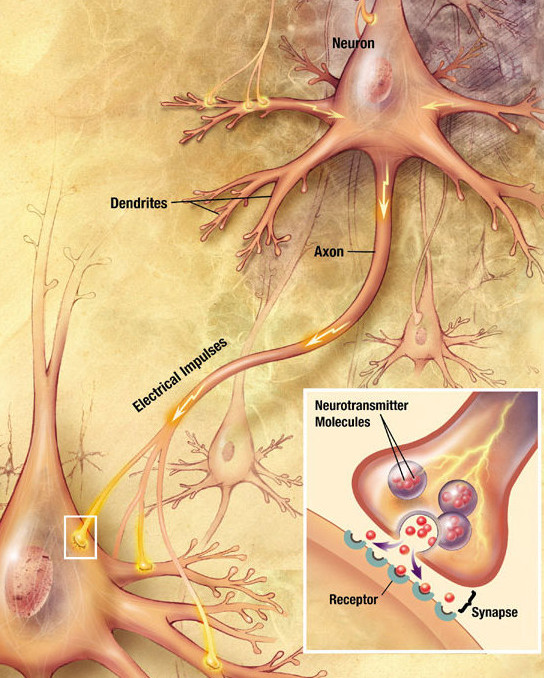
Chemical synapses are biological junctions through which neurons' signals can be sent to each other and to non-neuronal cells such as those in muscles or glands. Chemical synapses allow neurons to form circuits within the central nervous system. They are crucial to the biological computations that underlie perception and thought. They allow the nervous system to connect to and control other systems of the body. At a chemical synapse, one neuron releases neurotransmitter molecules into a small space (the synaptic cleft) that is adjacent to another neuron. The neurotransmitters are contained within small sacs called synaptic vesicles, and are released into the synaptic cleft by exocytosis. These molecules then bind to neurotransmitter receptors on the postsynaptic cell. Finally, the neurotransmitters are cleared from the synapse through one of several potential mechanisms including enzymatic degradation or re-uptake by specific transporters either on the presynaptic cell or o ... [...More Info...] [...Related Items...] OR: [Wikipedia] [Google] [Baidu] |
Presynaptic
In the nervous system, a synapse is a structure that allows a neuron (or nerve cell) to pass an electrical or chemical signal to another neuron or a target effector cell. Synapses can be classified as either chemical or electrical, depending on the mechanism of signal transmission between neurons. In the case of electrical synapses, neurons are coupled bidirectionally with each other through gap junctions and have a connected cytoplasmic milieu. These types of synapses are known to produce synchronous network activity in the brain, but can also result in complicated, chaotic network level dynamics. Therefore, signal directionality cannot always be defined across electrical synapses. Chemical synapses, on the other hand, communicate through neurotransmitters released from the presynaptic neuron into the synaptic cleft. Upon release, these neurotransmitters bind to specific receptors on the postsynaptic membrane, inducing an electrical or chemical response in the target neuron. T ... [...More Info...] [...Related Items...] OR: [Wikipedia] [Google] [Baidu] |
Îş-opioid Receptor
The Îş-opioid receptor or kappa opioid receptor, abbreviated KOR or KOP for its ligand ketazocine, is a G protein-coupled receptor that in humans is encoded by the ''OPRK1'' gene. The KOR is coupled to the G protein Gi/G0 and is one of four related receptors that bind opioid-like compounds in the brain and are responsible for mediating the effects of these compounds. These effects include altering nociception, consciousness, motor control, and mood. Dysregulation of this receptor system has been implicated in alcohol and drug addiction. The KOR is a type of opioid receptor that binds the opioid peptide dynorphin as the primary endogenous ligand (substrate naturally occurring in the body). In addition to dynorphin, a variety of natural alkaloids, terpenes and synthetic ligands bind to the receptor. The KOR may provide a natural addiction control mechanism, and therefore, drugs that target this receptor may have therapeutic potential in the treatment of addiction . There ... [...More Info...] [...Related Items...] OR: [Wikipedia] [Google] [Baidu] |


