|
Stratum Lucidum
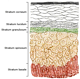
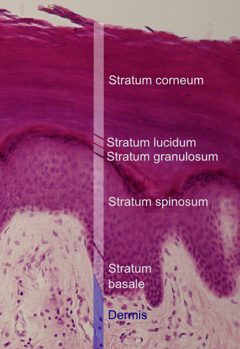
The stratum lucidum (Latin, 'clear layer') is a thin, clear layer of dead skin cells in the epidermis named for its translucent appearance under a microscope. It is readily visible by light microscopy only in areas of thick skin, which are found on the palms of the hands and the soles of the feet. Located between the stratum granulosum and stratum corneum layers, it is composed of three to five layers of dead, flattened keratinocytes.McGrath, J.A.; Eady, R.A.; Pope, F.M. (2004). ''Rook's Textbook of Dermatology'' (Seventh Edition). Blackwell Publishing. Pages 3.8. .Tortora, Gerard; Derrickson, Bryan; Principles of Anatomy and Physiology (2009)152 John Wiley & Sons Inc, Hoboken, NJ . The keratinocytes of the stratum lucidum do not feature distinct boundaries and are filled with eleidin, an intermediate form of keratin. They are surrounded by an oily substance that is the result of the exocytosis of lamellar bodies accumulated while the keratinocytes are moving through the st ... [...More Info...] [...Related Items...] OR: [Wikipedia] [Google] [Baidu] |
Epidermal Layers
The epidermis is the outermost of the three layers that comprise the skin, the inner layers being the dermis and Subcutaneous tissue, hypodermis. The epidermal layer provides a barrier to infection from environmental pathogens and regulates the amount of water released from the body into the atmosphere through transepidermal water loss. The epidermis is composed of stratified squamous epithelium, multiple layers of flattened cells that overlie a base layer (stratum basale) composed of Epithelium#Cell types, columnar cells arranged perpendicularly. The layers of cells develop from stem cells in the basal layer. The thickness of the epidermis varies from 31.2μm for the penis to 596.6μm for the Sole (foot), sole of the foot with most being roughly 90μm. Thickness does not vary between the sexes but becomes thinner with age. The human epidermis is an example of epithelium, particularly a stratified squamous epithelium. The word epidermis is derived through Latin , itself and . ... [...More Info...] [...Related Items...] OR: [Wikipedia] [Google] [Baidu] |
Keratin
Keratin () is one of a family of structural fibrous proteins also known as ''scleroproteins''. It is the key structural material making up Scale (anatomy), scales, hair, Nail (anatomy), nails, feathers, horn (anatomy), horns, claws, Hoof, hooves, and the outer layer of skin in vertebrates. Keratin also protects epithelial cells from damage or stress. Keratin is extremely insoluble in water and organic solvents. Keratin monomers assemble into bundles to form intermediate filaments, which are tough and form strong mineralization (biology), unmineralized epidermal appendages found in reptiles, birds, amphibians, and mammals. Excessive keratinization participate in fortification of certain tissues such as in horns of cattle and rhinos, and armadillos' osteoderm. The only other biology, biological matter known to approximate the toughness of keratinized tissue is chitin. Keratin comes in two types: the primitive, softer forms found in all vertebrates and the harder, derived forms fou ... [...More Info...] [...Related Items...] OR: [Wikipedia] [Google] [Baidu] |
Stratum Basale
The stratum basale (basal layer, sometimes referred to as ''stratum germinativum'') is the deepest layer of the five layers of the epidermis, the external covering of skin in mammals. The stratum basale is a single layer of columnar or cuboidal basal cells. The cells are attached to each other and to the overlying stratum spinosum cells by desmosomes and hemidesmosomes. The nucleus is large, ovoid and occupies most of the cell. Some basal cells can act like stem cells with the ability to divide and produce new cells, and these are sometimes called basal keratinocyte stem cells. Others serve to anchor the epidermis glabrous skin (hairless), and hyper-proliferative epidermis (from a skin disease).McGrath, J.A.; Eady, R.A.; Pope, F.M. (2004). ''Rook's Textbook of Dermatology'' (Seventh Edition). Blackwell Publishing. Pages 3.7. . They divide to form the keratinocytes of the stratum spinosum, which migrate superficially. Other types of cells found within the stratum basale ... [...More Info...] [...Related Items...] OR: [Wikipedia] [Google] [Baidu] |
Melanosomes
A melanosome is an organelle found in animal cells and is the site for synthesis, storage and transport of melanin, the most common light-absorbing pigment found in the animal kingdom. Melanosomes are responsible for color and photoprotection in animal cells and tissues. Melanosomes are synthesised in the skin in melanocyte cells, as well as the eye in choroidal melanocytes and retinal pigment epithelial (RPE) cells. In lower vertebrates, they are found in melanophores or chromatophores. Structure Melanosomes are relatively large organelles, measuring up to 500 nm in diameter. They are bound by a bilipid membrane and are, in general, rounded, sausage-like, or cigar-like in shape. The shape is constant for a given species and cell type. They have a characteristic ultrastructure on electron microscopy, which varies according to the maturity of the melanosome, and for research purposes a numeric staging system is sometimes used. Synthesis of melanin Melanosomes are dependen ... [...More Info...] [...Related Items...] OR: [Wikipedia] [Google] [Baidu] |
Mitosis
Mitosis () is a part of the cell cycle in eukaryote, eukaryotic cells in which replicated chromosomes are separated into two new Cell nucleus, nuclei. Cell division by mitosis is an equational division which gives rise to genetically identical cells in which the total number of chromosomes is maintained. Mitosis is preceded by the S phase of interphase (during which DNA replication occurs) and is followed by telophase and cytokinesis, which divide the cytoplasm, organelles, and cell membrane of one cell into two new cell (biology), cells containing roughly equal shares of these cellular components. The different stages of mitosis altogether define the mitotic phase (M phase) of a cell cycle—the cell division, division of the mother cell into two daughter cells genetically identical to each other. The process of mitosis is divided into stages corresponding to the completion of one set of activities and the start of the next. These stages are preprophase (specific to plant ce ... [...More Info...] [...Related Items...] OR: [Wikipedia] [Google] [Baidu] |
Stratum Spinosum
The stratum spinosum (or spinous layer/prickle cell layer) is a layer of the epidermis found between the stratum granulosum and stratum basale. This layer is composed of polyhedral keratinocytes. These are joined with desmosomes. Their spiny (Latin, spinosum) appearance is due to shrinking of the microfilament Microfilaments, also called actin filaments, are protein filaments in the cytoplasm of eukaryotic cells that form part of the cytoskeleton. They are primarily composed of polymers of actin, but are modified by and interact with numerous other ...s between desmosomes that occurs when stained with H&E. Keratinization begins in the stratum spinosum, although the actual keratinocytes begin in the stratum basale. They have large pale-staining nuclei as they are active in synthesizing fibrillar proteins, known as cytokeratin, which build up within the cells aggregating together forming tonofibrils. The tonofibrils go on to form the desmosomes, which allow for strong ... [...More Info...] [...Related Items...] OR: [Wikipedia] [Google] [Baidu] |
Lamellar Bodies
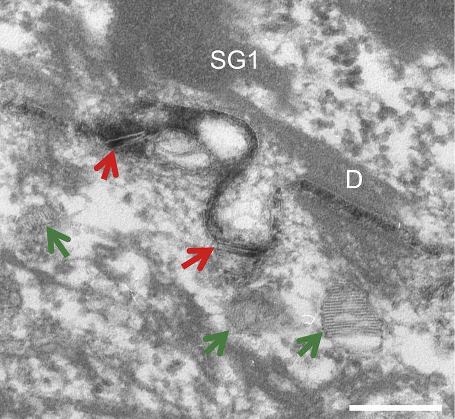
In cell biology, lamellar bodies (otherwise known as lamellar granules, membrane-coating granules (MCGs), keratinosomes or Odland bodies) are secretory organelles found in Alveolar cell, type II alveolar cells in the lungs, and in keratinocytes in the skin. They are oblong structures, appearing about 300-400 nm in width and 100-150 nm in length in transmission electron microscopy images. Lamellar bodies in the pulmonary alveolus, alveoli of the lungs fuse with the cell membrane and release pulmonary surfactant into the extracellular space. Role in lungs In alveolar cells the phosphatidylcholines (choline-based phospholipids) that are stored in the lamellar bodies serve as pulmonary surfactant after being exocytosis, released from the cell. In 1964, using transmission electron microscopy, which at that time was a relatively new tool for ultrastructural elucidation, John Balis identified the presence of lamellar bodies in type II alveolar cells, and further noted that u ... [...More Info...] [...Related Items...] OR: [Wikipedia] [Google] [Baidu] |
Exocytosis
Exocytosis is a term for the active transport process that transports large molecules from cell to the extracellular area. Hormones, proteins and neurotransmitters are examples of large molecules that can be transported out of the cell. Exocytosis is a crucial transport mechanism that enables polar molecules to flow through the cell membranes’ hydrophobic lipid bilayer. The transport process is essential to hormone secretion, immune response and neurotransmission. Both prokaryotes and eukaryotes undergo exocytosis. Prokaryotes secrete molecules and cellular waste through translocons that are localized to the cell membrane. In addition, they secrete molecules to other cells through specialized organs. Eukaryotes rely on multiple cellular processes to perform the exocytosis process. Eukaryotes have several organelles and a nucleus in the cytoplasm that are connected through multiple transport routes, that is formally known as the secretory pathway. This is a complex pathway with ... [...More Info...] [...Related Items...] OR: [Wikipedia] [Google] [Baidu] |
Eleidin
Eleidin is clear intracellular protein which is present in the stratum lucidum of the skin. Eleidin is a transformation product of the amino acid complex keratohyalin, the lifeless matter deposited in the form of minute granules within the protoplasm of living cells. Eleidin is then converted to keratin in the stratum corneum. Eleidin can be found in the vermilion border of the lip. The lip is thinly keratinized and has a high concentration of eleidin. The red appearance of the vermillion border is due to several factors, one of which is the transparent nature of eleidin showing the color of the red blood cell Red blood cells (RBCs), referred to as erythrocytes (, with -''cyte'' translated as 'cell' in modern usage) in academia and medical publishing, also known as red cells, erythroid cells, and rarely haematids, are the most common type of blood cel ...s beneath. References {{protein-stub Human proteins ... [...More Info...] [...Related Items...] OR: [Wikipedia] [Google] [Baidu] |
Keratinocytes
Keratinocytes are the primary type of cell found in the epidermis, the outermost layer of the skin. In humans, they constitute 90% of epidermal skin cells. Basal cells in the basal layer (''stratum basale'') of the skin are sometimes referred to as basal keratinocytes. Keratinocytes form a barrier against environmental damage by heat, UV radiation, water loss, pathogenic bacteria, fungi, parasites, and viruses. A number of structural proteins, enzymes, lipids, and antimicrobial peptides contribute to maintain the important barrier function of the skin. Keratinocytes differentiate from epidermal stem cells in the lower part of the epidermis and migrate towards the surface, finally becoming corneocytes and eventually being shed, which happens every 40 to 56 days in humans. Function The primary function of keratinocytes is the formation of a barrier against environmental damage by heat, UV radiation, dehydration, pathogenic bacteria, fungi, parasites, and viruses. Pa ... [...More Info...] [...Related Items...] OR: [Wikipedia] [Google] [Baidu] |
Stratum Corneum
The stratum corneum (Latin language, Latin for 'horny layer') is the outermost layer of the epidermis (skin), epidermis. Consisting of dead tissue, it protects underlying tissue from infection, dehydration, chemicals and mechanical stress. It is composed of 15–20 layers of flattened cells with no nuclei and cell organelles. Among its properties are mechanical shear, impact resistance, water flux and hydration regulation, microbial proliferation and invasion regulation, initiation of inflammation through cytokine activation and dendritic cell activity, and selective permeability to exclude toxins, irritants, and allergens. The cytoplasm of its cells shows filamentous keratin. These corneocytes are embedded in a lipid matrix composed of ceramides, cholesterol, and fatty acids. Desquamation is the process of cell shedding from the surface of the stratum corneum, balancing proliferating keratinocytes that form in the stratum basale. These cells migrate through the epidermis tow ... [...More Info...] [...Related Items...] OR: [Wikipedia] [Google] [Baidu] |


