|
Solitary Nucleus
The solitary nucleus (SN) (nucleus of the solitary tract, nucleus solitarius, or nucleus tractus solitarii) is a series of neurons whose cell bodies form a roughly vertical column of grey matter in the medulla oblongata of the brainstem. Their axons form the bulk of the enclosed solitary tract. The solitary nucleus can be divided into different parts including dorsomedial, dorsolateral, and ventrolateral subnuclei. The solitary nucleus receives general visceral and special visceral inputs from the facial nerve (CN VII), glossopharyngeal nerve (CN IX) and vagus nerve (CN X); it receives and relays stimuli related to taste and visceral sensation. It sends outputs to various parts of the brain, such as the hypothalamus, thalamus, and reticular formation, forming circuits that contribute to autonomic regulation. Cells along the length of the SN are arranged roughly in accordance with function; for instance, cells involved in taste are located in the rostral part, while those rec ... [...More Info...] [...Related Items...] OR: [Wikipedia] [Google] [Baidu] |
Cranial Nerve Nuclei
A cranial nerve nucleus is a collection of neuron cell bodies (gray matter) in the brain stem that is associated with one or more of the cranial nerves. Axons carrying information to and from the cranial nerves form a synapse first at these nuclei. Lesions occurring at these nuclei can lead to effects resembling those seen by the severing of nerve(s) they are associated with. All the nuclei except that of the trochlear nerve (CN IV) supply nerves of the same side of the body. Structure Motor and sensory In general, motor nuclei are closer to the front (ventral), and sensory nuclei and neurons are closer to the back (dorsal). This arrangement mirrors the arrangement of tracts in the spinal cord. * Close to the midline are the motor efferent nuclei, such as the oculomotor nucleus, which control skeletal muscle. Just lateral to this are the autonomic (or visceral) efferent nuclei. * There is a separation, called the sulcus limitans, and lateral to this are the sensory nuclei. Near ... [...More Info...] [...Related Items...] OR: [Wikipedia] [Google] [Baidu] |
Chorda Tympani
Chorda tympani is a branch of the facial nerve that carries gustatory (taste) sensory innervation from the front of the tongue and parasympathetic ( secretomotor) innervation to the submandibular and sublingual salivary glands. Chorda tympani has a complex course from the brainstem, through the temporal bone and middle ear, into the infratemporal fossa, and ending in the oral cavity. Structure Chorda tympani fibers emerge from the pons of the brainstem as part of the intermediate nerve of the facial nerve. The facial nerve exits the cranial cavity through the internal acoustic meatus and enters the facial canal. In the facial canal, the chorda tympani branches off the facial nerve and enters the lateral wall of the tympanic cavity inside the middle ear where it runs across the tympanic membrane (from posterior to anterior) and medial to the neck of the malleus. The chorda then exits the skull by descending through the petrotympanic fissure into the infrate ... [...More Info...] [...Related Items...] OR: [Wikipedia] [Google] [Baidu] |
Ventral Posteromedial Nucleus
The ventral posteromedial nucleus (VPM) is a nucleus within the ventral posterior nucleus of the thalamus and serves an analogous somatosensory relay role for the ascending trigeminothalamic tracts as its lateral neighbour the ventral posterolateral nucleus serves for dorsal column–medial lemniscus pathway 2nd-order neurons. The term "ventral posteromedial nucleus" was introduced by Le Gros Clark in 1930. Afferents and efferents Orofacial somatosensory The VPM receives second-order general somatic afferent fibers from the ventral trigeminal tract and the dorsal trigeminal tract which convey general somatic sensory information from the face and oral cavity (including touch, pressure, temperature, pain, and propriception). Proprioceptive synapses are situated anteriorly, ones mediating touch in the middle, and nociceptive ones posteriorly. Third-order neurons in turn project to the somatosensory cortex in the postcentral gyrus. Taste The VPM receives second-order tas ... [...More Info...] [...Related Items...] OR: [Wikipedia] [Google] [Baidu] |
Thalamus
The thalamus (: thalami; from Greek language, Greek Wikt:θάλαμος, θάλαμος, "chamber") is a large mass of gray matter on the lateral wall of the third ventricle forming the wikt:dorsal, dorsal part of the diencephalon (a division of the forebrain). Nerve fibers project out of the thalamus to the cerebral cortex in all directions, known as the thalamocortical radiations, allowing hub (network science), hub-like exchanges of information. It has several functions, such as the relaying of sensory neuron, sensory and motor neuron, motor signals to the cerebral cortex and the regulation of consciousness, sleep, and alertness. Anatomically, the thalami are paramedian symmetrical structures (left and right), within the vertebrate brain, situated between the cerebral cortex and the midbrain. It forms during embryonic development as the main product of the diencephalon, as first recognized by the Swiss embryologist and anatomist Wilhelm His Sr. in 1893. Anatomy The thalami ar ... [...More Info...] [...Related Items...] OR: [Wikipedia] [Google] [Baidu] |
Periaqueductal Gray
The periaqueductal gray (PAG), also known as the central gray, is a brain region that plays a critical role in autonomic function, motivated behavior and behavioural responses to threatening stimuli. PAG is also the primary control center for descending pain modulation. It has enkephalin-producing cells that suppress pain. The periaqueductal gray is the gray matter located around the cerebral aqueduct within the tegmentum of the midbrain. It projects to the nucleus raphe magnus, and also contains descending autonomic tracts. The ascending pain and temperature fibers of the spinothalamic tract send information to the PAG via the spinomesencephalic pathway (so-named because the fibers originate in the spine and terminate in the PAG, in the mesencephalon or midbrain). This region has been used as the target for brain-stimulating implants in patients with chronic pain. Role in analgesia Stimulation of the periaqueductal gray matter of the midbrain activates enkephalin-r ... [...More Info...] [...Related Items...] OR: [Wikipedia] [Google] [Baidu] |
Dorsal Longitudinal Fasciculus
The dorsal longitudinal fasciculus (DLF) is a distinctive nerve tract in the midbrain. It extends from the hypothalamus rostrally to the spinal cord caudally, and contains both descending and ascending fibers. Descending fibers arise in the hypothalamus to project directly or indirectly onto autonomic nuclei and lower motor neurons of the brainstem and spinal cord; the descending component is involved in controlling chewing, swallowing, salivation and gastrointestinal secretory function, and shivering. Among the ascending fibers is a serotonin pathway arising in the raphe nuclei. Anatomy Ascending fibers Fibres arising from the nuclei of the reticular formation ascend in the DLF to terminate in the hypothalamus. It conveys visceral information to the brain. Fibers arising from the parabrachial area pass in the DLF to convey taste and general visceral sensation from the nucleus tractus solitarii to the posterior nucleus and periventricular nuclei of the hypothalamus. A ... [...More Info...] [...Related Items...] OR: [Wikipedia] [Google] [Baidu] |
Hypothalamus
The hypothalamus (: hypothalami; ) is a small part of the vertebrate brain that contains a number of nucleus (neuroanatomy), nuclei with a variety of functions. One of the most important functions is to link the nervous system to the endocrine system via the pituitary gland. The hypothalamus is located below the thalamus and is part of the limbic system. It forms the Basal (anatomy), basal part of the diencephalon. All vertebrate brains contain a hypothalamus. In humans, it is about the size of an Almond#Nut, almond. The hypothalamus has the function of regulating certain metabolic biological process, processes and other activities of the autonomic nervous system. It biosynthesis, synthesizes and secretes certain neurohormones, called releasing hormones or hypothalamic hormones, and these in turn stimulate or inhibit the secretion of hormones from the pituitary gland. The hypothalamus controls thermoregulation, body temperature, hunger (physiology), hunger, important aspects o ... [...More Info...] [...Related Items...] OR: [Wikipedia] [Google] [Baidu] |
Aortic Arch
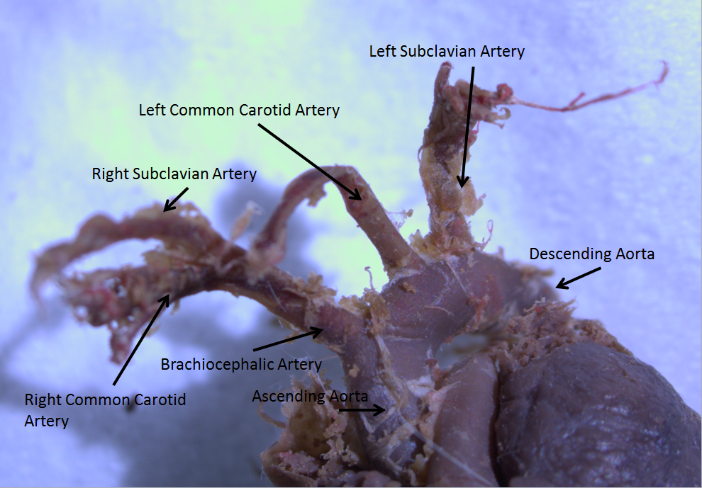
The aortic arch, arch of the aorta, or transverse aortic arch () is the part of the aorta between the ascending and descending aorta. The arch travels backward, so that it ultimately runs to the left of the trachea. Structure The aorta begins at the level of the upper border of the second/third sternocostal articulation of the right side, behind the ventricular outflow tract and pulmonary trunk. The right atrial appendage overlaps it. The first few centimeters of the ascending aorta and pulmonary trunk lies in the same pericardial sheath and runs at first upward, arches over the pulmonary trunk, right pulmonary artery, and right main bronchus to lie behind the right second coastal cartilage. The right lung and sternum lies anterior to the aorta at this point. The aorta then passes posteriorly and to the left, anterior to the trachea, and arches over left main bronchus and left pulmonary artery, and reaches to the left side of the T4 vertebral body. Apart from T4 vertebral ... [...More Info...] [...Related Items...] OR: [Wikipedia] [Google] [Baidu] |
Aortic Body
The aortic bodies are one of several small clusters of peripheral chemoreceptors located along the aortic arch. They are important in measuring partial pressures of oxygen and carbon dioxide in the blood, and blood pH. Structure The aortic bodies are collections of chemoreceptors present on the aortic arch. Most are located above the aortic arch, while some are located on the posterior side of the aortic arch between it and the pulmonary artery below. They consist of glomus cells and sustentacular cells. Some sources equate the "aortic bodies" and " paraaortic bodies", while other sources explicitly distinguish between the two. When a distinction is made, the "aortic bodies" are chemoreceptors which regulate the circulatory system, while the "paraaortic bodies" are the chromaffin cells which manufacture catecholamines. Function The aortic bodies measure partial gas pressures and the composition of arterial blood flowing past it. These changes may include: * oxygen par ... [...More Info...] [...Related Items...] OR: [Wikipedia] [Google] [Baidu] |
Carotid Sinus Nerve
The carotid branch of the glossopharyngeal nerve (carotid sinus nerve or Hering's nerve) is a small branch of the glossopharyngeal nerve (cranial nerve IX) that innervates the carotid sinus, and carotid body. Anatomy Course and relations It runs downward anterior to the internal carotid artery. It communicates with the vagus nerve and sympathetic trunk before dividing in the angle of the bifurcation of the common carotid artery to innervate the carotid body, and carotid sinus. Function It conveys information from the baroreceptors of the carotid sinus to the vasomotor center in the brainstem (in order to mediate blood pressure homeostasis), and from chemoreceptors of the carotid body (mainly conveying information about partial pressures In a mixture of gases, each constituent gas has a partial pressure which is the notional pressure of that constituent gas as if it alone occupied the entire volume of the original mixture at the same temperature. The total pressure of a ... [...More Info...] [...Related Items...] OR: [Wikipedia] [Google] [Baidu] |
Carotid Sinus
In human anatomy, the carotid sinus is a dilated area at the base of the internal carotid artery just superior to the bifurcation of the internal carotid and external carotid at the level of the superior border of thyroid cartilage. The carotid sinus extends from the bifurcation to the "true" internal carotid artery. The carotid sinuses are sensitive to pressure changes in the arterial blood at this level. They are two out of the four baroreception sites in humans and most mammals. Structure The carotid sinus is the reflex area of the carotid artery, consisting of baroreceptors which monitor blood pressure. Function The carotid sinus contains numerous baroreceptors which function as a "sampling area" for many homeostatic mechanisms for maintaining blood pressure. The carotid sinus baroreceptors are innervated by the carotid sinus nerve, which is a branch of the glossopharyngeal nerve (CN IX). The neurons which innervate the carotid sinus centrally project to the ... [...More Info...] [...Related Items...] OR: [Wikipedia] [Google] [Baidu] |


