|
Right Bundle Branch Block
A right bundle branch block (RBBB) is a heart block in the Bundle branches#Structure, right bundle branch of the Electrical conduction system of the heart, electrical conduction system. During a right bundle branch block, the right ventricle (heart), ventricle is not directly activated by impulses traveling through the right bundle branch. However, the left bundle branch still normally activates the left ventricle. These impulses can then travel through the myocardium of the left ventricle to the right ventricle and depolarize the right ventricle this way. As conduction through the myocardium is slower than conduction through the bundle of His-Purkinje fibres, the QRS complex is seen to be widened. The QRS complex often shows an extra deflection that reflects the rapid depolarisation of the left ventricle, followed by the slower depolarisation of the right ventricle. Incomplete right bundle branch block An incomplete right bundle branch block (IRBBB) is a conduction abnormalit ... [...More Info...] [...Related Items...] OR: [Wikipedia] [Google] [Baidu] |
Intraventricular Septum
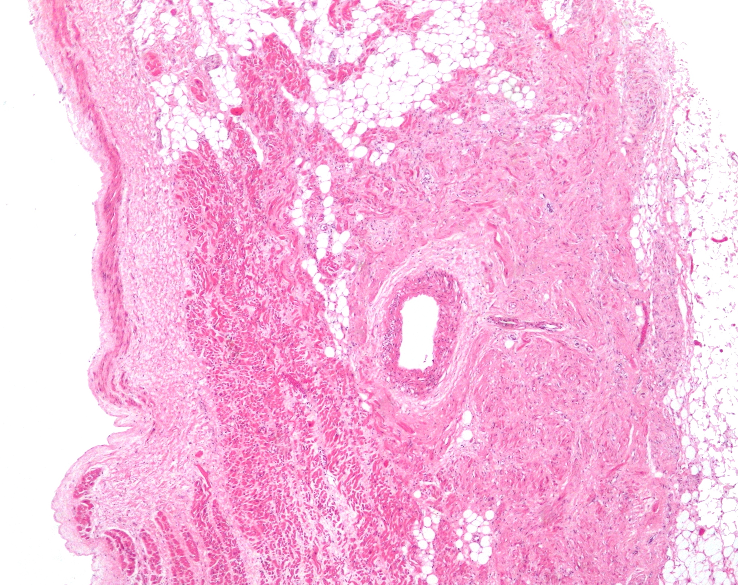
The interventricular septum (IVS, or ventricular septum, or during development septum inferius) is the stout wall separating the ventricle (heart), ventricles, the lower chambers of the heart, from one another. The interventricular septum is directed obliquely backward to the right and curved with the convexity toward the right ventricle; its margins correspond with the anterior interventricular sulcus, anterior and Posterior interventricular sulcus, posterior interventricular sulci. The lower part of the septum, which is the major part, is thick and muscular, and its much smaller upper part is thin and membraneous. During each cardiac cycle the interventricular septum contracts by shortening longitudinally and becoming thicker. Structure The interventricular septum is the stout wall separating the Ventricle (heart), ventricles, the lower chambers of the heart, from one another. The ventricular septum is directed obliquely backward to the right and curved with the convexity t ... [...More Info...] [...Related Items...] OR: [Wikipedia] [Google] [Baidu] |
Coronary Artery Disease
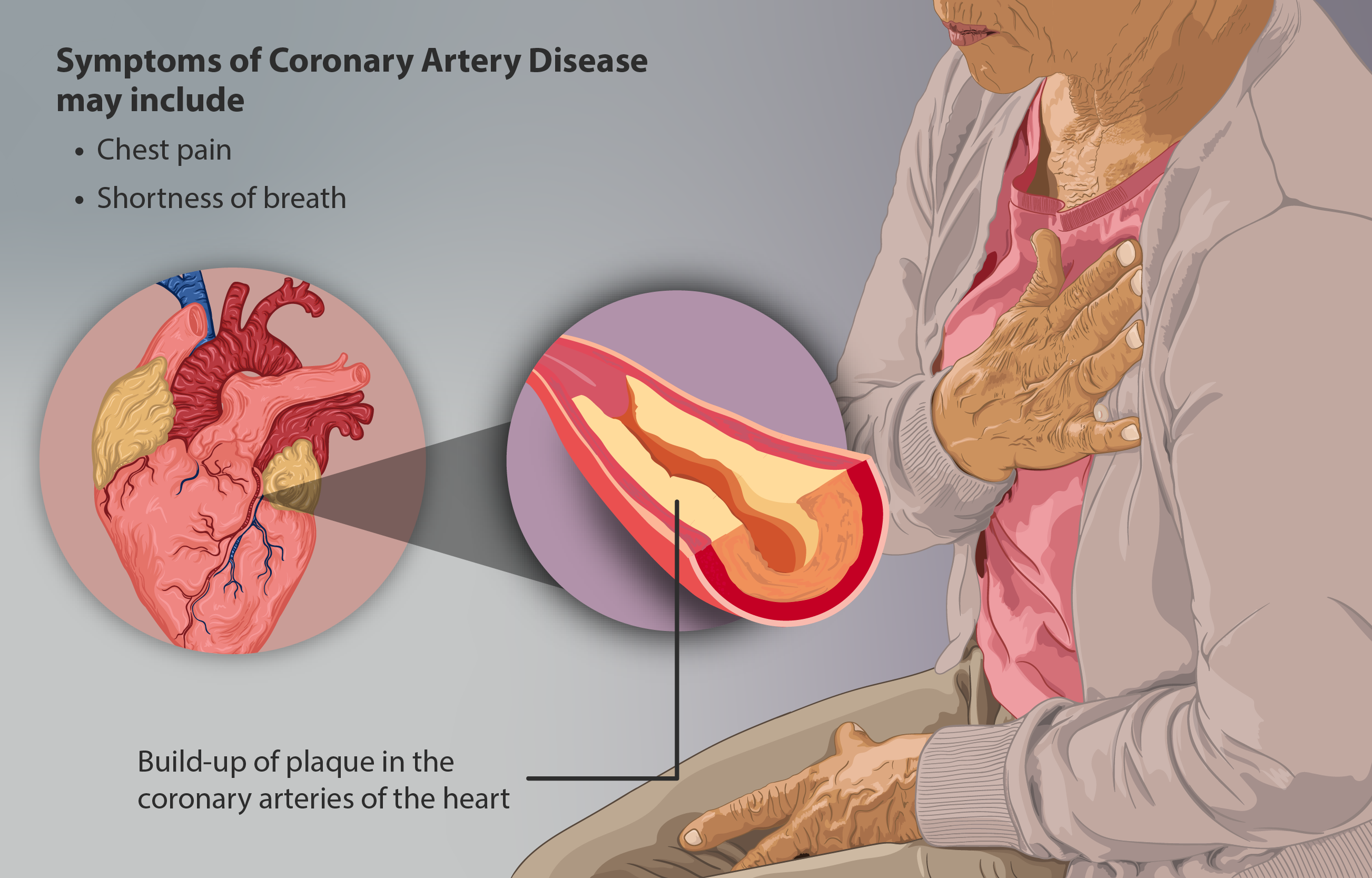
Coronary artery disease (CAD), also called coronary heart disease (CHD), or ischemic heart disease (IHD), is a type of cardiovascular disease, heart disease involving Ischemia, the reduction of blood flow to the cardiac muscle due to a build-up of atheromatous plaque in the Coronary arteries, arteries of the heart. It is the most common of the cardiovascular diseases. CAD can cause stable angina, unstable angina, myocardial ischemia, and myocardial infarction. A common symptom is angina, which is chest pain or discomfort that may travel into the shoulder, arm, back, neck, or jaw. Occasionally it may feel like heartburn. In stable angina, symptoms occur with exercise or emotional Psychological stress, stress, last less than a few minutes, and improve with rest. Shortness of breath may also occur and sometimes no symptoms are present. In many cases, the first sign is a Myocardial infarction, heart attack. Other complications include heart failure or an Heart arrhythmia, abnormal h ... [...More Info...] [...Related Items...] OR: [Wikipedia] [Google] [Baidu] |
Sinoatrial Node
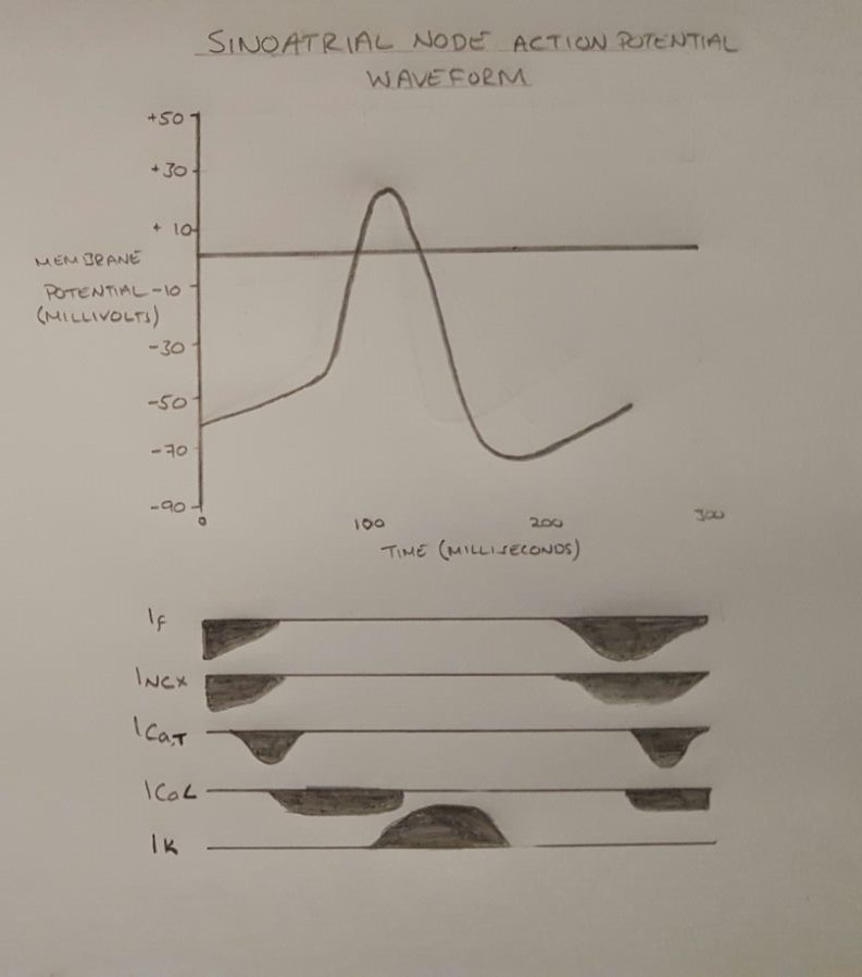
The sinoatrial node (also known as the sinuatrial node, SA node, sinus node or Keith–Flack node) is an ellipse, oval shaped region of special cardiac muscle in the upper back wall of the right atrium made up of Cell (biology), cells known as pacemaker cells. The sinus node is approximately 15 millimetre, mm long, 3 mm wide, and 1 mm thick, located directly below and to the side of the superior vena cava. These cells produce an Action potential, electrical impulse known as a cardiac action potential that travels through the electrical conduction system of the heart, causing it to muscle contraction, contract. In a healthy heart, the SA node continuously produces action potentials, setting the rhythm of the heart (sinus rhythm), and so is known as the heart's cardiac pacemaker, natural pacemaker. The rate of action potentials produced (and therefore the heart rate) is influenced by the nerves that supply it. Structure The sinoatrial node is an Ellipse, oval-shaped structure that ... [...More Info...] [...Related Items...] OR: [Wikipedia] [Google] [Baidu] |
Atrioventricular Node
The atrioventricular node (AV node, or Aschoff-Tawara node) electrically connects the heart's atria and ventricles to coordinate beating in the top of the heart; it is part of the electrical conduction system of the heart. The AV node lies at the lower back section of the interatrial septum near the opening of the coronary sinus, and conducts the normal electrical impulse from the atria to the ventricles. The AV node is quite compact (~1 x 3 x 5 mm).Full Size Picture triangle of-Koch.jpg Retrieved on 2008-12-22 Structure Location The AV node lies at the lower back section of the i ...[...More Info...] [...Related Items...] OR: [Wikipedia] [Google] [Baidu] |
Sinoatrial Node
The sinoatrial node (also known as the sinuatrial node, SA node, sinus node or Keith–Flack node) is an ellipse, oval shaped region of special cardiac muscle in the upper back wall of the right atrium made up of Cell (biology), cells known as pacemaker cells. The sinus node is approximately 15 millimetre, mm long, 3 mm wide, and 1 mm thick, located directly below and to the side of the superior vena cava. These cells produce an Action potential, electrical impulse known as a cardiac action potential that travels through the electrical conduction system of the heart, causing it to muscle contraction, contract. In a healthy heart, the SA node continuously produces action potentials, setting the rhythm of the heart (sinus rhythm), and so is known as the heart's cardiac pacemaker, natural pacemaker. The rate of action potentials produced (and therefore the heart rate) is influenced by the nerves that supply it. Structure The sinoatrial node is an Ellipse, oval-shaped structure that ... [...More Info...] [...Related Items...] OR: [Wikipedia] [Google] [Baidu] |
Electrical Conduction System Of The Heart
The cardiac conduction system (CCS, also called the electrical conduction system of the heart) transmits the Cardiac action potential, signals generated by the sinoatrial node – the heart's Cardiac pacemaker, pacemaker, to cause the heart muscle to Muscle contraction, contract, and pump blood through the body's circulatory system. The Cardiac pacemaker, pacemaking signal travels through the right atrium to the atrioventricular node, along the bundle of His, and through the bundle branches to Purkinje fibers in the Ventricle (heart), walls of the ventricles. The Purkinje fibers transmit the signals more rapidly to stimulate contraction of the ventricles. The conduction system consists of specialized Cardiomyocyte, heart muscle cells, situated within the myocardium. There is a cardiac skeleton, skeleton of fibrous tissue that surrounds the conduction system which can be seen on an ECG. Dysfunction of the conduction system can cause Heart arrhythmia, irregular heart rhythms includ ... [...More Info...] [...Related Items...] OR: [Wikipedia] [Google] [Baidu] |
Ventricular Remodeling
In cardiology, ventricular remodeling (or cardiac remodeling) refers to changes in the size, shape, structure, and function of the heart. This can happen as a result of exercise (physiological remodeling) or after injury to the heart muscle (pathological remodeling). The injury is typically due to acute myocardial infarction (usually transmural or ST segment elevation infarction), but may be from a number of causes that result in increased pressure or volume, causing pressure overload or volume overload (forms of strain) on the heart. Chronic hypertension, congenital heart disease with intracardiac shunting, and valvular heart disease may also lead to remodeling. After the insult occurs, a series of histopathological and structural changes occur in the left ventricular myocardium that lead to progressive decline in left ventricular performance. Ultimately, ventricular remodeling may result in diminished contractile ( systolic) function and reduced stroke volume. Physiologica ... [...More Info...] [...Related Items...] OR: [Wikipedia] [Google] [Baidu] |
Hypertension
Hypertension, also known as high blood pressure, is a Chronic condition, long-term Disease, medical condition in which the blood pressure in the artery, arteries is persistently elevated. High blood pressure usually does not cause symptoms itself. It is, however, a major risk factor for stroke, coronary artery disease, heart failure, atrial fibrillation, peripheral arterial disease, vision loss, chronic kidney disease, and dementia. Hypertension is a major cause of premature death worldwide. High blood pressure is classified as essential hypertension, primary (essential) hypertension or secondary hypertension. About 90–95% of cases are primary, defined as high blood pressure due to non-specific lifestyle and Genetics, genetic factors. Lifestyle factors that increase the risk include excess salt in the diet, overweight, excess body weight, smoking, physical inactivity and Alcohol (drug), alcohol use. The remaining 5–10% of cases are categorized as secondary hypertension, d ... [...More Info...] [...Related Items...] OR: [Wikipedia] [Google] [Baidu] |
Cardiomyopathy
Cardiomyopathy is a group of primary diseases of the heart muscle. Early on there may be few or no symptoms. As the disease worsens, shortness of breath, feeling tired, and swelling of the legs may occur, due to the onset of heart failure. An irregular heart beat and fainting may occur. Those affected are at an increased risk of sudden cardiac death. As of 2013, cardiomyopathies are defined as "disorders characterized by morphologically and functionally abnormal myocardium in the absence of any other disease that is sufficient, by itself, to cause the observed phenotype." Types of cardiomyopathy include hypertrophic cardiomyopathy, dilated cardiomyopathy, restrictive cardiomyopathy, arrhythmogenic right ventricular dysplasia, and Takotsubo cardiomyopathy (broken heart syndrome). In hypertrophic cardiomyopathy the heart muscle enlarges and thickens. In dilated cardiomyopathy the ventricles enlarge and weaken. In restrictive cardiomyopathy the ventricle stiffens. In ... [...More Info...] [...Related Items...] OR: [Wikipedia] [Google] [Baidu] |
Myocarditis
Myocarditis is inflammation of the cardiac muscle. Myocarditis can progress to inflammatory cardiomyopathy when there is associated ventricular remodeling and cardiac dysfunction due to chronic inflammation. Symptoms can include shortness of breath, chest pain, Exercise intolerance, decreased ability to exercise, and an irregular heartbeat. The duration of problems can vary from hours to months. Complications may include heart failure, due to dilated cardiomyopathy or cardiac arrest. Myocarditis is most often due to a viral infection. Other causes include bacterial infections, certain medications, toxins and autoimmune disorders. A diagnosis may be supported by an electrocardiogram (ECG), increased troponin, cardiac magnetic resonance imaging, heart MRI, and occasionally a heart biopsy. An echocardiogram, ultrasound of the heart is important to rule out other potential causes, such as valvular heart disease, heart valve problems. Treatment depends on both the severity and the c ... [...More Info...] [...Related Items...] OR: [Wikipedia] [Google] [Baidu] |
Rheumatic Heart Disease
Valvular heart disease is any cardiovascular disease process involving one or more of the four valves of the heart (the aortic and mitral valves on the left side of heart and the pulmonic and tricuspid valves on the right side of heart). These conditions occur largely as a consequence of aging,Burden of valvular heart diseases: a population-based study. Nkomo VT, Gardin JM, Skelton TN, Gottdiener JS, Scott CG, Enriquez-Sarano. Lancet. 2006 Sep;368(9540):1005-11. but may also be the result of congenital (inborn) abnormalities or specific disease or physiologic processes including rheumatic heart disease and pregnancy. Anatomically, the valves are part of the dense connective tissue of the heart known as the cardiac skeleton and are responsible for the regulation of blood flow through the heart and great vessels. Valve failure or dysfunction can result in diminished heart functionality, though the particular consequences are dependent on the type and severity of valvular dise ... [...More Info...] [...Related Items...] OR: [Wikipedia] [Google] [Baidu] |


