|
Remyelination
Remyelination is the process of propagating oligodendrocyte precursor cells to form oligodendrocytes to create new myelin sheaths on demyelinated axons in the Central nervous system (CNS). This is a process naturally regulated in the body and tends to be very efficient in a healthy CNS. The process creates a thinner myelin sheath than normal, but it helps to protect the axon from further damage, from overall degeneration, and proves to increase conductance once again. The processes underlying remyelination are under investigation in the hope of finding treatments for demyelinating diseases, such as multiple sclerosis. As of 2022 the status of possible remyelination acceleration is of trials only, with side effects of possible drugs one limiting issue. Function Remyelination is activated and regulated by a variety of factors surrounding lesion sites that control the migration and differentiation of Oligodendrocyte Precursor Cells. Remyelination looks different from developmental ... [...More Info...] [...Related Items...] OR: [Wikipedia] [Google] [Baidu] |
Oligodendrocyte Progenitor
Oligodendrocyte progenitor cells (OPCs), also known as oligodendrocyte precursor cells, NG2-glia, O2A cells, or polydendrocytes, are a subtype of glia in the central nervous system named for their essential role as precursors to oligodendrocytes and myelin. They are typically identified in the human by co-expression of PDGFRA and CSPG4. OPCs play a critical role in developmental and adult myelinogenesis. They give rise to oligodendrocytes, which then wrap around axons and provide electrical insulation by forming a myelin sheath. This enables faster action potential propagation and high fidelity transmission without a need for an increase in axonal diameter. The loss or lack of OPCs, and consequent lack of differentiated oligodendrocytes, is associated with a loss of myelination and subsequent impairment of neurological functions. In addition, OPCs express receptors for various neurotransmitters and undergo membrane depolarization when they receive synaptic inputs from neurons. ... [...More Info...] [...Related Items...] OR: [Wikipedia] [Google] [Baidu] |
Demyelinating Disease
A demyelinating disease refers to any disease affecting the nervous system where the myelin sheath surrounding neurons is damaged. This damage disrupts the transmission of signals through the affected nerves, resulting in a decrease in their conduction ability. Consequently, this reduction in conduction can lead to deficiencies in sensation, movement, cognition, or other functions depending on the nerves affected. Various factors can contribute to the development of demyelinating diseases, including genetic predisposition, infectious agents, autoimmune reactions, and other unknown factors. Proposed causes of demyelination include genetic predisposition, environmental factors such as viral infections or exposure to certain chemicals. Additionally, exposure to commercial insecticides like sheep dip, weed killers, and flea treatment preparations for pets, which contain organophosphates, can also lead to nerve demyelination. Chronic exposure to neuroleptic medications may also ... [...More Info...] [...Related Items...] OR: [Wikipedia] [Google] [Baidu] |
Multiple Sclerosis
Multiple sclerosis (MS) is an autoimmune disease resulting in damage to myelinthe insulating covers of nerve cellsin the brain and spinal cord. As a demyelinating disease, MS disrupts the nervous system's ability to Action potential, transmit signals, resulting in a range of signs and symptoms, including physical, cognitive disability, mental, and sometimes psychiatric problems. Symptoms include double vision, vision loss, eye pain, muscle weakness, and loss of Sensation (psychology), sensation or coordination. MS takes several forms, with new symptoms either occurring in isolated attacks (relapsing forms) or building up over time (progressive forms). In relapsing forms of MS, symptoms may disappear completely between attacks, although some permanent neurological problems often remain, especially as the disease advances. In progressive forms of MS, bodily function slowly deteriorates once symptoms manifest and will steadily worsen if left untreated. While its cause is unclear, ... [...More Info...] [...Related Items...] OR: [Wikipedia] [Google] [Baidu] |
Myelin
Myelin Sheath ( ) is a lipid-rich material that in most vertebrates surrounds the axons of neurons to insulate them and increase the rate at which electrical impulses (called action potentials) pass along the axon. The myelinated axon can be likened to an electrical wire (the axon) with insulating material (myelin) around it. However, unlike the plastic covering on an electrical wire, myelin does not form a single long sheath over the entire length of the axon. Myelin ensheaths part of an axon known as an internodal segment, in multiple myelin layers of a tightly regulated internodal length. The ensheathed segments are separated at regular short unmyelinated intervals, called nodes of Ranvier. Each node of Ranvier is around one micrometre long. Nodes of Ranvier enable a much faster rate of conduction known as saltatory conduction where the action potential recharges at each node to jump over to the next node, and so on till it reaches the axon terminal. At the terminal the ... [...More Info...] [...Related Items...] OR: [Wikipedia] [Google] [Baidu] |
LINGO1
Leucine-rich repeat and Immunoglobulin-like domain-containing protein 1 also known as LINGO-1 is a protein which is encoded by the ''LINGO1'' gene in humans. It belongs to the family of leucine-rich repeat proteins which are known for playing key roles in the biology of the central nervous system. LINGO-1 is a functional component of the Nogo (neurite outgrowth inhibitor) receptor also known as the reticulon 4 receptor. It has been suggested that LINGO-1 antagonists such as BIIB033 could significantly improve and regulate survival after neural injury caused by the protein. Structure The human LINGO-1 is a single-pass type 1 transmembrane protein of 614 amino acids. It contains a signal sequence of 34 residues, followed by a LRR (leucine-rich repeat) domain, an Ig (immunoglobulin-like) domain, a stalk domain, a transmembrane region and a short cytoplasmic tail. As a transmembrane protein, it can mostly be found on the cell membrane. The LINGO-1 structure has been shown to be ... [...More Info...] [...Related Items...] OR: [Wikipedia] [Google] [Baidu] |
Oligodendrocyte
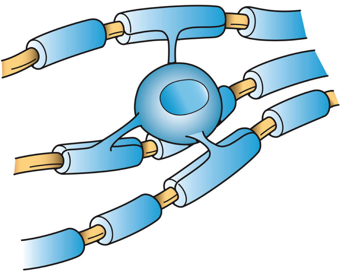
Oligodendrocytes (), also known as oligodendroglia, are a type of neuroglia whose main function is to provide the myelin sheath to neuronal axons in the central nervous system (CNS). Myelination gives metabolic support to, and insulates the axons of most vertebrates. A single oligodendrocyte can extend its Cellular extensions, processes to cover up to 40 axons, that can include multiple adjacent axons. The myelin sheath is segmented along the axon's length at gaps known as the nodes of Ranvier. In the peripheral nervous system the myelination of axons is carried out by Schwann cells. Oligodendrocytes are found exclusively in the CNS, which comprises the brain and spinal cord. They are the most widespread cell lineage, including oligodendrocyte progenitor cells, pre-myelinating cells, and mature myelinating oligodendrocytes in the CNS white matter. Non-myelinating oligodendrocytes are found in the grey matter surrounding and lying next to neuronal cell bodies. They are known as neu ... [...More Info...] [...Related Items...] OR: [Wikipedia] [Google] [Baidu] |
Schwann Cell
Schwann cells or neurolemmocytes (named after German physiologist Theodor Schwann) are the principal glia of the peripheral nervous system (PNS). Glial cells function to support neurons and in the PNS, also include Satellite glial cell, satellite cells, olfactory ensheathing cells, enteric glia and glia that reside at sensory nerve endings, such as the Pacinian corpuscle. The two types of Schwann cells are Myelin, myelinating and Nonmyelinating Schwann cell, nonmyelinating. Myelinating Schwann cells wrap around axons of motor and sensory neurons to form the myelin sheath. The Schwann cell promoter is present in the Upstream and downstream (DNA), downstream region of the human dystrophin gene that gives shortened Transcription (biology), transcript that are again synthesized in a tissue-specific manner. During the development of the PNS, the regulatory mechanisms of myelination are controlled by feedforward interaction of specific genes, influencing transcriptional cascades and sh ... [...More Info...] [...Related Items...] OR: [Wikipedia] [Google] [Baidu] |
SEMA3A
Semaphorin-3A is a protein that in humans is encoded by the ''SEMA3A'' gene. Function The ''SEMA3A'' gene is a member of the semaphorin family and encodes a protein with an Ig-like C2-type (immunoglobulin-like) domain, a PSI domain and a Sema domain. This secreted Semaphorin-3A protein can function as either a chemorepulsive agent, inhibiting axonal outgrowth, or as a chemoattractive agent, stimulating the growth of apical dendrites. In both cases, the protein is vital for normal neuronal pattern development. Semaphorin-3A is secreted by neurons and surrounding tissue to guide migrating cells and axons in the developing nervous system. Axon pathfinding is the process by which neurons follow very precise paths, send out axons, and react to specific chemical environments to reach the correct endpoint. The guidance is critical for the precise formation of neurons and the surrounding vasculature. Guidance cues, such as Sema3A, induce the collapse and paralysis of neuronal gr ... [...More Info...] [...Related Items...] OR: [Wikipedia] [Google] [Baidu] |
TCF4
Transcription factor 4 (TCF-4) also known as immunoglobulin transcription factor 2 (ITF-2) is a protein that in humans is encoded by the TCF4 gene located on chromosome 18q21.2. Function TCF4 proteins act as transcription factors which will bind to the immunoglobulin enhancer mu-E5/kappa-E2 motif. TCF4 activates transcription by binding to the E-box (5’-CANNTG-3’) found usually on SSTR2-INR, or somatostatin receptor 2 initiator element. TCF4 is primarily involved in neurological development of the fetus during pregnancy by initiating neural differentiation by binding to DNA. It is found in the central nervous system, somites, and gonadal ridge during early development. Later in development it will be found in the thyroid, thymus, and kidneys while in adulthood TCF4 it is found in lymphocytes, muscles, mature neurons, and gastrointestinal system. Clinical significance Mutations in TCF4 cause Pitt-Hopkins Syndrome (PTHS). These mutations cause TCF4 proteins to not bi ... [...More Info...] [...Related Items...] OR: [Wikipedia] [Google] [Baidu] |
Growth Factor
A growth factor is a naturally occurring substance capable of stimulating cell proliferation, wound healing, and occasionally cellular differentiation. Usually it is a secreted protein or a steroid hormone. Growth factors are important for regulating a variety of cellular processes. Growth factors typically act as signaling molecules between cells. Examples are cytokines and hormones that bind to specific receptors on the surface of their target cells. They often promote cell differentiation and maturation, which varies between growth factors. For example, epidermal growth factor (EGF) enhances osteogenic differentiation ( osteogenesis or bone formation), while fibroblast growth factors and vascular endothelial growth factors stimulate blood vessel differentiation ( angiogenesis). Comparison to cytokines ''Growth factor'' is sometimes used interchangeably among scientists with the term '' cytokine.'' Historically, cytokines were associated with hematopoietic (b ... [...More Info...] [...Related Items...] OR: [Wikipedia] [Google] [Baidu] |
Axon Guidance
Axon guidance (also called axon pathfinding) is a subfield of neural development concerning the process by which neurons send out axons to reach their correct targets. Axons often follow very precise paths in the nervous system, and how they manage to find their way so accurately is an area of ongoing research. Axon growth takes place from a region called the growth cone and reaching the axon target is accomplished with relatively few guidance molecules. Growth cone receptors respond to the guidance cues. Mechanisms Growing axons have a highly motile structure at the growing tip called the growth cone, which responds to signals in the extracellular environment that instruct the axon in which direction to grow. These signals, called guidance cues, can be fixed in place or diffusible; they can attract or repel axons. Growth cones contain receptors that recognize these guidance cues and interpret the signal into a chemotropic response. The general theoretical framework is that wh ... [...More Info...] [...Related Items...] OR: [Wikipedia] [Google] [Baidu] |


