|
Proprioceptor
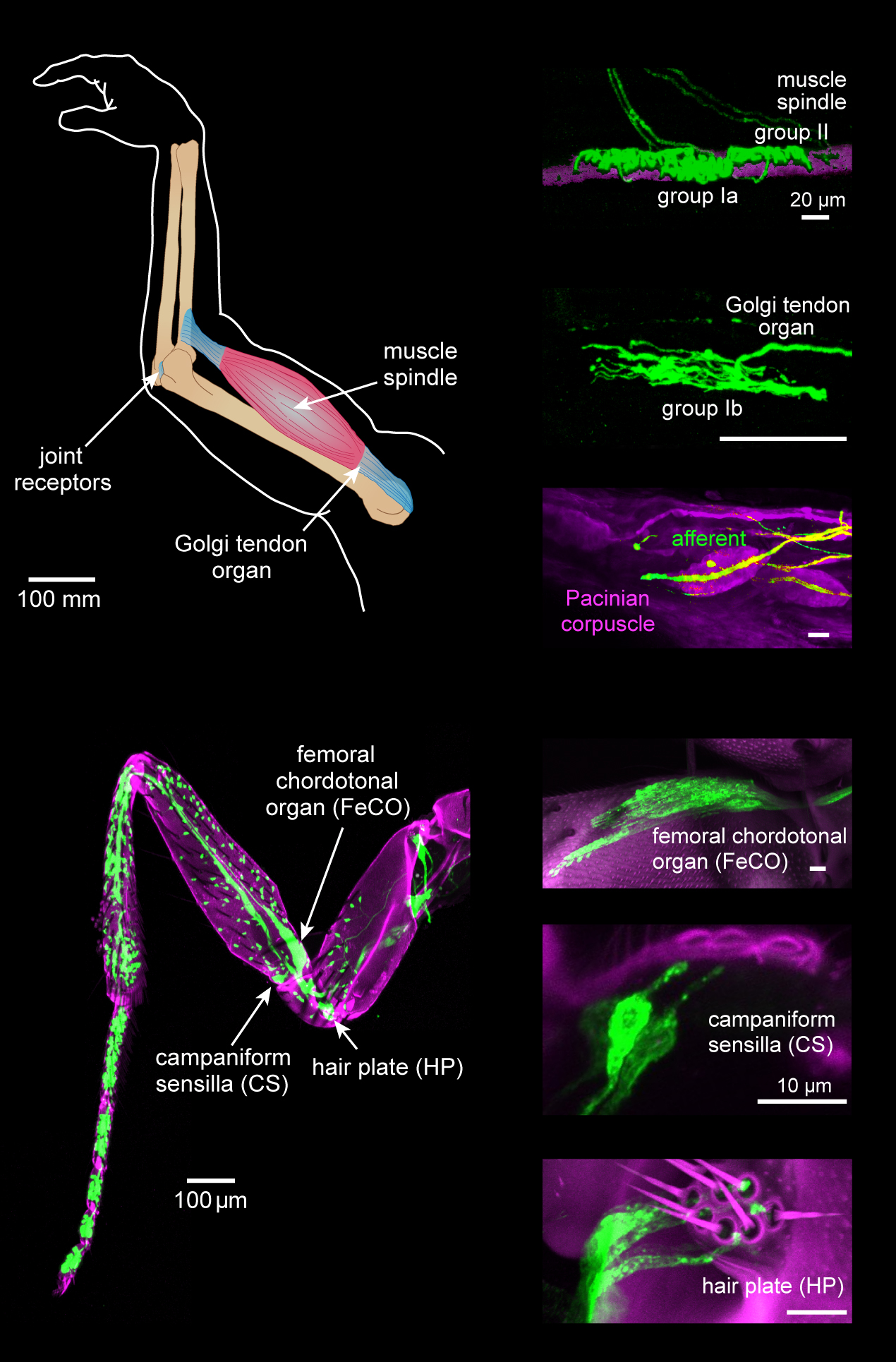
Proprioception ( ), also referred to as kinaesthesia (or kinesthesia), is the sense of self-movement, force, and body position. It is sometimes described as the "sixth sense". Proprioception is mediated by proprioceptors, mechanosensory neurons located within muscles, tendons, and joints. Most animals possess multiple subtypes of proprioceptors, which detect distinct kinematic parameters, such as joint position, movement, and load. Although all mobile animals possess proprioceptors, the structure of the sensory organs can vary across species. Proprioceptive signals are transmitted to the central nervous system, where they are integrated with information from other sensory systems, such as the visual system and the vestibular system, to create an overall representation of body position, movement, and acceleration. In many animals, sensory feedback from proprioceptors is essential for stabilizing body posture and coordinating body movement. System overview In vertebrates, limb ve ... [...More Info...] [...Related Items...] OR: [Wikipedia] [Google] [Baidu] |
Proprioception Image-01
Proprioception ( ), also referred to as kinaesthesia (or kinesthesia), is the sense of self-movement, force, and body position. It is sometimes described as the "sixth sense". Proprioception is mediated by proprioceptors, mechanosensory neurons located within muscles, tendons, and joints. Most animals possess multiple subtypes of proprioceptors, which detect distinct kinematic parameters, such as joint position, movement, and load. Although all mobile animals possess proprioceptors, the structure of the sensory organs can vary across species. Proprioceptive signals are transmitted to the central nervous system, where they are integrated with information from other sensory systems, such as the visual system and the vestibular system, to create an overall representation of body position, movement, and acceleration. In many animals, sensory feedback from proprioceptors is essential for stabilizing body posture and coordinating body movement. System overview In vertebrates, limb ve ... [...More Info...] [...Related Items...] OR: [Wikipedia] [Google] [Baidu] |
Hair Plate
Hair-plates are a type of proprioceptor found in the folds of insect joints. They consist of a cluster of hairs, in which each hair is innervated by a single mechanosensory neuron. Functionally, hair-plates operate as "limit-detectors" by signaling the extreme ranges of motion of a joint. Structure and Neuron Anatomy Hair-plates typically consist of a cluster of individual sensory hairs, in which each hair is innervated by a single sensory neuron (Fig. 1 & 2). The number of sensory hairs can vary across hair-plates as well as the length of hairs within a hair-plate. Hair-plates are often positioned within folds of cuticle, so that hairs are deflected during joint movement. They are located on different body parts, including the legs, neck, and antennae. On the legs of insects, hair-plates are typically found at the proximal joints (i.e. thorax-coxa, coxa-trochanter, and trochanter-femur joints) and vary in number for the front, middle, and hind legs. Unlike the front legs, the nu ... [...More Info...] [...Related Items...] OR: [Wikipedia] [Google] [Baidu] |
Campaniform Sensilla
Campaniform sensilla are a class of mechanoreceptors found in insects, which respond to local stress and strain within the animal's cuticle. Campaniform sensilla function as proprioceptors that detect mechanical load as resistance to muscle contraction, similar to mammalian Golgi tendon organs. Sensory feedback from campaniform sensilla is integrated in the control of posture and locomotion. Structure Each campaniform sensillum consists of a flexible dome, which is embedded in a spongy socket within the cuticle and innervated by the dendrites of a single bipolar sensory neuron (see schematic cross-section). Campaniform sensilla are often oval-shaped with long axes of about 5-10 µm (see SEM). Campaniform sensilla are distributed across the body surface of many insects. The fruit fly ''Drosophila melanogaster'', for example, has over 680 sensilla. Campaniform sensilla are located in regions where stress is likely to be high, including on the legs, antennae, wings, and ha ... [...More Info...] [...Related Items...] OR: [Wikipedia] [Google] [Baidu] |
Type Ia Sensory Fiber
A type Ia sensory fiber, or a primary afferent fiber is a type of afferent nerve fiber. It is the sensory fiber of a stretch receptor called the muscle spindle found in muscles, which constantly monitors the rate at which a muscle stretch changes. The information carried by type Ia fibers contributes to the sense of proprioception. Function of muscle spindles For the body to keep moving properly and with finesse, the nervous system has to have a constant input of sensory data coming from areas such as the muscles and joints. In order to receive a continuous stream of sensory data, the body has developed special sensory receptors called proprioceptors. Muscle spindles are a type of proprioceptor, and they are found inside the muscle itself. They lie parallel with the contractile fibers. This gives them the ability to monitor muscle length with precision. Types of sensory fibers This change in length of the spindle is transduced (transformed into electric membrane potential ... [...More Info...] [...Related Items...] OR: [Wikipedia] [Google] [Baidu] |
Muscle
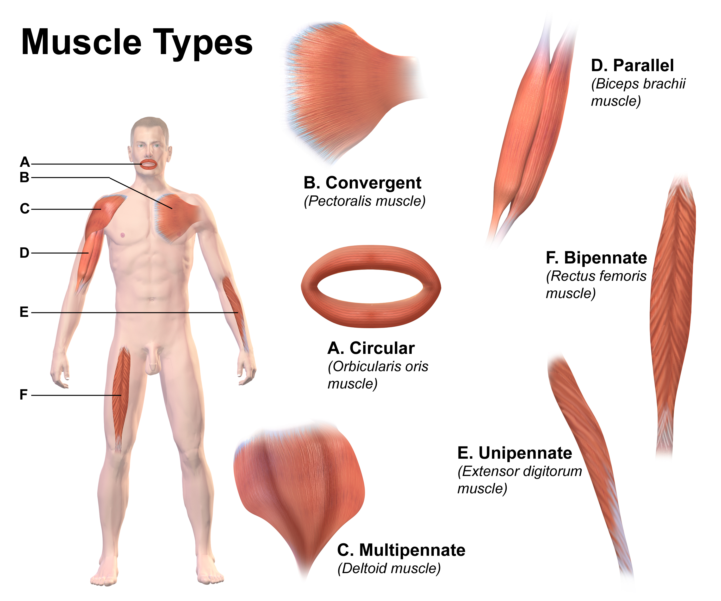
Skeletal muscles (commonly referred to as muscles) are organs of the vertebrate muscular system and typically are attached by tendons to bones of a skeleton. The muscle cells of skeletal muscles are much longer than in the other types of muscle tissue, and are often known as muscle fibers. The muscle tissue of a skeletal muscle is striated – having a striped appearance due to the arrangement of the sarcomeres. Skeletal muscles are voluntary muscles under the control of the somatic nervous system. The other types of muscle are cardiac muscle which is also striated and smooth muscle which is non-striated; both of these types of muscle tissue are classified as involuntary, or, under the control of the autonomic nervous system. A skeletal muscle contains multiple fascicles – bundles of muscle fibers. Each individual fiber, and each muscle is surrounded by a type of connective tissue layer of fascia. Muscle fibers are formed from the fusion of developmental myoblasts ... [...More Info...] [...Related Items...] OR: [Wikipedia] [Google] [Baidu] |
Golgi Tendon Organ
The Golgi tendon organ (GTO) (also called Golgi organ, tendon organ, neurotendinous organ or neurotendinous spindle) is a proprioceptor – a type of sensory receptor that senses changes in muscle tension. It lies at the interface between a muscle and its tendon known as the musculotendinous junction also known as the myotendinous junction. It provides the sensory component of the Golgi tendon reflex. The Golgi tendon organ is one of several eponymous terms named after the Italian physician Camillo Golgi. Structure The body of the Golgi tendon organ is made up of braided strands of collagen (intrafusal fasciculi) that are less compact than elsewhere in the tendon and are encapsulated. The capsule is connected in series (along a single path) with a group of muscle fibers () at one end, and merge into the tendon proper at the other. Each capsule is about long, has a diameter of about , and is perforated by one or more afferent type Ib sensory nerve fibers ( Aɑ fiber), whic ... [...More Info...] [...Related Items...] OR: [Wikipedia] [Google] [Baidu] |
Skeletal Muscle
Skeletal muscles (commonly referred to as muscles) are organs of the vertebrate muscular system and typically are attached by tendons to bones of a skeleton. The muscle cells of skeletal muscles are much longer than in the other types of muscle tissue, and are often known as muscle fibers. The muscle tissue of a skeletal muscle is striated – having a striped appearance due to the arrangement of the sarcomeres. Skeletal muscles are voluntary muscles under the control of the somatic nervous system. The other types of muscle are cardiac muscle which is also striated and smooth muscle which is non-striated; both of these types of muscle tissue are classified as involuntary, or, under the control of the autonomic nervous system. A skeletal muscle contains multiple fascicles – bundles of muscle fibers. Each individual fiber, and each muscle is surrounded by a type of connective tissue layer of fascia. Muscle fibers are formed from the fusion of developmental myobla ... [...More Info...] [...Related Items...] OR: [Wikipedia] [Google] [Baidu] |
Ventral Spinocerebellar Tract
The spinocerebellar tract is a nerve tract originating in the spinal cord and terminating in the same side (ipsilateral) of the cerebellum. Origins of proprioceptive information Proprioceptive information is obtained by Golgi tendon organs and muscle spindles. * Golgi tendon organs consist of a fibrous capsule enclosing tendon fascicles and bare nerve endings that respond to tension in the tendon by causing action potentials in type Ib afferents. These fibers are relatively large, myelinated, and quickly conducting. * Muscle spindles monitor the length within muscles and send information via faster Ia afferents. These axons are larger and faster than type Ib (from both nuclear bag fibers and nuclear chain fibers) and type II afferents (solely from nuclear chain fibers). All of these neurons are sensory (first order, or primary) and have their cell bodies in the dorsal root ganglia. They pass through Rexed laminae layers I-VI of the posterior grey column (dorsal horn) to ... [...More Info...] [...Related Items...] OR: [Wikipedia] [Google] [Baidu] |
Dorsal Spinocerebellar Tract
The spinocerebellar tract is a nerve tract originating in the spinal cord and terminating in the same side (ipsilateral) of the cerebellum. Origins of proprioceptive information Proprioceptive information is obtained by Golgi tendon organs and muscle spindles. * Golgi tendon organs consist of a fibrous capsule enclosing tendon fascicles and bare nerve endings that respond to tension in the tendon by causing action potentials in type Ib afferents. These fibers are relatively large, myelinated, and quickly conducting. * Muscle spindles monitor the length within muscles and send information via faster Ia afferents. These axons are larger and faster than type Ib (from both nuclear bag fibers and nuclear chain fibers) and type II afferents (solely from nuclear chain fibers). All of these neurons are sensory (first order, or primary) and have their cell bodies in the dorsal root ganglia. They pass through Rexed laminae layers I-VI of the posterior grey column (dorsal horn) t ... [...More Info...] [...Related Items...] OR: [Wikipedia] [Google] [Baidu] |
Stretch Reflex
The stretch reflex (myotatic reflex), or more accurately "muscle stretch reflex", is a muscle contraction in response to stretching within the muscle. The reflex functions to maintain the muscle at a constant length. The term deep tendon reflex is often used by many health workers and students to refer to this reflex. "Tendons have little to do with the response, other than being responsible for mechanically transmitting the sudden stretch from the reflex hammer to the muscle spindle. In addition, some muscles with stretch reflexes have no tendons (e.g., "jaw jerk" of the masseter muscle)". As an example of a spinal reflex, it results in a fast response that involves an afferent signal into the spinal cord and an efferent signal out to the muscle. The stretch reflex can be a monosynaptic reflex which provides automatic regulation of skeletal muscle length, whereby the signal entering the spinal cord arises from a change in muscle length or velocity. It can also include a polysyna ... [...More Info...] [...Related Items...] OR: [Wikipedia] [Google] [Baidu] |
Joint Capsule
In anatomy, a joint capsule or articular capsule is an envelope surrounding a synovial joint. Each joint capsule has two parts: an outer fibrous layer or membrane, and an inner synovial layer or membrane. Membranes Each capsule consists of two layers or membranes: * an outer (fibrous membrane, ''fibrous stratum'') composed of avascular white fibrous tissue * an inner ('''', ''synovial stratum'') which is a secreting layer On the inside of the capsule, articular cartilage covers the end surfaces of the bones that articulate within th ...[...More Info...] [...Related Items...] OR: [Wikipedia] [Google] [Baidu] |


